
All the branches and course of facial nerve mandibular nerve and maxillary nerve
Reading File
Now let me search the medical library for all three nerves simultaneously.
Finding Sources
Finding Sources
Finding Sources
Finding Sources
Finding Sources
Finding Sources
Excellent - found great sources in Gray's Anatomy for Students and Scott-Brown's. Let me read all three in parallel.
Reading File
Reading File
Reading File
Now let me read the detailed branches from Gray's Anatomy block 5 for both mandibular and maxillary, and also fetch the facial nerve section from Scott-Brown's:
Reading File
Reading File
Reading File
Now let me read more of the mandibular nerve section from Gray's and the facial nerve course from the facial nerve block:
Reading File
Reading File
Reading File
Let me get the facial nerve course from Cummings and the maxillary branches from Gray's block 5:
Reading File
Reading File
I now have comprehensive data from Gray's Anatomy for Students, Cummings Otolaryngology, and Scott-Brown's. Let me now fetch the key anatomical diagrams to include in the answer.
Loading Image
Loading Image
Loading Image
Excellent - I now have all the information and anatomical diagrams needed. Let me also get one more facial nerve diagram:
Finding Sources
Reading File
Reading File
I now have comprehensive information from multiple authoritative sources. Let me compile the complete answer.
Facial Nerve (CN VII), Maxillary Nerve (V2), and Mandibular Nerve (V3) - Branches and Course
FACIAL NERVE (CN VII)
Fiber Types
The facial nerve carries four fiber types (Gray's Anatomy for Students):
- GSA - general somatic afferent: sensation from part of the external acoustic meatus and deeper auricle
- SA - special afferent: taste from the anterior 2/3 of tongue
- GVE - general visceral efferent (parasympathetic): secretomotor to lacrimal gland, submandibular and sublingual glands, nasal and palatal glands
- BE - branchial efferent (motor): muscles of facial expression, stapedius, stylohyoid, posterior belly of digastric
Course
The facial nerve attaches to the lateral brainstem between the pons and medulla oblongata. It consists of:
- A large motor root (BE fibers)
- A smaller sensory root (the intermediate nerve / nervus intermedius) carrying taste (SA), parasympathetic (GVE), and somatic sensory (GSA) fibers
Intracranial course: Both roots cross the posterior cranial fossa and enter the internal acoustic meatus (IAC). Within the IAC the nerve lacks a fibrous sheath and is surrounded by thin arachnoid.
Intratemporal course within the fallopian (facial) canal (Cummings Otolaryngology):
| Segment | Key Features |
|---|---|
| Labyrinthine segment | First, shortest, narrowest - travels superior to cochlea; opens into geniculate fossa |
| Geniculate ganglion | Cell bodies of sensory neurons; gives off Greater Superficial Petrosal Nerve (GSPN) anteriorly; first genu here |
| Tympanic (horizontal) segment | Runs along medial wall of anterior attic; most common site of bony dehiscence |
| Second genu | At pyramidal eminence; turns inferior into mastoid segment |
| Mastoid (vertical) segment | Most variable path; gives off chorda tympani and branch to stapedius |
The nerve exits the skull through the stylomastoid foramen.
Branches
Before entering the parotid gland, immediately after exiting the stylomastoid foramen (Scott-Brown's):
- Posterior auricular nerve - supplies the occipital belly of occipitofrontalis
- Branch to posterior belly of digastric
- Branch to stylohyoid
Within the parotid gland - the main trunk divides into upper (zygomaticotemporal) and lower (cervicofacial) divisions, which further branch to form the "pes anserinus" (five terminal groups):
| Terminal Branch | Muscles Supplied | Area |
|---|---|---|
| Temporal | Frontalis, orbicularis oculi (superior), corrugator supercilii, auricular muscles | Emerges from upper border of parotid |
| Zygomatic | Orbicularis oculi (inferior), zygomaticus | Anterior border of parotid |
| Buccal | Buccinator, zygomaticus major/minor, levator labii superioris, nasalis | Anterior border of parotid |
| Marginal mandibular | Depressor anguli oris, depressor labii inferioris, mentalis | Lower border of parotid |
| Cervical | Platysma | Lower border of parotid |
Note: Cross-innervation (anastomosis) between branches is most pronounced between the zygomatic and buccal branches, and least for the marginal mandibular branch - which is why marginal mandibular nerve injury causes the most persistent deficits.
Intratemporal Branches (from within the fallopian canal)
- Greater superficial petrosal nerve (GSPN) - from geniculate ganglion; preganglionic parasympathetic to pterygopalatine ganglion → lacrimal gland and nasal/palatine glands; also carries taste from palate
- Nerve to stapedius - from mastoid segment
- Chorda tympani - from mastoid segment; carries taste (SA) from anterior 2/3 of tongue + preganglionic parasympathetic to submandibular ganglion → submandibular and sublingual glands
MAXILLARY NERVE (CN V2)
Nature
Purely sensory. The middle division of the trigeminal nerve.
Course
V2 passes in the dura of the lateral wall of the cavernous sinus, just inferior to V1. It exits the cranial cavity through the foramen rotundum and enters the pterygopalatine fossa. It then enters the inferior orbital fissure, passes along the orbital floor first in a groove then in a canal (infraorbital canal), and exits onto the face via the infraorbital foramen as the infraorbital nerve (Scott-Brown's Otorhinolaryngology).
Branches
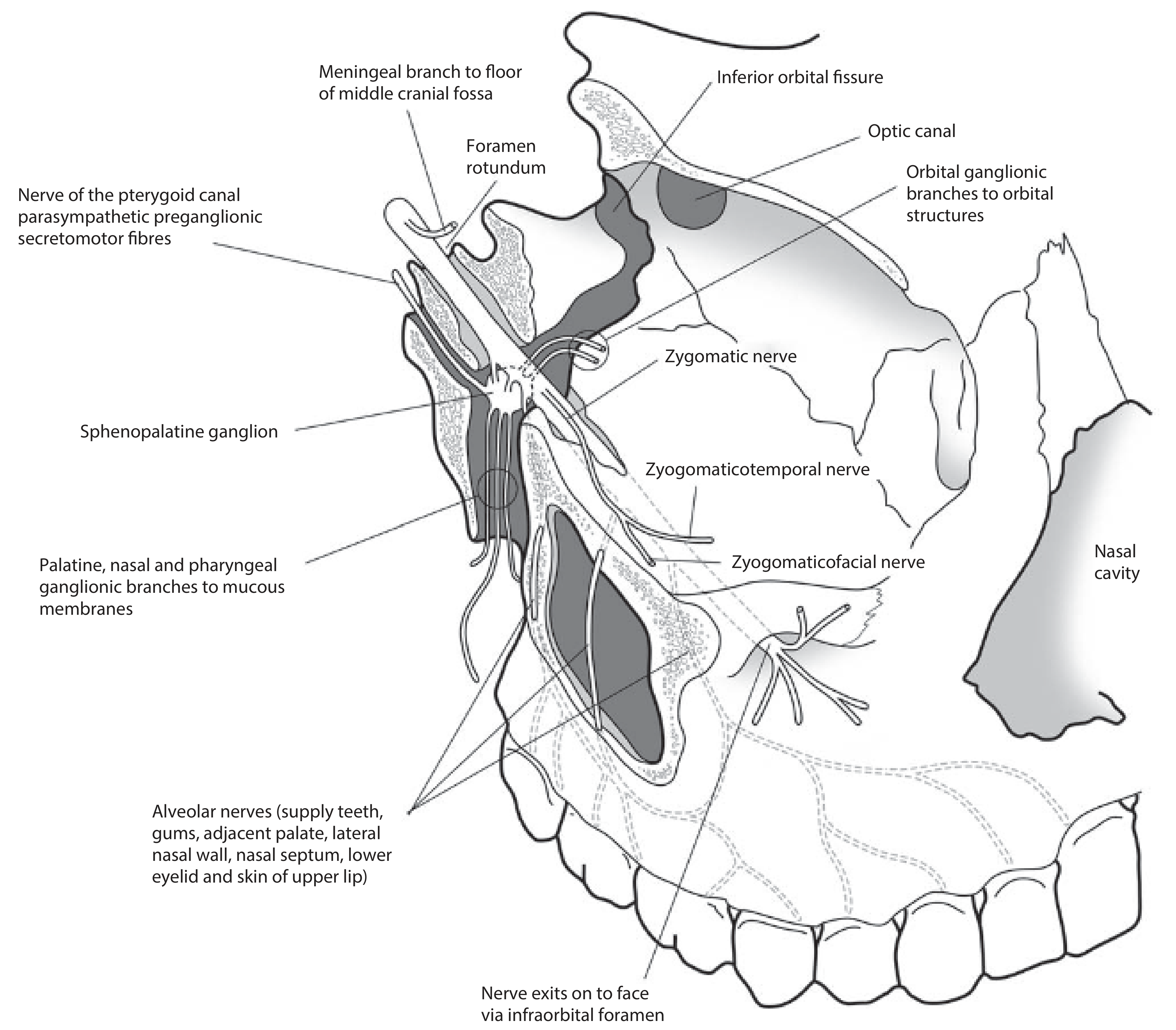
1. Meningeal branches - to the floor of the middle cranial fossa
2. Ganglionic (pterygopalatine) branches - two branches connecting to the pterygopalatine (sphenopalatine) ganglion in the pterygopalatine fossa. They carry postganglionic parasympathetic secretomotor fibers destined for the lacrimal gland. The ganglion output travels via:
- Orbital branches - to orbital structures
- Palatine nerves (greater and lesser) - to palate mucosa
- Nasal branches - to nasal cavity mucosa
- Pharyngeal branch - to nasopharynx
3. Zygomatic nerve - enters orbit via inferior orbital fissure, divides into:
- Zygomaticotemporal nerve - cutaneous sensation from temporal area; carries secretomotor fibers from pterygopalatine ganglion to the lacrimal gland (via a communicating branch to the lacrimal nerve)
- Zygomaticofacial nerve - penetrates the zygomatic bone; cutaneous sensation from the prominence of the cheek
4. Alveolar nerves (form the superior dental plexus):
- Posterior superior alveolar nerve - branches before infraorbital foramen; supplies the upper molar teeth and adjacent gingiva
- Middle superior alveolar nerve - supplies upper premolars
- Anterior superior alveolar nerve (largest) - branches from infraorbital nerve inside the canal; supplies incisors and canines, lateral nasal wall, nasal septum, lower eyelid, and upper lip
5. Infraorbital nerve (terminal branch) - exits via infraorbital foramen; supplies:
- Skin of the cheek, midlateral nose and lateral alar
- Lower eyelid
- Mucous membrane of cheek and upper lip
Sensory territory of V2 in summary: Dura of middle cranial fossa, nasopharynx, palate, nasal cavity, upper teeth and gingiva, maxillary sinus, skin of lower eyelid, side of nose, cheek, and upper lip (Gray's Anatomy for Students).
MANDIBULAR NERVE (CN V3)
Nature
Both motor and sensory - the only division of the trigeminal nerve with a motor component. It is also the largest division.
Course
The sensory root drops from the trigeminal ganglion through the foramen ovale into the infratemporal fossa, between tensor veli palatini and the upper head of lateral pterygoid. The small motor root passes medial to the trigeminal ganglion in the cranial cavity, then also passes through the foramen ovale and immediately joins the sensory root outside the skull (Gray's Anatomy for Students).
All branches originate in the infratemporal fossa.
Division and Branches
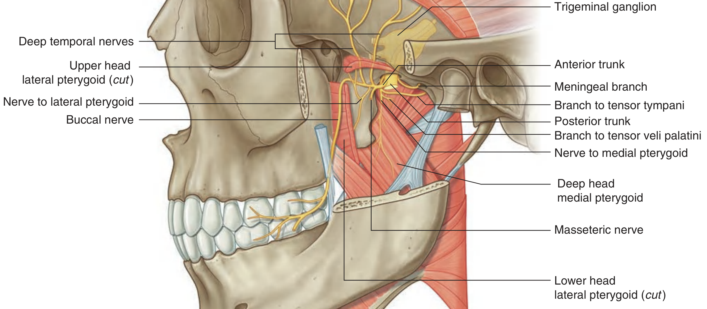
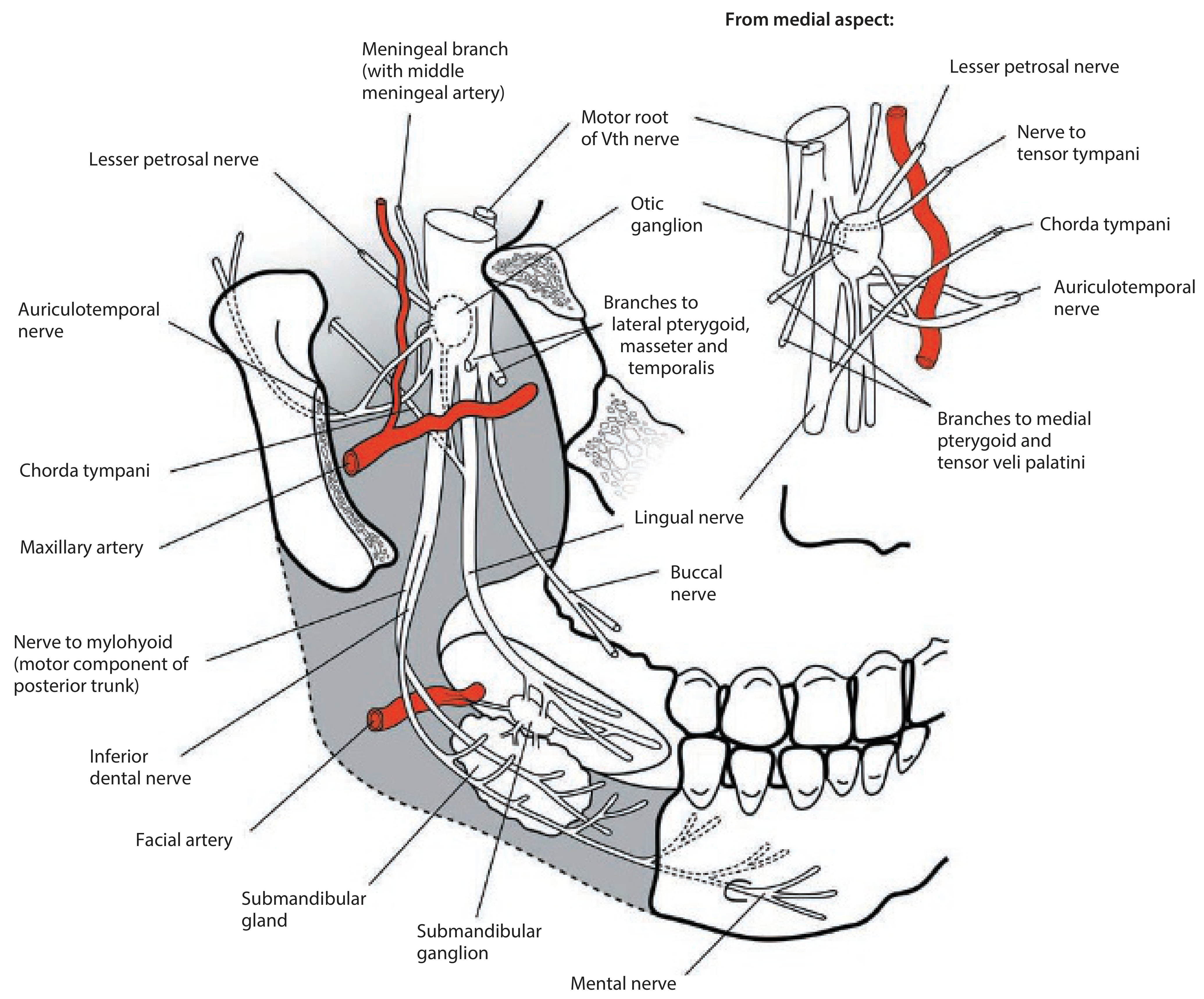
Branches before division (from main trunk):
-
Meningeal branch (nervus spinosus) - re-enters the skull with the middle meningeal artery through the foramen spinosum; sensory to dura of middle cranial fossa and mastoid air cells
-
Nerve to medial pterygoid - descends to supply medial pterygoid; gives two small branches:
- Branch to tensor veli palatini
- Branch to tensor tympani
V3 then divides into anterior and posterior trunks:
Anterior Trunk (predominantly motor, one sensory branch)
| Branch | Nature | Supply |
|---|---|---|
| Masseteric nerve | Motor | Passes through the mandibular notch → masseter muscle |
| Deep temporal nerves (anterior and posterior) | Motor | Pass over lateral pterygoid → temporalis muscle |
| Nerve to lateral pterygoid | Motor | Lateral pterygoid muscle |
| Buccal nerve | Predominantly sensory | Passes between upper and lower heads of lateral pterygoid, descends anterior to masseter → skin over buccinator, buccal mucosa, buccal gingivae of lower molars |
Posterior Trunk (predominantly sensory, one motor branch)
| Branch | Nature | Supply |
|---|---|---|
| Auriculotemporal nerve | Sensory + secretomotor | Passes behind the TMJ; supplies skin over tragus, helix, external auditory meatus, tympanic membrane, and temporal scalp via superficial temporal branches; carries secretomotor (postganglionic parasympathetic from otic ganglion via lesser petrosal nerve) to parotid gland |
| Lingual nerve | Sensory + carries chorda tympani | General sensation from presulcal tongue, floor of mouth, and lower gingiva; joined by chorda tympani (CN VII) in the infratemporal fossa, which adds taste from anterior 2/3 of tongue; attached to submandibular ganglion (which relays parasympathetic fibers to submandibular and sublingual glands) |
| Inferior alveolar nerve | Sensory (+ one motor branch) | Enters mandibular foramen → runs through mandibular canal → exits as mental nerve (at mental foramen) supplying lower lip and chin skin; gives the incisive nerve to incisors and canines inside the canal; branches to all lower teeth and gingivae; gives off nerve to mylohyoid (motor) just before entering the foramen → mylohyoid and anterior belly of digastric |
Motor Summary for V3
| Muscle | Branch |
|---|---|
| Temporalis | Deep temporal nerves (anterior trunk) |
| Masseter | Masseteric nerve (anterior trunk) |
| Lateral pterygoid | Nerve to lateral pterygoid (anterior trunk) |
| Medial pterygoid | Nerve to medial pterygoid (main trunk) |
| Tensor veli palatini | Via nerve to medial pterygoid |
| Tensor tympani | Via nerve to medial pterygoid |
| Mylohyoid | Nerve to mylohyoid (from inferior alveolar) |
| Anterior belly of digastric | Nerve to mylohyoid (from inferior alveolar) |
Sources
- Gray's Anatomy for Students - Mandibular nerve [V3], Maxillary nerve [V2], Facial nerve sections
- Scott-Brown's Otorhinolaryngology Head & Neck Surgery Vol 2 - The maxillary nerve (V2), The mandibular nerve (V3)
- Cummings Otolaryngology Head and Neck Surgery - Facial nerve anatomy and intratemporal course
- Scott-Brown's Otorhinolaryngology Vol 1 - Facial nerve intraparotid branching patterns
Explain me interior of skull in best way possible and all mention all the formens and structures passing through them
Finding Sources
Finding Sources
Finding Sources
Finding Sources
Reading File
Finding Sources
Reading File
Reading File
Reading File
Reading File
Now let me fetch the key anatomical diagrams - the three cranial fossa images from Gray's:
Loading Image
Loading Image
Loading Image
Excellent - all three cranial fossa diagrams are perfect. Now let me get the remaining detailed content on the middle cranial fossa foramina and the jugular foramen/hypoglossal canal:
Reading File
Reading File
There's a summary foramina diagram - let me fetch it:
Loading Image
I now have all the information and diagrams needed for a comprehensive, well-illustrated answer. Let me compile it.
Interior of the Skull - The Three Cranial Fossae and All Foramina
The interior of the skull base (endocranium) is divided into three stepped fossae - anterior, middle, and posterior - each lying at a progressively lower level. They support and protect the brain, and are riddled with foramina (openings) and fissures through which nerves, arteries, and veins pass.
ANTERIOR CRANIAL FOSSA
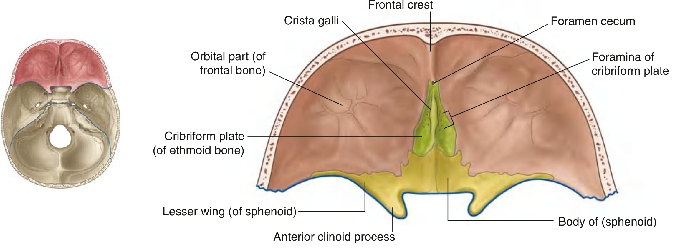
Bones Forming It
Formed by parts of three bones:
- Frontal bone - anteriorly and laterally (orbital plate forms both the ACF floor and roof of orbit)
- Ethmoid bone - midline (cribriform plate)
- Sphenoid bone - posteriorly: body (midline) and lesser wings (laterally)
Contents
Houses the frontal lobes of the cerebral hemispheres. Lies above the nasal cavity and orbits.
Key Midline Landmarks
- Frontal crest - small midline ridge; attachment for the falx cerebri
- Foramen cecum - just behind the frontal crest, between frontal and ethmoid bones; may transmit emissary veins (nasal cavity to superior sagittal sinus); in the fetus transmits the dural sinus
- Crista galli - prominent wedge from ethmoid bone; another attachment for the falx cerebri
- Cribriform plate - sieve-like structure on either side of crista galli
Foramina of the Anterior Cranial Fossa
| Foramen/Opening | Location | Structures Passing Through |
|---|---|---|
| Foramen cecum | Between frontal and ethmoid bones | Emissary vein (nasal cavity ↔ superior sagittal sinus) |
| Foramina of the cribriform plate | Lateral to crista galli, ethmoid bone | CN I - Olfactory nerve fibers (from nasal mucosa to olfactory bulb); anterior ethmoidal nerve and artery also pass through |
Boundary with Middle Fossa
The anterior edge of the prechiasmatic sulcus (a smooth groove crossing the sphenoid body between the two optic canals) marks the posterior boundary of the ACF in the midline. The free posterior edge of the lesser wings of the sphenoid forms the lateral posterior boundary, overhanging the MCF.
MIDDLE CRANIAL FOSSA
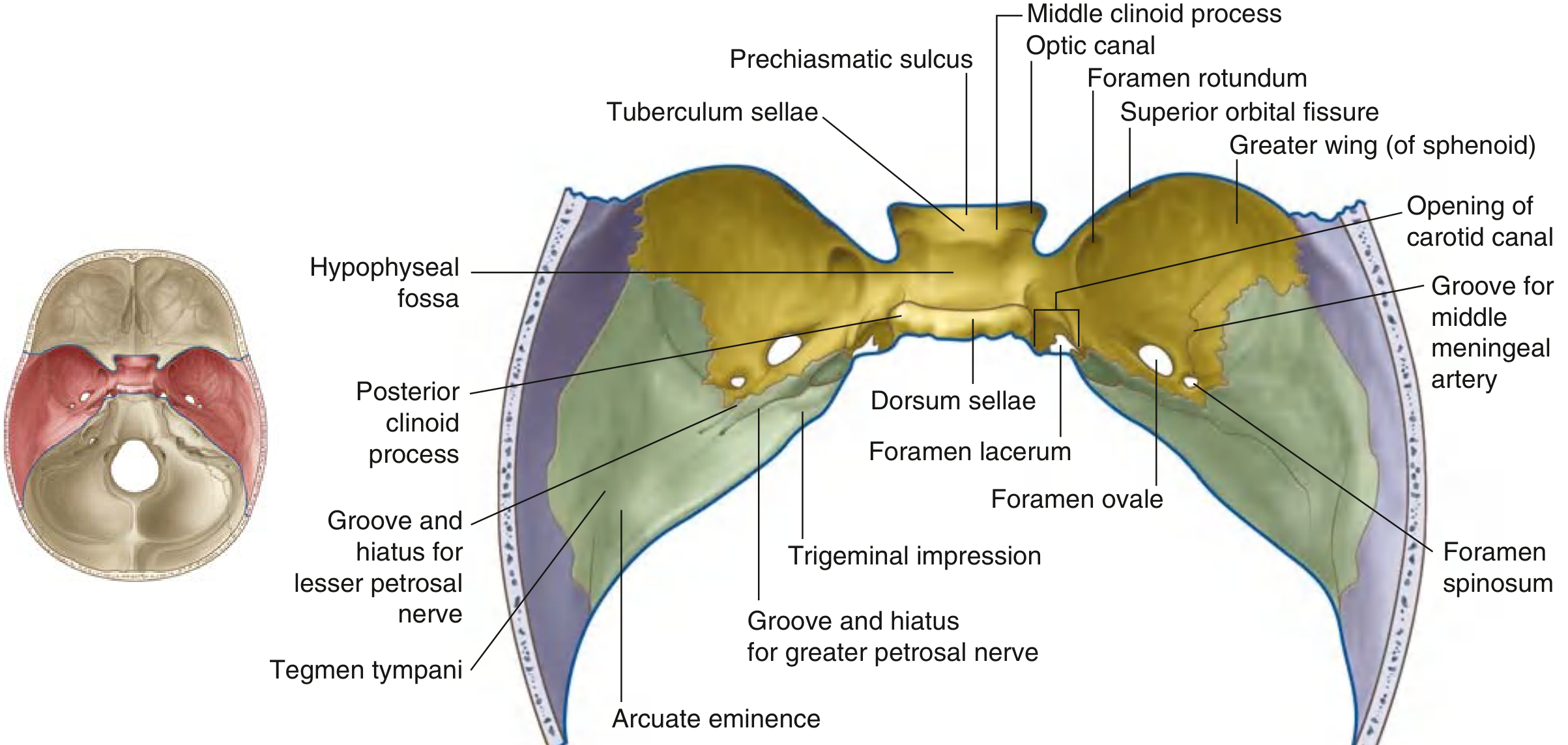
Bones Forming It
- Sphenoid bone - body (midline) and greater wings (lateral floors)
- Temporal bone - squamous and petrous parts (posterior part of lateral floor)
Contents
The central elevated midline region contains the pituitary gland. The large lateral depressions house the temporal lobes of the brain.
Key Midline Landmarks (Sella Turcica)
- Prechiasmatic sulcus - groove stretching between optic canals; accommodates the optic chiasma
- Tuberculum sellae - posterior edge of prechiasmatic sulcus; sometimes has middle clinoid processes projecting laterally
- Hypophyseal (pituitary) fossa - deep central depression; contains the pituitary gland
- Dorsum sellae - large posterior bony ridge projecting upward; its lateral upper corners are the posterior clinoid processes (attachment for tentorium cerebelli)
- Anterior clinoid processes - medial ends of lesser wings; attachment for tentorium cerebelli
Key Lateral Landmarks (Petrous Part of Temporal Bone)
- Trigeminal impression - shallow depression near apex of petrous bone; houses the trigeminal (Gasserian/semilunar) ganglion (in Meckel's cave)
- Groove and hiatus for greater petrosal nerve - small groove ending in a hiatus; transmits the GSPN (branch of CN VII from geniculate ganglion)
- Groove and hiatus for lesser petrosal nerve - smaller, anterolateral; transmits the lesser petrosal nerve (from tympanic plexus, CN IX fibers → otic ganglion)
- Arcuate eminence - rounded bony prominence caused by underlying anterior semicircular canal
- Tegmen tympani - thin bony plate anterior and lateral to arcuate eminence; roof of the middle ear cavity
- Opening of carotid canal - rounded opening posteromedial to foramen ovale; internal carotid artery enters the cranial cavity here
Foramina of the Middle Cranial Fossa
| Foramen/Opening | Bones | Communicates With | Structures Passing Through |
|---|---|---|---|
| Optic canal | Sphenoid (lesser wing) | MCF ↔ Orbit | CN II (Optic nerve) + Ophthalmic artery |
| Superior orbital fissure | Between greater and lesser wings of sphenoid | MCF ↔ Orbit | CN III (Oculomotor), CN IV (Trochlear), CN V1 (Ophthalmic), CN VI (Abducent), Superior ophthalmic vein |
| Foramen rotundum | Greater wing of sphenoid | MCF ↔ Pterygopalatine fossa | CN V2 (Maxillary nerve) |
| Foramen ovale | Greater wing of sphenoid | MCF ↔ Infratemporal fossa | CN V3 (Mandibular nerve), Lesser petrosal nerve, accessory meningeal artery, emissary vein |
| Foramen spinosum | Greater wing of sphenoid (posterolateral to F. ovale) | MCF ↔ Infratemporal fossa | Middle meningeal artery (and accompanying veins), meningeal branch of V3 (nervus spinosus) |
| Foramen lacerum | Junction of sphenoid, temporal, and occipital bones | MCF ↔ neck (theoretical) | Filled with fibrocartilage in life - no structures pass all the way through; the internal carotid artery traverses its superior opening; greater and deep petrosal nerves form the nerve of the pterygoid canal here |
| Carotid canal opening | Petrous temporal bone | MCF ↔ neck | Internal carotid artery (enters skull base, passes through carotid canal, exits superiorly into cranial cavity near foramen lacerum); sympathetic plexus |
| Hiatus for greater petrosal nerve | Anterior surface of petrous temporal | Within MCF | Greater petrosal nerve (branch of CN VII) exits to travel along floor of MCF |
| Hiatus for lesser petrosal nerve | Anterior surface of petrous temporal | Within MCF | Lesser petrosal nerve (from CN IX via tympanic plexus) exits |
The arrangement from anteromedial to posterolateral in the floor of the MCF is a clinically useful sequence to memorize: optic canal → superior orbital fissure → foramen rotundum → foramen ovale → foramen spinosum → foramen lacerum → carotid canal opening
POSTERIOR CRANIAL FOSSA
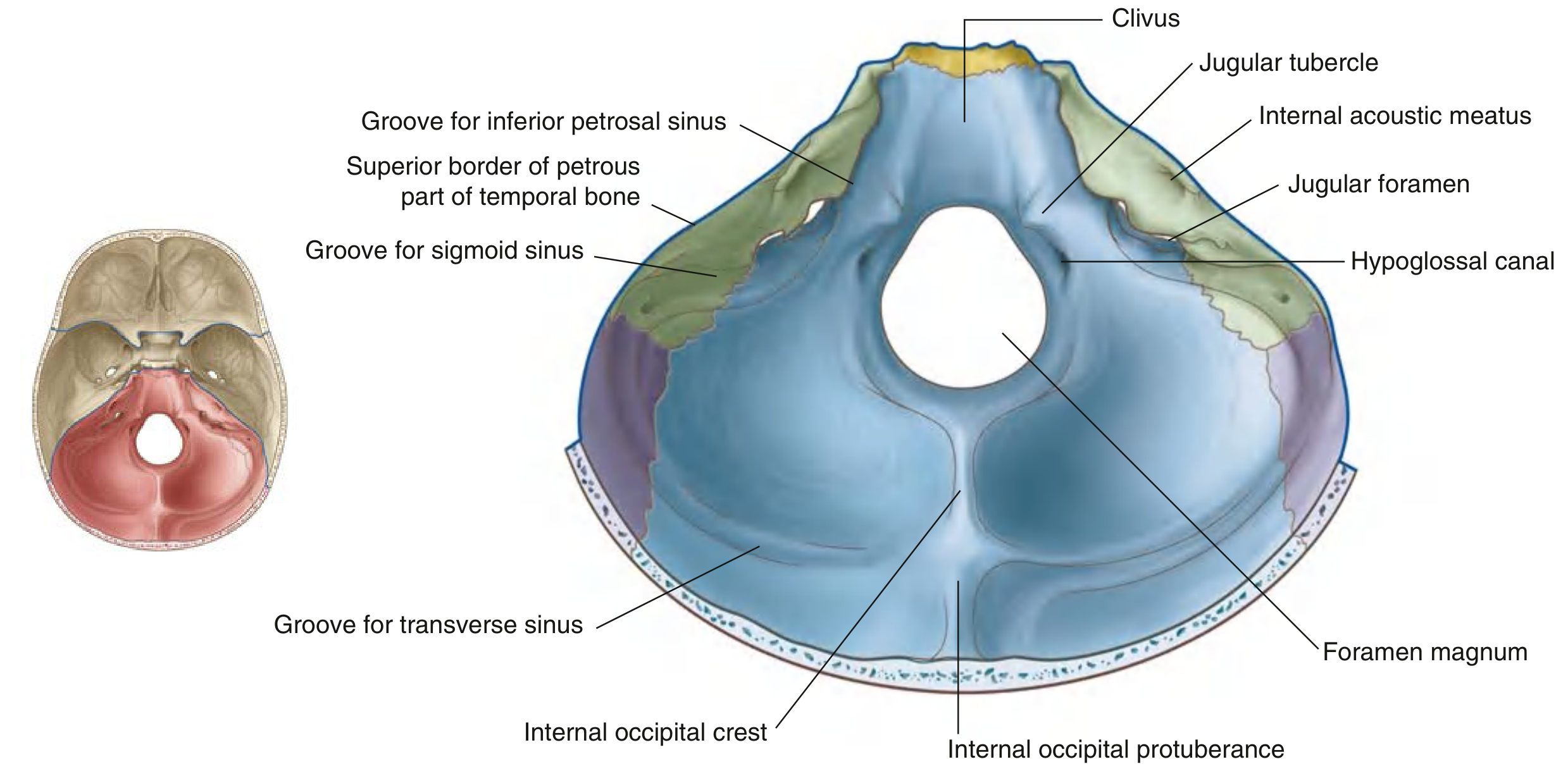
Bones Forming It
The largest and deepest of the three fossae:
- Occipital bone - the dominant bone (basilar, lateral, and squamous parts)
- Temporal bone (petromastoid part) - anterolateral walls
- Small contributions from sphenoid and parietal bones
Contents
Houses the brainstem (midbrain, pons, medulla) and cerebellum.
Key Landmarks
- Clivus - a slope of bone extending upward from the foramen magnum; formed by the basisphenoid superiorly and basiocciput inferiorly. Lies anterior to the pons and brainstem.
- Jugular tubercle - rounded mound of occipital bone medial to jugular foramen
- Internal occipital crest - runs from foramen magnum upward in midline; attachment for falx cerebelli
- Internal occipital protuberance - where internal occipital crest meets the transverse sinus grooves
- Groove for transverse sinus - extends laterally from internal occipital protuberance
- Groove for sigmoid sinus - continuation of transverse groove, curves inferiorly toward jugular foramen
- Groove for inferior petrosal sinus - between basilar occipital and petrous temporal
Foramina of the Posterior Cranial Fossa
| Foramen/Opening | Location | Structures Passing Through |
|---|---|---|
| Internal acoustic meatus (IAC) | Posterior surface of petrous temporal bone | CN VII (Facial nerve) + nervus intermedius, CN VIII (Vestibulocochlear nerve), Labyrinthine artery and vein |
| Jugular foramen | Between temporal and occipital bones | Anterior part: inferior petrosal sinus; Middle part: CN IX (Glossopharyngeal), CN X (Vagus), CN XI (Accessory); Posterior part: Internal jugular vein (continuation of sigmoid sinus), meningeal branches of occipital and ascending pharyngeal arteries |
| Hypoglossal canal | Anterolateral wall of foramen magnum, in occipital condyle | CN XII (Hypoglossal nerve), meningeal branch of ascending pharyngeal artery, emissary vein |
| Foramen magnum | Centre of PCF floor, occipital bone | Spinal cord / medulla oblongata (lower medulla continues as spinal cord), Vertebral arteries (both), anterior and posterior spinal arteries, spinal roots of CN XI (Accessory nerve) (ascending to enter cranial cavity), meninges |
| Condylar canal | Posterolateral to hypoglossal canal (when present) | Emissary vein (posterior cranial fossa ↔ suboccipital venous plexus) |
| Mastoid foramen | Posterior part of mastoid temporal bone (when present) | Emissary vein + meningeal branch of occipital artery |
MASTER SUMMARY - ALL FORAMINA AT A GLANCE
| Foramen | Fossa | Structures Transmitted |
|---|---|---|
| Foramen cecum | Anterior | Emissary vein (nasal cavity → superior sagittal sinus) |
| Cribriform plate foramina | Anterior | CN I (Olfactory nerve fibers); anterior ethmoidal nerve and artery |
| Optic canal | Middle | CN II; Ophthalmic artery |
| Superior orbital fissure | Middle | CN III, IV, V1, VI; Superior ophthalmic vein |
| Foramen rotundum | Middle | CN V2 (Maxillary) |
| Foramen ovale | Middle | CN V3 (Mandibular); Lesser petrosal nerve; Accessory meningeal artery; Emissary vein |
| Foramen spinosum | Middle | Middle meningeal artery + veins; Nervus spinosus (meningeal branch V3) |
| Foramen lacerum | Middle | Filled with cartilage - ICA traverses its superior aspect; Greater + deep petrosal nerves join to form nerve of pterygoid canal here |
| Carotid canal | Middle | Internal carotid artery; Sympathetic plexus |
| Hiatus for GSPN | Middle | Greater superficial petrosal nerve (CN VII branch) |
| Hiatus for lesser petrosal nerve | Middle | Lesser petrosal nerve (CN IX → otic ganglion) |
| Internal acoustic meatus | Posterior | CN VII, CN VIII; Labyrinthine artery and vein |
| Jugular foramen | Posterior | CN IX, X, XI; Internal jugular vein; Inferior petrosal sinus |
| Hypoglossal canal | Posterior | CN XII; Emissary vein; Meningeal branch of ascending pharyngeal artery |
| Foramen magnum | Posterior | Spinal cord/medulla; Vertebral arteries; Anterior + posterior spinal arteries; Spinal roots of CN XI |
| Condylar canal | Posterior | Emissary vein (when present) |
| Mastoid foramen | Posterior (temporal) | Emissary vein; Meningeal branch of occipital artery |
| Stylomastoid foramen | External (not seen from interior) | CN VII exits the skull here after leaving the IAC and traversing the facial canal |
MEMORY AIDS
Cranial nerves through foramina - "Some Clever Friends" helps with the SOF:
- Superior Orbital Fissure: CN III, IV, V1, VI (and superior ophthalmic vein) - "Oh, To Touch And Feel Very Soft" - Oculomotor, Trochlear, (V1), Abducens
Middle fossa sequence medial → lateral:
Optic canal → SOF → Foramen Rotundum → Foramen Ovale → Foramen Spinosum
Structures at the foramen magnum: The medulla, two vertebrals, two spinal arteries, and spinal XI roots
Jugular foramen CNs: IX, X, XI - the three lowest cranial nerves before XII ("Nine Ten Eleven in the Jugular")
Foramen lacerum trick: Nothing passes through it entirely - the ICA passes over its superior opening (the foramen is plugged by cartilage in life)
Sources: Gray's Anatomy for Students (Figs. 8.25-8.28 and text, pp. 1002-1007); K.J. Lee's Essential Otolaryngology (foramina list); Cummings Otolaryngology Head and Neck Surgery (middle cranial base).
Please add best diagram that has all above content labelled
The Fig. 8.28 summary diagram I already retrieved is exactly what you need - it shows the entire skull base (both superior/interior view and inferior view) with every foramen labeled and the structures passing through each one. Let me display it prominently again, and also fetch the individual fossa diagrams together for a complete visual set.
Loading Image
Finding Sources
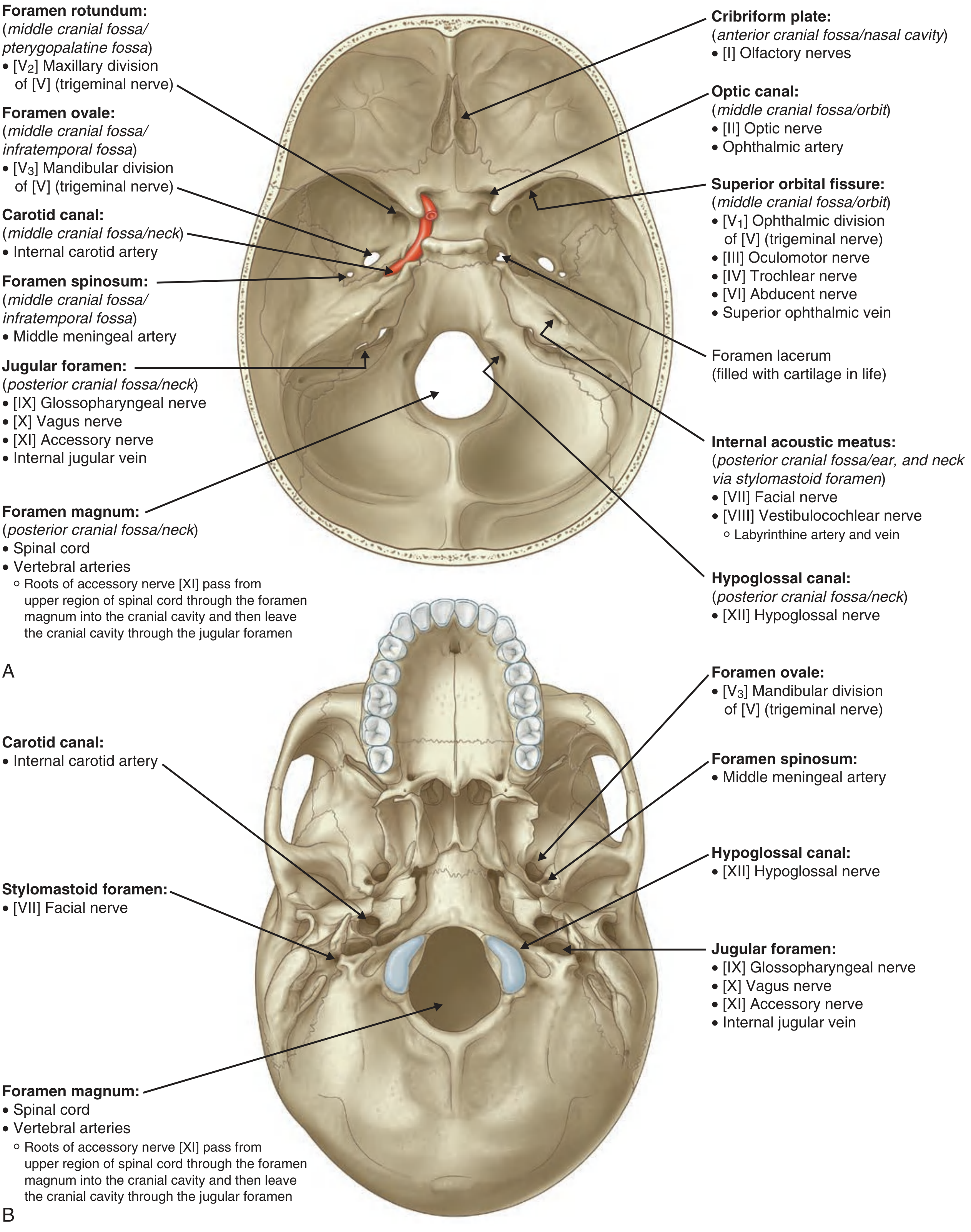
Here is the best single diagram from Gray's Anatomy for Students (Fig. 8.28) that covers everything discussed - all foramina labeled with the structures passing through them, shown from both the interior (superior) view (Panel A) and the external (inferior) view (Panel B):
What this diagram shows:
Panel A (Interior/Superior view of skull base):
- Cribriform plate → CN I (Olfactory nerves)
- Optic canal → CN II + Ophthalmic artery
- Superior orbital fissure → CN III, IV, V1, VI + Superior ophthalmic vein
- Foramen rotundum → CN V2 (Maxillary)
- Foramen ovale → CN V3 (Mandibular)
- Foramen spinosum → Middle meningeal artery
- Carotid canal → Internal carotid artery
- Foramen lacerum → Filled with cartilage in life
- Internal acoustic meatus → CN VII + CN VIII + Labyrinthine artery/vein
- Jugular foramen → CN IX, X, XI + Internal jugular vein
- Hypoglossal canal → CN XII
- Foramen magnum → Spinal cord, Vertebral arteries, Spinal roots of CN XI
Panel B (Inferior/External view of skull base):
- Confirms the same foramina from below, with the addition of the Stylomastoid foramen (CN VII exit) visible only from the external surface
This is the single most comprehensive labeled diagram of the skull base available in the textbook library, directly from Gray's Anatomy for Students.
This is a shared conversation. Sign in to Orris to start your own chat.