
80 year old male presents with problems of anxiety and stress whenever he doesn’t remember something, he keeps on asking and writing names of things and continuously poke people to tell names whenever he forgets what could be the reason
anxiety memory complaints elderly dementia
Alzheimer disease dementia stages cognitive decline
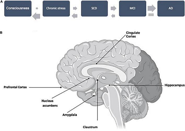
This composite educational graphic details the Alzheimer's Disease (AD) continuum and associated neuroanatomy. Section A presents a flowchart of the AD continuum across five stages: Consciousness, Chronic Stress, Subjective Cognitive Decline (SCD), Mild Cognitive Impairment (MCI), and AD. Thick forward-pointing arrows indicate the progression towards dementia, while thinner reverse arrows suggest potential reversibility in early stages (prior to reaching the AD stage). Section B is a labeled anatomical diagram of a medial sagittal section of the human brain. Key structures highlighted include the Prefrontal Cortex, Cingulate Cortex, Nucleus Accumbens, Amygdala, Claustrum, and Hippocampus. The diagram illustrates the spatial relationship between these cortical and subcortical regions involved in emotion, consciousness, and memory, emphasizing their role in the pathophysiological progression from early cognitive symptoms to clinically diagnosed Alzheimer's Disease. This resource serves to visualize the transition from emotional and behavioral changes to cognitive decline and irreversible neurodegeneration.
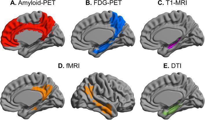
This infographic presents a multimodal imaging signature of Subjective Cognitive Decline (SCD) in the context of Alzheimer's disease progression. It displays five distinct neuroimaging modalities mapped onto a sagittal brain template. (A) Amyloid-PET highlights amyloid accumulation in red within the medial prefrontal cortex, cingulate cortex, and precuneus. (B) FDG-PET illustrates hypometabolism in blue, primarily affecting the medial temporal lobe and precuneus. (C) T1-MRI identifies structural changes in purple, specifically reduced volume in the hippocampus and thinning of the entorhinal cortex. (D) fMRI depicts abnormal functional connectivity in orange, focusing on the posterior Default Mode Network (DMN) and related cortical regions. (E) Diffusion Tensor Imaging (DTI) shows decreased fractional anisotropy (FA) in green, indicating white matter microstructural deficits in the hippocampal and parahippocampal areas. This comparative visual serves as a summary of molecular, structural, and functional biomarkers used to identify early neuroanatomical vulnerability in preclinical stages of dementia.
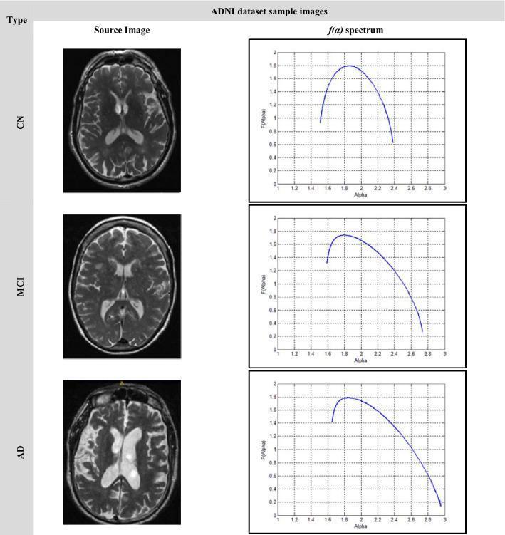
This infographic illustrates the neuroanatomical and multifractal changes across three stages of cognitive decline in the ADNI dataset: Cognitively Normal (CN), Mild Cognitive Impairment (MCI), and Alzheimer's Disease (AD). The visual is organized into a matrix comparing T2-weighted axial brain MRI slices (Source Image) with their corresponding singularity spectrum plots (f(α) spectrum). Clinical Imaging Findings: - CN: Shows normal brain volume with well-defined sulci and standard ventricular size. - MCI: Exhibits subtle cortical atrophy and mild ventricular enlargement. - AD: Demonstrates significant global cerebral atrophy, marked widening of the sulci, and pronounced ventriculomegaly, indicating substantial loss of brain tissue and increased cerebrospinal fluid (CSF) volume. Multifractal Analysis: The f(α) spectrum plots quantify structural complexity. As the disease progresses from CN to AD, the curves shift to the right (higher Alpha values) and become significantly broader. This widening indicates increased structural heterogeneity and irregularity within the brain tissue, serving as a mathematical signature for the pathological progression of Alzheimer's dementia.
Clinical Assessment: 80-Year-Old Male with Anxiety, Repetitive Questioning, and Memory Aids
What the Symptoms Tell Us
| Symptom | Clinical Significance |
|---|---|
| Anxiety when forgetting something | Classic emotional response to early memory loss - patients in early AD often remain acutely attuned to their own deficits |
| Constantly asking people for names | "Repetitive questioning" - a hallmark behavioral feature of early dementia listed in every diagnostic framework |
| Writing down names as a coping strategy | Compensatory behavior; the patient is aware of his memory problem (preserved insight - early stage) |
| Poking/persistently pressing people | Compulsive/repetitive behavior pattern seen in MCI and early dementia |
Most Likely Diagnosis: Alzheimer's Disease / MCI
"The cognitive changes of AD tend to follow a characteristic pattern, beginning with memory impairment... Depression, social withdrawal, and anxiety occur in early disease stages and may represent a prodrome before cognitive symptoms are apparent."
"Most [patients] remain acutely attuned to their deficits in early disease stages."
Chronic Stress → Subjective Cognitive Decline (SCD) → Mild Cognitive Impairment (MCI) → Alzheimer's Disease
Why the Anxiety Specifically?
- Awareness of memory failure - Early AD patients who retain insight become distressed when they notice gaps. The brain is losing episodic memory storage but the emotional centers (amygdala) remain intact, so the fear and distress are very real.
- Loss of control - Forgetting names (a form of anomia - word-finding difficulty) is one of the earliest and most distressing symptoms of AD.
- Compensatory behavior - Writing names down and repeatedly asking others are adaptive coping strategies that signal the patient knows something is wrong and is trying to compensate.
"In the dementia trajectory, most patients experience changes in mood, personality, thoughts, and perceptions, leading to behavioral changes including wandering, repetitive questioning, paranoia, delusions, hallucinations, agitation, aggression, apathy, disinhibition, irritability..."
Differential Diagnoses to Consider
| Condition | Key Distinguishing Features |
|---|---|
| Vascular Cognitive Impairment | Stepwise decline, history of strokes/TIAs, white matter changes on MRI |
| Frontotemporal Dementia (FTD) | Prominent personality/behavior change, language problems; memory relatively spared early |
| Lewy Body Dementia (DLB) | Visual hallucinations, fluctuating cognition, parkinsonism, REM sleep behavior disorder |
| Depression ("Pseudodementia") | Memory complaints with low mood, hopelessness; responds to antidepressants |
| Medication-induced | Sedatives, anticholinergics, benzodiazepines can cause memory loss in elderly |
| Thyroid dysfunction / B12 deficiency | Reversible causes - always check labs |
What Should Be Done
- Formal cognitive testing: Mini-Mental State Examination (MMSE), Montreal Cognitive Assessment (MoCA)
- Blood tests: TSH, B12, folate, CBC, metabolic panel, RPR (to rule out reversible causes)
- Brain MRI: Look for hippocampal atrophy (AD pattern), white matter lesions (vascular), or focal atrophy (FTD)
- Review all medications: Several common drugs cause memory problems in the elderly
- Neuropsychological assessment if available
- Caregiver education and structured environment (reduce environmental stressors, simplify routines)
Key Takeaway
Treatment of alzheimers in this case
Alzheimer disease treatment pharmacotherapy
lecanemab donanemab Alzheimer disease 2024 2025
PMID: 40194268
cholinesterase inhibitor mechanism Alzheimer acetylcholine synapse
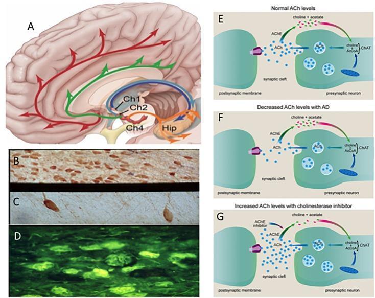
A multi-panel educational composite illustrating the cholinergic system in health and Alzheimer’s Disease (AD). (A) Sagittal anatomical diagram of the human brain showing cholinotrophic projection systems from cholinergic cell groups Ch1 (medial septal), Ch2 (diagonal band), and Ch4 (nucleus basalis) to the neocortex and hippocampus (Hip). (B-D) Photomicrographs comparing basal forebrain neurons: (B) healthy aged control with high neuron density; (C) AD sample showing significant neuronal loss and atrophy; (D) Thioflavin-S fluorescent staining identifying neurofibrillary tangles in p75NTR neurons. (E-G) Pathophysiology diagrams of the cholinergic synapse: (E) normal acetylcholine (ACh) synthesis via ChAT and degradation via AChE; (F) reduced ACh levels typical of AD pathology; and (G) the therapeutic mechanism of acetylcholinesterase inhibitors (AChEIs), which block AChE to increase synaptic ACh availability. The content highlights the neuroanatomical basis, histopathological changes, and pharmacological interventions relevant to dementia and neurodegeneration.
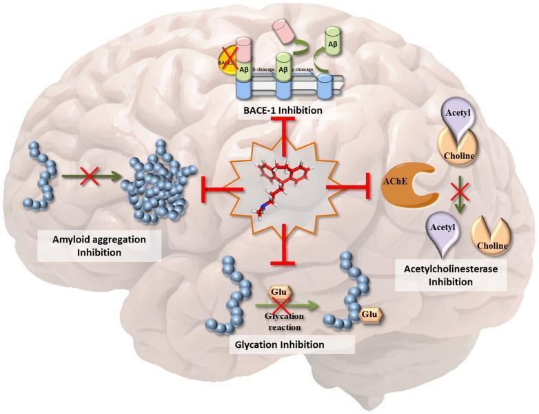
This medical infographic illustrates the multi-target-directed ligand (MTDL) mechanism of the drug protriptyline in the context of Alzheimer's Disease (AD) pathogenesis. Set against the anatomical backdrop of a human brain, the central 3D molecular structure of protriptyline is shown radiating inhibitory T-bars towards four primary pathological targets. 1) BACE-1 Inhibition: Depicts the blockade of beta-secretase-mediated cleavage of amyloid precursor protein, preventing Amyloid-beta (Aβ) formation. 2) Amyloid Aggregation Inhibition: Shows a red 'X' blocking the transition of Aβ monomers (blue spheres) into neurotoxic oligomeric aggregates. 3) Acetylcholinesterase (AChE) Inhibition: Illustrates the prevention of acetylcholine breakdown into acetyl and choline, maintaining neurotransmitter levels. 4) Glycation Inhibition: Demonstrates the inhibition of glucose (Glu) reacting with Aβ peptides, a process that normally exacerbates amyloid toxicity. The diagram emphasizes the drug's therapeutic potential to concurrently address enzymatic dysfunction, protein misfolding, and metabolic complications associated with neurodegeneration.
Treatment of Alzheimer's Disease in This Patient (80-Year-Old Male, Early Stage)
Step 1 - Confirm the Stage First
| Stage | MMSE Score | Key Features | Drug Approach |
|---|---|---|---|
| Mild (early) | 21-26 | Aware of deficits, anxiety, writing aids, repetitive questioning | Cholinesterase inhibitor (AChEI) |
| Moderate | 10-20 | Needs help with daily tasks, word-finding fails | AChEI + Memantine |
| Severe | <10 | Bedridden, incontinent, mute | AChEI + Memantine (continue) |
A. Neurotransmitter-Based (Symptomatic) Therapies
1. Cholinesterase Inhibitors (AChEIs) - FIRST LINE for Mild AD
| Drug | Dose | Notes |
|---|---|---|
| Donepezil (Aricept) | Start 5 mg/day; increase to 10 mg/day after 4-6 weeks | Once daily, easy for elderly; also approved for severe AD |
| Rivastigmine (Exelon) | Start 1.5 mg BD; target 6 mg BD, or 9.5 mg/day patch | Patch preferred in elderly - better tolerability, avoids GI side effects |
| Galantamine (Razadyne) | Start 4 mg BD; target 24 mg/day ER | Also has nicotinic receptor agonist activity |
- Nausea, diarrhea, abdominal cramps (GI cholinergic effects)
- Bradycardia - use cautiously if cardiac conduction disease
- Altered sleep / vivid dreams
- Muscle cramps
2. Memantine (Namenda) - For Moderate-to-Severe Stage
- Dose: Start 5 mg/day, increase by 5 mg weekly; target 10 mg twice daily
- Used in combination with AChEI in moderate-severe AD (combination is superior to either alone)
- Side effects: constipation, dizziness, headache, somnolence
"A common approach to AD drug therapy is to initiate a cholinesterase inhibitor for a patient diagnosed with mild AD dementia and to add memantine when patients enter the moderate stage of disease." - Harrison's Principles of Internal Medicine 22E
B. Disease-Modifying Therapies (Anti-Amyloid Antibodies) - New & Important
| Drug | Status | Efficacy |
|---|---|---|
| Lecanemab (Leqembi) | FDA-approved 2023 (early AD) | ~27% slowing of decline on CDR-SoB |
| Donanemab (Kisunla) | FDA-approved 2024 (early AD) | Best-ranked in 2025 network meta-analysis; ~35% slowing, NNT=10 |
| Aducanumab (Aduhelm) | Approved but controversial; withdrawn from many markets | Less effective than above two |
- ARIA-E (brain edema): 13% with lecanemab, 24% with donanemab
- ARIA-H (microbleeds): 17% with lecanemab, 31% with donanemab
- Most cases are asymptomatic but severe cases can cause seizures, stroke-like episodes, or rarely death
- Risk is highest in ApoE ε4 homozygotes (FDA black box warning)
- Age 80+ increases ARIA risk substantially
- If on anticoagulants or antiplatelets, risk increases further
- Uncontrolled hypertension is a contraindication
- ApoE ε4 genotyping is recommended before starting
Per the 2025 network meta-analysis (PMID 40194268): "Special caution is required regarding cerebral edema and microbleeding... which may outweigh the benefits" - the NNT for harm (edema) was 8 for donanemab vs. NNT of 10 for benefit. Shared decision-making with family is essential.
C. Treating This Patient's Anxiety Specifically
- Structured daily routine - reduces uncertainty and anxiety triggers
- Reassurance and validation techniques (don't argue or correct)
- Allow the compensatory writing behavior - it helps him function
- Involve family/caregivers in consistent responses to repeated questions
- Reduce environmental stimuli (noise, overstimulation)
- Cognitive-behavioral support
- SSRIs (sertraline, escitalopram) - first choice for anxiety/depression in AD (well-tolerated in elderly)
- Avoid benzodiazepines - they worsen memory and fall risk in elderly
- AChEIs themselves can reduce anxiety and behavioral symptoms over time
D. General Management in an 80-Year-Old
| Area | Intervention |
|---|---|
| Vascular risk factors | Control hypertension, diabetes, dyslipidemia - reduces vascular contribution |
| Nutrition | Mediterranean diet; ensure adequate B12, folate |
| Exercise | Even light walking - reduces rate of cognitive decline |
| Sleep | Treat sleep apnea if present; melatonin for circadian disruption |
| Hearing/Vision | Correct sensory deficits - they worsen cognitive performance |
| Medication review | Stop anticholinergics, benzodiazepines, unnecessary sedatives |
| Caregiver education | Crucial - delays institutionalization, reduces caregiver burden |
| Advance directives | Discuss while patient still has capacity |
Treatment Summary for This Patient
STEP 1: Start Donepezil 5 mg/day → increase to 10 mg/day at 6 weeks
STEP 2: If moderate stage reached → ADD Memantine 5 mg/day → titrate to 10 mg BD
STEP 3: Treat anxiety → Structured routine + SSRI if needed (avoid benzodiazepines)
STEP 4: Consider anti-amyloid therapy (lecanemab/donanemab) only if:
        - Early confirmed AD with biomarker evidence (amyloid PET or CSF)
        - ApoE genotyping done, not ε4 homozygote
        - No anticoagulation, no uncontrolled HTN
        - Patient/family understand ARIA risks
STEP 5: Vascular risk factor control + lifestyle modification + caregiver support