
Give Me Simple image Form easy to Understand notes On Pulmonary Function test
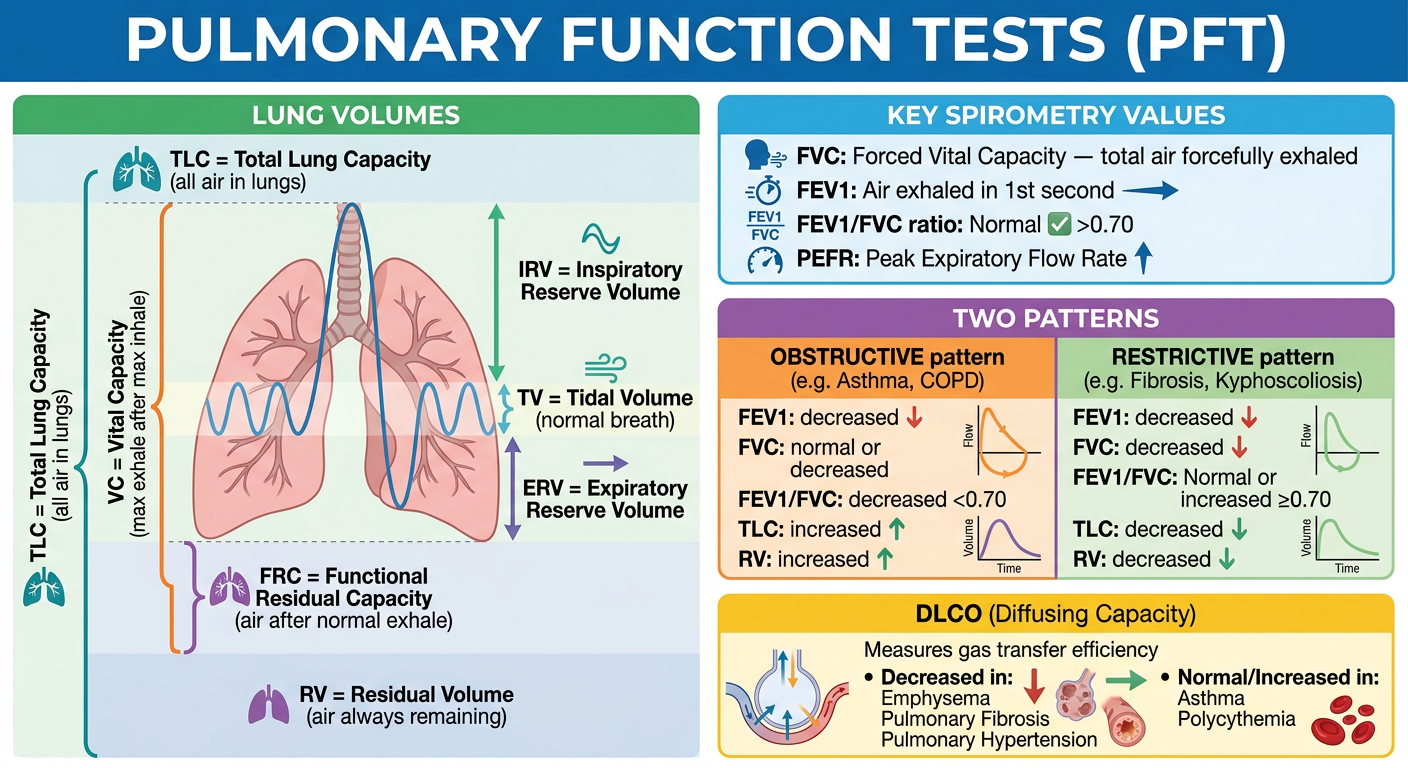
Medical study notes infographic on Pulmonary Function Tests (PFT). Clean, colorful, easy to understand layout with the following sections clearly organized: 1. TITLE: "Pulmonary Function Tests (PFT)" in large bold text at top 2. Section: LUNG VOLUMES (with a simple lung diagram showing): - TLC = Total Lung Capacity (all air in lungs) - VC = Vital Capacity (max exhale after max inhale) - FRC = Functional Residual Capacity (air after normal exhale) - RV = Residual Volume (air always remaining) - ERV = Expiratory Reserve Volume - IRV = Inspiratory Reserve Volume - TV = Tidal Volume (normal breath) 3. Section: KEY SPIROMETRY VALUES in a colored box: - FVC: Forced Vital Capacity — total air forcefully exhaled - FEV1: Air exhaled in 1st second - FEV1/FVC ratio: Normal >0.70 - PEFR: Peak Expiratory Flow Rate 4. Section: TWO PATTERNS (side-by-side comparison table): OBSTRUCTIVE pattern (e.g. Asthma, COPD): - FEV1: decreased ↓ - FVC: normal or decreased - FEV1/FVC: decreased <0.70 - TLC: increased ↑, RV: increased ↑ RESTRICTIVE pattern (e.g. Fibrosis, Kyphoscoliosis): - FEV1: decreased ↓ - FVC: decreased ↓ - FEV1/FVC: Normal or increased ≥0.70 - TLC: decreased ↓, RV: decreased ↓ 5. Section: DLCO (Diffusing Capacity): - Measures gas transfer efficiency - Decreased in: Emphysema, Pulmonary Fibrosis, Pulmonary Hypertension - Normal/Increased in: Asthma, Polycythemia Use bright colors, icons, arrows, simple diagrams. Medical student-friendly. White background. Professional but easy to read.
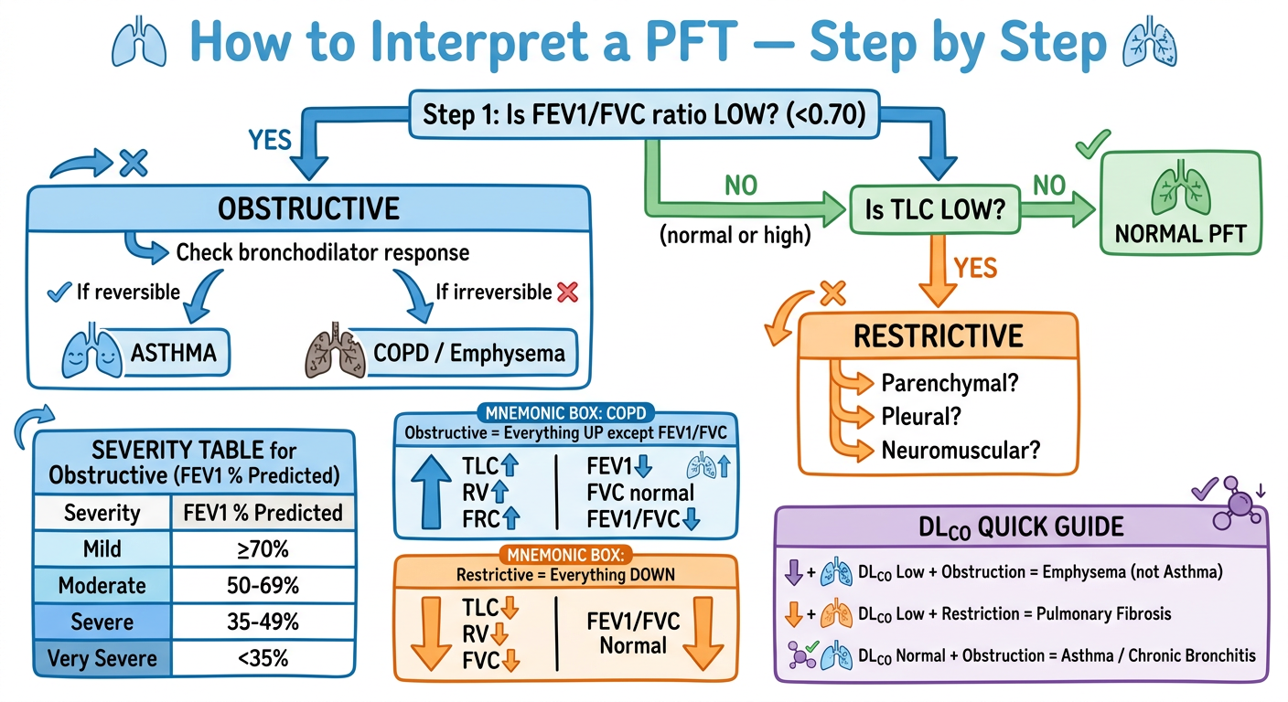
Medical study notes infographic Part 2: "PFT Interpretation Algorithm & Clinical Mnemonics". Clean colorful layout for medical students. 1. TITLE: "How to Interpret a PFT — Step by Step" 2. FLOWCHART (top to bottom): Step 1: Is FEV1/FVC ratio LOW? (<0.70) YES → OBSTRUCTIVE → Check bronchodilator response - If reversible → ASTHMA - If irreversible → COPD / Emphysema NO (normal or high) → Is TLC LOW? YES → RESTRICTIVE → Parenchymal? Pleural? Neuromuscular? NO → NORMAL PFT 3. SEVERITY TABLE for Obstructive (FEV1 % Predicted): - Mild: ≥70% - Moderate: 50-69% - Severe: 35-49% - Very Severe: <35% 4. MNEMONIC BOX: "COPD Obstructive = Everything UP except FEV1/FVC" TLC↑ RV↑ FRC↑ | FEV1↓ FVC normal | FEV1/FVC↓ 5. MNEMONIC BOX: "Restrictive = Everything DOWN" TLC↓ RV↓ FVC↓ | FEV1/FVC Normal 6. DLCO QUICK GUIDE: - DLCO Low + Obstruction = Emphysema (not Asthma) - DLCO Low + Restriction = Pulmonary Fibrosis - DLCO Normal + Obstruction = Asthma / Chronic Bronchitis Use arrows, color coding (blue for obstructive, orange for restrictive, green for normal). Simple icons. White background. Student-friendly medical notes style.
🫁 Card 1 — Lung Volumes + Key Values + Patterns
🔍 Card 2 — Interpretation Algorithm + Mnemonics + DLCO Guide
Quick Text Summary to Go With the Images
| Test | What it Measures |
|---|---|
| FVC | Total air forcefully exhaled |
| FEV1 | Air exhaled in 1st second |
| FEV1/FVC | The single most important ratio (normal >0.70) |
| TLC | Total lung capacity |
| DLCO | How well gas crosses the lung membrane |
- Obstructive (Asthma, COPD) - FEV1/FVC low (<0.70), TLC/RV high
- Restrictive (Fibrosis, Obesity) - FEV1/FVC normal/high, TLC/RV low
- Low DLCO + Obstruction = Emphysema (not asthma)
- Low DLCO + Restriction = Pulmonary fibrosis
- Normal DLCO + Obstruction = Asthma or Chronic Bronchitis