
Rewrite the whole content as it is plus add maximum number of high yield images along the content from your side # Physiology of the Cerebral Cortex, Intellectual Functions of the Brain, Learning, and Memory --- ## PART I: ANATOMY AND ORGANIZATION OF THE CEREBRAL CORTEX ### Gross Structure The cerebral cortex is a **2-5 mm thick** layer of gray matter covering the entire surface of the cerebral hemispheres. It contains approximately **100 billion neurons**. The total surface area is approximately **2,200 cm²**, but about two-thirds of this area is hidden within the sulci and fissures. ### Histological Layers of the Cortex The cerebral cortex is composed of **six layers** (from superficial to deep): **Layer I – Molecular (Plexiform) Layer:** - The most superficial layer - Contains very few cell bodies - Primarily composed of dendrites and axons running horizontally (tangentially) - Contains dendrites of cells from deeper layers and axons from other cortical areas and thalamic nuclei - Cajal-Retzius cells are found here during development **Layer II – External Granular Layer:** - Contains small pyramidal cells and numerous stellate (granule) cells - Densely packed small neurons - Dendrites extend into Layer I, axons project to deeper layers **Layer III – External Pyramidal Layer:** - Contains medium-sized pyramidal cells (larger than Layer II) - **This is the primary source of cortico-cortical (association and commissural) fibers** - Axons project to the contralateral hemisphere via the corpus callosum and to ipsilateral association areas **Layer IV – Internal Granular Layer:** - Contains densely packed stellate (granule) cells - **This is the primary RECEIVING layer for thalamocortical afferents** (specific sensory input from thalamus) - Very prominent in primary sensory cortices (visual cortex, somatosensory cortex) — hence called **"granular cortex" or "koniocortex"** - Contains a dense band of horizontally running fibers called the **external band of Baillarger** - In the primary visual cortex (area 17), this band is so prominent it is visible to the naked eye and is called the **Line (Stria) of Gennari** — hence the term **"striate cortex"** **Layer V – Internal Pyramidal (Ganglionic) Layer:** - Contains the **largest pyramidal cells**, including the **giant cells of Betz** in the primary motor cortex - **Primary source of corticospinal, corticobulbar, corticostriatal, and corticopontine fibers** - Contains the **internal band of Baillarger** - This is the main OUTPUT layer for subcortical projections (other than thalamus) **Layer VI – Multiform (Polymorphic/Fusiform) Layer:** - Deepest layer, blends with subcortical white matter - Contains fusiform (spindle-shaped) cells and modified pyramidal cells - **Primary source of corticothalamic fibers** (feedback projections to the thalamus) ### Types of Cortex Based on Layering 1. **Homotypical Cortex (6 clear layers):** Association areas — most of the cortex 2. **Heterotypical Cortex:** - **Granular (Koniocortex):** Layer IV is very thick. Found in primary sensory areas (S1, V1, A1). Receives massive thalamic input. - **Agranular:** Layer IV is thin or absent; Layers III and V are thick. Found in primary motor cortex (M1) and premotor areas. Heavy output function. ### Cell Types in the Cortex 1. **Pyramidal Cells:** - Most abundant (approximately 75%) - Excitatory neurons using **glutamate** - Have a triangular cell body with a single large apical dendrite extending toward the surface and multiple basal dendrites - Long axons that project to distant cortical and subcortical areas - Found in layers II, III, V, and VI - The largest are the **Betz cells** of layer V in the precentral gyrus (primary motor cortex), measuring up to **120 μm** 2. **Stellate (Granule) Cells:** - Small, star-shaped neurons - Two types: - **Spiny stellate cells:** Excitatory, use glutamate. Found mainly in Layer IV. - **Smooth stellate cells (aspiny):** Inhibitory, use **GABA**. - Local circuit interneurons 3. **Other Interneurons:** - **Basket cells:** Inhibitory (GABAergic), synapse on cell bodies of pyramidal cells - **Chandelier cells:** Inhibitory, synapse specifically on the axon initial segment of pyramidal cells — very powerful inhibition - **Double bouquet cells:** Vertically oriented inhibitory interneurons - **Martinotti cells:** Found in deep layers, send axons to layer I ### Cortical Columns — The Functional Unit The cortex is organized into **vertical columns** approximately **300-600 μm in diameter**, each containing about **10,000 neurons**. This concept was championed by **Vernon Mountcastle** (for somatosensory cortex) and **David Hubel and Torsten Wiesel** (for visual cortex). Each column functions as an elementary processing unit: - All neurons within a column respond to the same type of stimulus - Information flows vertically within columns - Columns are interconnected horizontally **In the somatosensory cortex:** Each column responds to a single modality (touch, pressure, vibration) from a specific skin location. **In the visual cortex:** - **Orientation columns** — neurons respond to a specific edge orientation - **Ocular dominance columns** — neurons preferentially respond to input from one eye - A **hypercolumn** contains a complete set of orientation columns and a pair of ocular dominance columns --- ## PART II: FUNCTIONAL AREAS OF THE CEREBRAL CORTEX The cortex is divided into functional areas based on several classification systems: ### Brodmann's Classification Based on cytoarchitecture (histological structure), Korbinian Brodmann divided the cortex into **52 numbered areas**. Key areas include: | Brodmann Area | Location | Function | |---|---|---| | 4 | Precentral gyrus | Primary motor cortex | | 6 | Anterior to area 4 | Premotor and supplementary motor area | | 3, 1, 2 | Postcentral gyrus | Primary somatosensory cortex | | 5, 7 | Superior parietal lobule | Somatosensory association area | | 17 | Banks of calcarine sulcus | Primary visual cortex | | 18, 19 | Surrounding area 17 | Visual association areas | | 41, 42 | Superior temporal gyrus (Heschl's gyrus) | Primary auditory cortex | | 22 | Superior temporal gyrus | Auditory association area (Wernicke's area in dominant hemisphere) | | 44, 45 | Inferior frontal gyrus | Broca's area (dominant hemisphere) | | 9, 10, 11, 12 | Prefrontal cortex | Higher executive functions | | 39 | Angular gyrus | Reading, writing, calculation | | 40 | Supramarginal gyrus | Language integration | | 28 | Parahippocampal gyrus | Entorhinal cortex (memory gateway) | ### Functional Classification The cerebral cortex can be divided into: 1. **Primary areas** (idiotypic cortex) 2. **Secondary/Unimodal association areas** (homotypical) 3. **Tertiary/Multimodal (heteromodal) association areas** --- ## PART III: SENSORY AREAS OF THE CORTEX ### A. Primary Somatosensory Cortex (S1) — Brodmann Areas 3, 1, 2 **Location:** Postcentral gyrus and the posterior wall of the central sulcus **Thalamic Input:** Ventral posterolateral (VPL) nucleus (body) and Ventral posteromedial (VPM) nucleus (face) of the thalamus **Organization:** - **Somatotopic organization** — the famous **"Sensory Homunculus"** described by Wilder Penfield - Body is represented **upside down**: foot and leg on the medial surface (extending into the interhemispheric fissure), trunk and arm on the lateral convexity, face and tongue at the lateral end near the Sylvian fissure - The **pharynx and intra-abdominal regions** are represented most laterally (within the Sylvian fissure) - **Contralateral representation** — each hemisphere receives sensory information from the opposite side of the body (except some bilateral representation for the face) **The Homunculus:** - The size of cortical representation is proportional to the **density of sensory innervation** (receptor density), NOT to the physical size of the body part - The **lips, face, thumb, and fingers** have disproportionately large representation - The **trunk, back, and proximal limbs** have very small representation **Subdivision of S1:** - **Area 3a:** Receives proprioceptive input (from muscle spindles) - **Area 3b:** Receives cutaneous tactile input — this is considered the TRUE primary somatosensory cortex - **Area 1:** Processes texture information - **Area 2:** Processes size and shape information (stereognosis) **Cortical Columns in S1:** - Rapidly adapting (RA) columns and slowly adapting (SA) columns alternate - Each column responds to one modality from one point on the body #### Clinical Correlates — S1 Lesions: **Cortical lesion of postcentral gyrus:** - **Contralateral loss of fine touch, proprioception, vibration, two-point discrimination, and stereognosis** - Pain and temperature are relatively preserved (because these pathways have significant processing at thalamic and subcortical levels) - **Astereognosis** (inability to recognize objects by touch) - **Agraphesthesia** (inability to recognize letters/numbers traced on skin) - **Loss of two-point discrimination** - Positive phenomena: **Focal sensory seizures** (Jacksonian sensory seizures) — tingling/numbness that may march along the body following the homunculus **Cortical Sensory Syndrome (parietal lobe syndrome):** - Combined loss of discriminative sensations with preserved crude touch and pain - Sensory inattention/extinction on double simultaneous stimulation ### B. Somatosensory Association Cortex (S2 and Areas 5, 7) **Secondary Somatosensory Cortex (S2):** - Located in the **superior wall of the lateral sulcus** (parietal operculum) - Receives bilateral input - Involved in tactile learning, tactile memory, and attention to somatosensory stimuli **Posterior Parietal Cortex (Areas 5 and 7):** - **Area 5:** Integrates somatosensory information for spatial awareness of the body and limb position - **Area 7:** Integrates visual and somatosensory information for visuospatial processing and hand-eye coordination #### Clinical Correlate — Posterior Parietal Lobe Lesions: **Non-dominant (usually right) parietal lobe lesions:** - **Contralateral hemispatial neglect (hemineglect)** — patient ignores the left side of space and their own left body. They may not dress the left side, not eat food on the left side of the plate, not draw the left side of a clock - **Anosognosia** — denial of illness, especially denial of left-sided weakness - **Asomatognosia** — unawareness or denial of one's own body parts on the affected side - **Constructional apraxia** — inability to draw or copy figures - **Dressing apraxia** — difficulty putting on clothes **Dominant (usually left) parietal lobe lesions:** - **Gerstmann syndrome** (lesion of angular gyrus, area 39): 1. **Agraphia** (inability to write) 2. **Acalculia** (inability to calculate) 3. **Finger agnosia** (inability to recognize individual fingers) 4. **Left-right disorientation** (inability to distinguish left from right) **Bilateral parietal lobe lesions:** - **Balint syndrome:** 1. **Simultanagnosia** — inability to perceive more than one object at a time in the visual field 2. **Optic ataxia** — inability to accurately reach for objects under visual guidance 3. **Oculomotor apraxia** — inability to voluntarily direct gaze to objects in the peripheral visual field ### C. Primary Visual Cortex (V1) — Brodmann Area 17 **Location:** Walls of the **calcarine sulcus** on the medial surface of the occipital lobe. Extends slightly onto the lateral surface at the occipital pole. **Thalamic Input:** Lateral geniculate nucleus (LGN) of the thalamus → via optic radiations (geniculocalcarine tract) **Histological Features:** - Classic **6-layered** cortex with extremely thick **Layer IV** (granular/koniocortex) - Layer IV is subdivided into **4a, 4b, 4cα, and 4cβ** - **Layer 4cα** receives input from **magnocellular layers** of LGN (motion, depth) - **Layer 4cβ** receives input from **parvocellular layers** of LGN (color, form) - Contains the **Line of Gennari** (stria of Gennari) — a dense band of myelinated fibers in layer 4b, visible to the naked eye → hence called **"striate cortex"** **Retinotopic Organization:** - Point-to-point mapping of the retina onto the cortex - **Contralateral visual field** is represented in each hemisphere - The **macula** (central vision) has an enormous cortical representation — about 50% of V1 is devoted to the central 10 degrees of the visual field → this is called **cortical magnification** - **Upper visual field** → represented on the **lower** bank of the calcarine sulcus (lingual gyrus) - **Lower visual field** → represented on the **upper** bank (cuneus) - **Peripheral visual field** → represented anteriorly - **Central (macular) visual field** → represented posteriorly at the occipital pole **Ocular Dominance Columns:** - Alternating columns of neurons preferentially responsive to left or right eye - Input from the two eyes remains segregated in Layer IV but converges in other layers - Basis of binocular vision and depth perception **Orientation Columns:** - Within each ocular dominance column, neurons are arranged in a systematic sequence of preferred stimulus orientations (e.g., vertical, 10°, 20°, etc.) - Described by Hubel and Wiesel **Types of Neurons:** - **Simple cells:** Respond to bars of light with a specific orientation at a specific position. Found mainly in Layer IV. - **Complex cells:** Respond to bars of light with a specific orientation that can be anywhere within a larger receptive field. Can detect movement direction. Found in Layers II, III, V, VI. - **Hypercomplex (end-stopped) cells:** Respond to bars/edges of a specific length, orientation, and position. Detect corners and curvatures. #### Clinical Correlates — V1 Lesions: - **Complete bilateral destruction** → **Cortical blindness** — pupillary reflexes are intact (because they use the pretectal pathway, not the cortex), but the patient cannot see - **Unilateral destruction** → **Contralateral homonymous hemianopia** (loss of the opposite visual field in both eyes) with **macular sparing** (because the macular region has dual blood supply from both posterior cerebral artery and middle cerebral artery, and has massive bilateral representation) - **Upper bank lesion (cuneus)** → Contralateral inferior quadrantanopia - **Lower bank lesion (lingual gyrus)** → Contralateral superior quadrantanopia **Anton syndrome (cortical blindness with anosognosia):** - Patient is cortically blind (bilateral V1 destruction, typically from bilateral posterior cerebral artery occlusion) - But the patient **denies being blind** and confabulates visual experiences - May bump into objects while insisting they can see **Riddoch phenomenon:** - Patient with V1 lesion can perceive moving objects in the "blind" field but cannot perceive stationary objects - Mediated by alternative visual pathways (superior colliculus → pulvinar → V5/MT) ### D. Visual Association Areas (V2-V5 and beyond) **Location:** Areas 18, 19 and temporal regions **Two Streams of Visual Processing:** 1. **Dorsal Stream ("Where/How" pathway):** - V1 → V2 → V3 → V5/MT (middle temporal area) → Posterior parietal cortex (area 7) - Processes **motion, spatial location, depth, visually guided actions** - Dominated by magnocellular input - **V5/MT (Middle Temporal area):** Specialized for motion detection 2. **Ventral Stream ("What" pathway):** - V1 → V2 → V4 → Inferotemporal cortex (IT) - Processes **object recognition, face recognition, color** - Dominated by parvocellular input - **V4:** Color processing - **Fusiform face area (FFA):** Specialized for face recognition (in fusiform gyrus) - **Parahippocampal place area:** Scene and place recognition #### Clinical Correlates — Visual Association Area Lesions: **Visual Agnosia:** Inability to recognize objects by sight despite intact visual acuity and intellectual function - **Apperceptive agnosia:** Cannot form a complete percept of the object. Cannot copy drawings. (Bilateral parieto-occipital lesions) - **Associative agnosia:** Can perceive and copy the object but cannot identify/name it. Can still identify it through other senses (touch, sound). (Bilateral ventral occipitotemporal lesions) **Prosopagnosia:** Inability to recognize familiar faces (including their own in a mirror). Can recognize people by voice or other cues. **Lesion:** Bilateral (or right-sided) fusiform gyrus / inferotemporal cortex. **Achromatopsia (Cerebral):** Loss of color vision due to cortical damage. **Lesion:** V4 area (bilateral or unilateral causing contralateral hemi-achromatopsia). Distinguished from retinal color blindness because patient may recall colors from before the lesion and may not be able to perceive any colors at all. **Akinetopsia (Cerebral motion blindness):** Inability to perceive motion. Objects appear frozen or jump from one position to another (like a strobe light effect). **Lesion:** Bilateral V5/MT area. Patient can see static objects but cannot see a car approaching or water pouring into a glass. ### E. Primary Auditory Cortex (A1) — Brodmann Areas 41, 42 **Location:** **Heschl's gyrus** (transverse temporal gyri) — located on the superior surface of the temporal lobe, buried within the lateral sulcus **Thalamic Input:** Medial geniculate nucleus (MGN) of the thalamus **Organization:** - **Tonotopic organization** — different frequencies mapped systematically - **Low frequencies** → anterolateral - **High frequencies** → posteromedial - Receives **bilateral input** (but predominantly contralateral) - Each ear projects to both auditory cortices #### Clinical Correlates — A1 Lesions: - **Unilateral lesion** → Difficulty in **sound localization** on the contralateral side, slight decrease in hearing in the contralateral ear, but NO significant deafness (because of bilateral representation) - **Bilateral destruction** → **Cortical deafness** (very rare) ### F. Auditory Association Cortex — Area 22 **Location:** Superior temporal gyrus, surrounding A1 **Wernicke's area** (posterior part of area 22, typically in the left/dominant hemisphere) — critical for **language comprehension** (discussed in detail later) #### Clinical Correlate: **Auditory agnosia (Word deafness):** - Pure word deafness — inability to understand spoken language despite intact hearing and ability to read, write, and speak - Lesion: Bilateral or dominant temporal lobe, disconnecting Wernicke's area from auditory input **Auditory agnosia for sounds:** - Cannot recognize non-verbal sounds (e.g., a dog barking, telephone ringing) - Usually non-dominant temporal lobe lesion **Amusia:** Loss of ability to perceive or produce music. Usually right (non-dominant) temporal lobe lesion. ### G. Gustatory Cortex **Primary gustatory cortex:** **Anterior insula** and **frontal operculum** (Brodmann area 43) - Thalamic input from VPM nucleus - Somatotopic and gustotopic organization ### H. Vestibular Cortex **Location:** Area at the junction of the **intraparietal sulcus and the posterior end of the lateral sulcus** (parieto-insular vestibular cortex, PIVC) - Receives input from the vestibular nuclei via the thalamus - Provides conscious perception of body position and movement in space ### I. Olfactory Cortex **Primary olfactory cortex:** **Piriform cortex** (in the temporal lobe), part of the uncus - Unique: The ONLY sensory system that reaches the cortex **WITHOUT a thalamic relay** (olfactory bulb → olfactory tract → piriform cortex directly) - Also projects to entorhinal cortex, amygdala, and orbitofrontal cortex #### Clinical Correlate: **Uncinate fits:** Temporal lobe seizures involving the uncus producing **olfactory hallucinations** (usually unpleasant smells — cacosmia) often accompanied by lip-smacking, chewing movements, and a **dreamy state**. Can be a sign of temporal lobe tumors. --- ## PART IV: MOTOR AREAS OF THE CORTEX ### A. Primary Motor Cortex (M1) — Brodmann Area 4 **Location:** **Precentral gyrus** and anterior wall of the central sulcus **Histological Features:** - **Agranular cortex** — Layer IV is very thin/absent - Layer V contains the **giant Betz cells** (giant pyramidal neurons, up to 120 μm) - Betz cells constitute only about 3-5% of corticospinal tract fibers; the majority come from other pyramidal neurons in M1 and other areas **Organization:** - **Somatotopic organization** — **Motor Homunculus** (Penfield and Rasmussen) - Body represented **upside down**: - Lower extremity → medial surface (interhemispheric fissure) - Upper extremity → lateral convexity - Face, tongue, larynx → most lateral (near Sylvian fissure) - The size of cortical representation is proportional to the **fineness and complexity of movement** (dexterity), NOT physical size - **Hands, fingers, lips, tongue, and larynx** have enormous representation - **Trunk and proximal limbs** have small representation - **Contralateral representation** — each hemisphere controls the opposite side of the body **Functions:** - Controls **voluntary fine, skilled, fractionated movements** especially of distal extremities - Encodes **force, direction, and speed** of movement - Individual neurons code for the direction of movement; a **population code** (population vector) determines the overall movement direction **Output:** - Corticospinal tract (pyramidal tract) — passes through internal capsule → cerebral peduncle → basis pontis → medullary pyramids - At the pyramidal decussation: 85-90% cross → **lateral corticospinal tract** (controls distal limb muscles) - 10-15% remain uncrossed → **anterior corticospinal tract** (controls axial and proximal muscles, eventually crosses at segmental level) - Corticobulbar fibers → to cranial nerve motor nuclei #### Clinical Correlates — M1 Lesions: **Upper Motor Neuron (UMN) Lesion Signs:** 1. **Contralateral spastic paralysis** (initially flaccid in acute phase — "cerebral shock") 2. **Hyperreflexia** — exaggerated deep tendon reflexes 3. **Spasticity** — velocity-dependent increase in muscle tone (clasp-knife phenomenon) 4. **Clonus** — rhythmic involuntary contractions 5. **Babinski sign** positive — extension (dorsiflexion) of the great toe with fanning of other toes on stroking the lateral sole 6. **Loss of fine, skilled movements** — particularly of fingers and hands 7. **No significant muscle atrophy** (only mild disuse atrophy) 8. **No fasciculations** 9. **Absent superficial reflexes** (abdominal, cremasteric) **Jacksonian Motor Seizures:** - Irritative lesions of M1 cause focal seizures that begin in one body part (e.g., thumb) and **"march"** to adjacent body parts following the motor homunculus - May secondarily generalize to a full tonic-clonic seizure - Named after John Hughlings Jackson **Todd's Paralysis:** - Transient postictal (after seizure) weakness or paralysis of the body parts involved in a focal motor seizure - Usually resolves within 24-48 hours - Important to distinguish from stroke ### B. Premotor Cortex (PMC) — Brodmann Area 6 (Lateral Part) **Location:** Lateral surface of the frontal lobe, anterior to M1 **Functions:** - **Planning and programming of movements** based on external sensory cues - Controls **proximal and trunk muscles** for postural stabilization - Role in **learning new motor programs** - Contains **mirror neurons** — neurons that fire both when performing an action AND when observing someone else perform the same action (also in inferior parietal lobule). Important for: - Imitation and motor learning - Understanding others' actions and intentions - Empathy - Language evolution (possibly) **Connections:** Receives input from posterior parietal cortex (spatial information) and prefrontal cortex (executive planning). Projects to M1, reticulospinal tract, and spinal cord. #### Clinical Correlates: - **Premotor cortex lesion** → difficulty in planning complex sequences of movements - Loss of ability to use visual cues to guide movement - Mirror neuron dysfunction has been hypothesized (controversially) to play a role in **autism spectrum disorder** ### C. Supplementary Motor Area (SMA) — Brodmann Area 6 (Medial Part) **Location:** Medial surface of the frontal lobe, anterior to leg area of M1, on the superior frontal gyrus **Functions:** - **Planning and initiation of self-generated (internally cued) movements** — movements initiated by internal will rather than external stimuli - **Bimanual coordination** — coordinating movements of both hands - **Motor sequencing** — planning sequential movements - **Mental rehearsal of movement** — SMA is active even when you THINK about making a movement without actually doing it **Bereitschaftspotential (Readiness Potential):** - A slowly rising negative electrical potential recorded over the SMA beginning approximately **1-1.5 seconds BEFORE** a voluntary self-initiated movement - Discovered by Kornhuber and Deecke (1965) - Indicates the cortical preparation for voluntary movement - Starts bilaterally over SMA, then lateralizes to contralateral M1 just before movement #### Clinical Correlates: **SMA Syndrome (SMA Lesion):** - **Transient akinesia** (inability to initiate voluntary movements) — especially contralateral - **Mutism** (if dominant hemisphere SMA is affected) - Difficulty with bimanual coordination - Patients recover relatively well over weeks to months - Can occur after surgical resection of SMA (e.g., for tumor or epilepsy surgery) **Alien Hand Syndrome (Anarchic Hand):** - One hand performs purposeful actions that the patient denies initiating or controlling - The hand seems to "have a mind of its own" - Patient may need to physically restrain the alien hand with the other hand - Occurs with lesions of **medial frontal cortex (SMA) and/or corpus callosum** (anterior cerebral artery territory) - Can also occur with callosal lesions causing intermanual conflict ### D. Frontal Eye Field (FEF) — Brodmann Area 8 **Location:** Middle frontal gyrus, anterior to premotor cortex **Function:** - Controls **voluntary saccadic eye movements** to the contralateral side - Involved in visual search and attention #### Clinical Correlates: **Destructive Lesion (e.g., stroke):** - Eyes **deviate TOWARD** the side of the lesion (away from the contralateral hemiplegic side) - "The patient looks at his lesion" (or "looks away from the paralyzed side") - Because the intact FEF on the opposite side drives the eyes toward the lesioned side **Irritative Lesion (seizure focus):** - Eyes **deviate AWAY** from the side of the lesion (toward the contralateral side) - Because the stimulated FEF drives eyes contralaterally ### E. Broca's Area — Brodmann Areas 44, 45 **Location:** Inferior frontal gyrus (**pars opercularis** = area 44, **pars triangularis** = area 45) of the **dominant hemisphere** (left in ~95% of right-handers and ~70% of left-handers) **Function:** - **Motor programming of speech** — plans the sequential movements of lips, tongue, larynx, and pharynx needed for speech production - Also involved in grammar and syntax processing - Connected to Wernicke's area via the **arcuate fasciculus** #### Clinical Correlate — Broca's Aphasia (Motor/Expressive/Non-fluent Aphasia): **Features:** - **Non-fluent speech** — halting, effortful, telegraphic speech with very few words - **Relatively preserved comprehension** — patient understands spoken and written language - **Impaired repetition** - Speech output is limited to short phrases, often nouns and verbs, lacking prepositions, articles, and conjunctions (agrammatic) - Example: Asked "How are you doing?" patient may say "Fine...fine" or when describing a picture: "Dog...cat...chase" - **Patient is aware of deficit and is frustrated** - Often accompanied by right-sided hemiparesis (especially face and arm) because of proximity to motor cortex - Writing is also impaired (agraphia) **Etiology:** Usually stroke in the territory of the **superior division of the left middle cerebral artery** --- ## PART V: ASSOCIATION AREAS OF THE CORTEX Association areas constitute the MAJORITY of the human cerebral cortex and are responsible for the highest-order cognitive functions. They integrate information from multiple sensory and motor areas. ### A. Prefrontal Association Cortex (Areas 9, 10, 11, 12, 46, 47) **Location:** Anterior part of the frontal lobe, anterior to motor and premotor areas This is the most highly developed region in humans compared to other primates (approximately 29% of total cortical surface). **Subdivisions and Functions:** **1. Dorsolateral Prefrontal Cortex (DLPFC) — Areas 9, 46:** - **Working memory** — the ability to hold information in mind temporarily and manipulate it (e.g., mental arithmetic, remembering a phone number while dialing) - **Executive functions:** - Planning and strategizing - Problem-solving - Cognitive flexibility (ability to shift between tasks/strategies) - Abstract thinking - Judgment and decision-making - Temporal ordering (sequencing events in time) - **Set shifting** — ability to change behavior when rules change - **Sustained attention and concentration** **2. Orbitofrontal Cortex (OFC) — Areas 11, 12, 47:** - **Emotional regulation** and social behavior - **Impulse control** and inhibition of inappropriate behavior - **Personality and social conduct** - Reward-based decision making - Processing of punishment and reward signals - Integration of sensory information with emotional and motivational states **3. Medial Prefrontal Cortex — Area 10 (Frontopolar):** - **Self-referential processing** — thinking about oneself - **Theory of mind** — understanding others' mental states, beliefs, and intentions - Moral reasoning - Default mode network (active during rest/daydreaming) **4. Ventrolateral Prefrontal Cortex — Areas 44, 45, 47:** - Language processing (left hemisphere) - Response inhibition - Working memory for objects #### Clinical Correlates — Prefrontal Lesions: **Frontal Lobe Syndrome (Prefrontal Syndrome):** This is one of the most clinically important and frequently tested topics. The prefrontal cortex modulates behavior, personality, and higher cognitive functions. **A. Dorsolateral Prefrontal Syndrome (Dysexecutive Syndrome):** - Impaired working memory - Difficulty with **planning, organizing, problem-solving** - **Perseveration** — inability to shift from one task/response to another; patient repeats the same response even when it's no longer appropriate - Demonstrated by: - **Wisconsin Card Sorting Test** — patient continues sorting by the old rule even after the rule changes - **Trail Making Test** — difficulty connecting numbered/lettered dots in alternating sequence - Reduced **verbal fluency** — when asked to list words starting with "F" in one minute, patient produces very few - **Apathy** and reduced motivation (abulia) - Difficulty with abstract thinking — tends toward concrete interpretations of proverbs - Impaired temporal ordering (cannot correctly sequence past events) **B. Orbitofrontal Syndrome (Disinhibited/Sociopathic Syndrome):** - **Disinhibition** — inappropriate social behavior, tactlessness - **Personality change** — typically becomes more impulsive, irresponsible - **Emotional lability** — inappropriate joking (**Witzelsucht** — tendency to make inappropriate jokes and puns), euphoria, or irritability - **Impaired judgment** and poor decision-making - **Loss of social awareness** — cannot read social cues - May exhibit **sexual inappropriateness, aggression, or criminal behavior** - **Lack of concern for consequences** of actions - **Utilization behavior** — compulsive use of objects placed in front of them (e.g., if you place a comb, they comb their hair; if you place glasses, they put them on) **The Classic Case — Phineas Gage (1848):** - A railway construction foreman who survived an iron tamping rod passing through his left frontal lobe (orbitofrontal cortex) - Before the accident: responsible, capable, well-liked - After the accident: "fitful, irreverent, indulging at times in the grossest profanity, manifesting but little deference for his fellows, impatient of restraint or advice... a child in his intellectual capacity and manifestations, he has the animal passions of a strong man" - This case was historically crucial in establishing the role of the frontal lobes in personality and social behavior **C. Medial Prefrontal/Cingulate Syndrome (Akinetic/Apathetic Syndrome):** - **Abulia** — profound lack of will, motivation, and spontaneous action - **Akinetic mutism** (severe cases) — patient is awake and appears alert but makes no spontaneous movements or speech - Reduced emotional responsiveness - Usually bilateral medial frontal lesions (anterior cerebral artery territory) **Frontal Release Signs (Primitive Reflexes):** When the frontal lobe is damaged, inhibitory control over primitive brainstem reflexes is lost, and these reflexes re-emerge: 1. **Grasp reflex** — stroking the palm causes involuntary grasping 2. **Rooting reflex** — stroking the cheek causes the head to turn toward the stimulus 3. **Sucking reflex** — touching the lips causes sucking movements 4. **Palmomental reflex** — stroking the palm causes contraction of the ipsilateral mentalis (chin) muscle 5. **Snout reflex** — tapping the lips causes lip puckering 6. **Glabellar tap (Myerson's sign)** — repeated tapping on the glabella causes persistent blinking (normally, blinking habituates after a few taps) **Other Frontal Lobe Signs:** - **Gegenhalten (paratonia)** — involuntary resistance to passive movement that increases with the speed of movement (as opposed to clasp-knife spasticity or lead-pipe rigidity) - **Motor impersistence** — inability to sustain a voluntary act (e.g., cannot keep eyes closed, cannot keep tongue protruded) - **Echopraxia** — involuntary imitation of movements of others - **Incontinence** — medial frontal lesions can cause urinary incontinence (parasagittal meningioma, anterior cerebral artery infarct) ### B. Parieto-Occipito-Temporal Association Cortex **Location:** Junction of parietal, occipital, and temporal lobes **Functions:** - Integration of somatosensory, visual, and auditory information - Spatial awareness and body schema - Language comprehension - Reading, writing, and calculation - Naming objects **Key Areas:** **1. Wernicke's Area — Posterior part of Area 22 (dominant hemisphere):** - **Comprehension of spoken language** - Connected to Broca's area via the **arcuate fasciculus** - Part of the broader language network **2. Angular Gyrus — Area 39 (dominant hemisphere):** - **Integration of visual information with language** - Critical for **reading** (converting written symbols to language) - Cross-modal association area (visual → auditory → language) **3. Supramarginal Gyrus — Area 40:** - Phonological processing - Integration for language production #### Clinical Correlates: **Wernicke's Aphasia (Sensory/Receptive/Fluent Aphasia):** - **Fluent speech** — normal or even excessive speech output, normal prosody and rhythm - **But the speech is MEANINGLESS** — filled with: - **Paraphasias:** Substitution of incorrect words - **Phonemic (literal) paraphasias:** Substituting similar-sounding words or syllables ("spork" for "fork") - **Semantic (verbal) paraphasias:** Substituting related words ("knife" for "fork") - **Neologisms:** Made-up words ("flurken") - **Jargon:** Speech becomes an unintelligible stream of neologisms and paraphasias (jargon aphasia) - **Severely impaired comprehension** — patient does not understand spoken or written language - **Impaired repetition** - Patient is **unaware of the deficit** (anosognosia for the language deficit) — they don't realize their speech makes no sense - **No hemiparesis** (Wernicke's area is distant from the motor cortex) - Can be mistaken for psychiatric illness (psychosis) because of fluent but nonsensical speech **Etiology:** Stroke in the territory of the **inferior division of the left middle cerebral artery** **Conduction Aphasia:** - **Lesion:** Arcuate fasciculus (disconnects Broca's from Wernicke's) - **Fluent speech** with frequent phonemic paraphasias - **Good comprehension** - **Severely impaired repetition** — this is the hallmark - Patient is aware of errors and attempts to self-correct (conduite d'approche) **Global Aphasia:** - **Lesion:** Large lesion of the dominant hemisphere involving both Broca's and Wernicke's areas and the arcuate fasciculus (usually complete MCA territory infarction) - **Non-fluent speech** (may only produce stereotyped utterances like "tan-tan") - **Impaired comprehension** - **Impaired repetition** - Usually accompanied by right hemiplegia, right hemianopia, right hemisensory loss **Transcortical Motor Aphasia:** - **Lesion:** Anterior/superior to Broca's area (supplementary motor area or connections) - **Non-fluent speech** - **Good comprehension** - **INTACT repetition** — this distinguishes it from Broca's aphasia - Patient can repeat perfectly but has difficulty with spontaneous speech **Transcortical Sensory Aphasia:** - **Lesion:** Posterior to Wernicke's area (watershed area between PCA and MCA) - **Fluent, paraphasic speech** - **Impaired comprehension** - **INTACT repetition** — this distinguishes it from Wernicke's aphasia - Patient can repeat sentences without understanding them (echolalia) **Mixed Transcortical Aphasia (Isolation Aphasia):** - **Lesion:** Large watershed infarction isolating the perisylvian language zone - **Non-fluent speech** - **Impaired comprehension** - **INTACT repetition** (only thing preserved) - The language zone is intact but disconnected from surrounding cortex **Summary Table of Aphasias:** | Type | Fluency | Comprehension | Repetition | Lesion | |------|---------|---------------|------------|--------| | Broca's | Non-fluent | Good | Impaired | Inferior frontal (area 44,45) | | Wernicke's | Fluent | Impaired | Impaired | Superior temporal (area 22) | | Conduction | Fluent | Good | Impaired | Arcuate fasciculus | | Global | Non-fluent | Impaired | Impaired | Large perisylvian | | Transcortical Motor | Non-fluent | Good | Intact | Anterior/superior to Broca's | | Transcortical Sensory | Fluent | Impaired | Intact | Posterior to Wernicke's | | Mixed Transcortical | Non-fluent | Impaired | Intact | Watershed zones | **Anomic Aphasia:** - Difficulty with **word-finding** (naming) is the primary deficit - Fluent speech with circumlocutions (talking around a word they can't find) - Good comprehension and repetition - Can result from lesions in various areas; not well-localizing - Can be a residual deficit as other aphasias improve **Alexia (Acquired inability to read):** 1. **Alexia with agraphia:** Cannot read or write. **Lesion:** Dominant angular gyrus (area 39). Essentially a visual-linguistic disconnection plus loss of the writing center. 2. **Alexia without agraphia (Pure alexia/Pure word blindness):** Can write but cannot read (even their own writing!). **Lesion:** Left occipital cortex (destroying visual input) + splenium of corpus callosum (preventing right visual cortex from sending information to left language areas). The patient can still write because Wernicke's-to-motor connections are intact. Classic disconnection syndrome. **Agraphia:** Isolated writing impairment. Can occur from lesions of Exner's writing area (posterior part of the middle frontal gyrus/area 6) in the dominant hemisphere, or as part of aphasia syndromes. ### C. Limbic Association Cortex **Location:** Cingulate gyrus, parahippocampal gyrus, orbitofrontal cortex **Functions:** - Emotion and motivation - Memory (especially declarative memory formation) - Integration of emotional significance with sensory information - Discussed in detail in the limbic system and memory sections below --- ## PART VI: CEREBRAL DOMINANCE AND LATERALIZATION ### Concept of Cerebral Dominance The two hemispheres are NOT functionally equivalent. Each hemisphere has specialized functions, though they work together via the **corpus callosum** (the largest commissure, containing ~200-300 million fibers). ### Left Hemisphere (Dominant in Most People) Dominant for: - **Language** — speech production, comprehension, reading, writing - **Logical/analytical processing** - **Mathematical calculations** - **Sequential/temporal processing** - **Categorical perception** (classifying objects into categories) - **Fine motor control of the dominant hand** **Language dominance:** - Left hemisphere dominant in **96% of right-handers** and **70% of left-handers** - Overall, the left hemisphere is dominant for language in about **95%** of the population - Determined by the **Wada test** (intracarotid amobarbital test) — injection of sodium amobarbital into one carotid artery temporarily anesthetizes that hemisphere, and language function is tested - Now increasingly replaced by functional MRI ### Right Hemisphere (Non-dominant in Most People) Dominant for: - **Visuospatial processing** — spatial orientation, mental rotation, spatial relationships - **Facial recognition** (prosopagnosia results from right hemisphere damage) - **Music perception and production** (especially melody, pitch, timbre) - **Emotional processing:** - **Emotional prosody** — understanding and expressing emotion through tone of voice - Processing emotional content of faces and situations - **Attention** — especially spatial attention (hence right hemisphere lesions cause left hemineglect) - **Holistic/gestalt processing** — perceiving the "whole picture" rather than details - **Body schema and spatial awareness** - **Constructional abilities** — drawing, copying complex figures #### Clinical Correlate: **Aprosodia:** - **Lesion:** Right (non-dominant) hemisphere, particularly right homologues of Broca's and Wernicke's areas - **Motor aprosodia** (right frontal): Cannot express emotion through tone of voice — speech is monotone, flat - **Sensory aprosodia** (right temporal): Cannot understand emotional tone of others' speech ### Anatomical Asymmetries 1. **Planum temporale:** Area of the superior temporal gyrus posterior to Heschl's gyrus. **Larger on the left** in 65% of brains. Corresponds to Wernicke's area. One of the most consistent anatomical asymmetries. 2. **Sylvian fissure:** Longer on the left, angles up more steeply on the right. 3. **Heschl's gyrus:** Often two gyri on the right, one on the left. 4. **Frontal lobe:** Right frontal lobe slightly larger and protrudes further anteriorly (**right frontal petalia**). 5. **Occipital lobe:** Left occipital lobe slightly larger and protrudes further posteriorly (**left occipital petalia**). This pattern is called **Yakovlevian torque**. ### Split-Brain Studies Pioneered by **Roger Sperry** (Nobel Prize, 1981) and **Michael Gazzaniga**, studying patients who had undergone **corpus callosotomy** (cutting the corpus callosum) for intractable epilepsy. **Key Findings:** 1. **Object in left visual field** (goes to right hemisphere) → patient cannot **name** the object (right hemisphere has no language output) but can **pick it out by touch with the left hand** (right hemisphere controls left hand) 2. **Object in right visual field** (goes to left hemisphere) → patient can **name** the object normally 3. **Object placed in left hand** (right hemisphere) → patient cannot name it verbally but can demonstrate its use or pick out a matching object with the same hand 4. **Object placed in right hand** (left hemisphere) → patient can name it normally 5. **Left hand may act autonomously** — intermanual conflict; each hemisphere controls its hand independently 6. **Right hemisphere can understand simple language** — can follow simple commands presented to the left visual field **This demonstrated that:** - Language production is lateralized to the left hemisphere - The right hemisphere has spatial, emotional, and some language comprehension abilities - Without the corpus callosum, each hemisphere operates independently --- ## PART VII: INTELLECTUAL FUNCTIONS OF THE BRAIN ### A. Attention **Types:** 1. **Alertness/Arousal:** Maintained by the reticular activating system (RAS) 2. **Selective attention:** Ability to focus on relevant stimuli while ignoring irrelevant ones. Involves prefrontal cortex, parietal cortex, and thalamus. 3. **Sustained attention (vigilance):** Maintaining focus over prolonged periods. Right frontal and parietal cortex. 4. **Divided attention:** Attending to multiple tasks simultaneously. Prefrontal cortex. **Neural Substrate of Attention:** - **Right parietal cortex** — spatial attention (dominant role). Attends to BOTH sides of space, whereas the left parietal cortex primarily attends to the right side. This is why right parietal lesions cause profound left neglect. - **Prefrontal cortex** — executive attention, working memory, sustained attention - **Anterior cingulate cortex** — conflict monitoring, error detection - **Thalamic reticular nucleus** — gates sensory information to the cortex - **Ascending reticular activating system** — maintains overall arousal #### Clinical Correlate: **ADHD (Attention Deficit Hyperactivity Disorder):** - Impaired sustained and selective attention, hyperactivity, impulsivity - Dysfunction of prefrontal cortex and its dopaminergic/noradrenergic innervation - Treated with stimulants (methylphenidate, amphetamines) that increase dopamine and norepinephrine in prefrontal circuits ### B. Language Already covered extensively above. Key additional points: **Language Development:** - **Critical period** for language acquisition: Birth to approximately 12 years (puberty) - After this period, acquiring native-like language proficiency becomes extremely difficult - This is demonstrated by cases of feral children (e.g., Genie, a girl isolated until age 13, who never acquired normal language despite intensive training) - **Evidence for critical period:** Children who learn a second language before puberty typically achieve native-like proficiency; those who learn after puberty usually retain an accent and make grammatical errors **Bilingualism and the Brain:** - Early bilinguals (learned both languages in childhood) — both languages tend to share the same cortical representation in Broca's area - Late bilinguals — the two languages have spatially distinct representations in Broca's area (Wernicke's area representation is similar) ### C. Higher Cognitive Functions **1. Abstract Thinking:** - Ability to understand concepts, metaphors, and relationships beyond the literal - Tested by asking proverb interpretation - "A stitch in time saves nine" → Concrete answer: "If you fix a hole early, you don't need more stitches" (literal). Abstract answer: "Taking care of problems early prevents bigger problems later." - Impaired in frontal lobe lesions and dementia **2. Judgment:** - Ability to make appropriate decisions in social and practical situations - Tested by hypothetical scenarios: "What would you do if you found a stamped, addressed envelope on the ground?" → "Mail it" - Impaired in frontal lobe lesions **3. Insight:** - Awareness of one's own cognitive and behavioral deficits - Often impaired in frontal lobe lesions, psychiatric conditions, and dementia **4. Planning and Problem-Solving:** - Executive functions dependent on dorsolateral prefrontal cortex - Tested by Tower of London/Hanoi, Wisconsin Card Sorting Test --- ## PART VIII: LEARNING AND MEMORY This is one of the most complex and fascinating areas of neuroscience. ### Definitions - **Learning:** The process of acquiring new information or modifying existing knowledge, behaviors, skills, values, or preferences - **Memory:** The process of encoding, storing, and retrieving information ### Classification of Memory #### A. By Duration: **1. Immediate Memory (Sensory Memory/Ultra-short-term Memory):** - Duration: Milliseconds to seconds (< 1 second for visual, < 4 seconds for auditory) - Capacity: Large but rapidly decaying - Types: - **Iconic memory** (visual) — lasts ~200-500 milliseconds - **Echoic memory** (auditory) — lasts ~3-4 seconds - Not consciously accessible for long - Acts as a buffer for incoming sensory information - If attention is directed to it, information transfers to short-term memory **2. Short-Term Memory (STM) / Working Memory:** - Duration: **Seconds to minutes** (approximately 15-30 seconds without rehearsal) - Capacity: **7 ± 2 items** (Miller's Magic Number, 1956) — approximately 7 chunks of information - Can be maintained by rehearsal (maintenance rehearsal keeps information in STM; elaborative rehearsal transfers it to LTM) - **Working Memory** (Baddeley's Model) is an expanded concept of STM that includes: - **Phonological loop:** Stores and rehearses verbal/acoustic information. Has two components: - Phonological store (inner ear) — holds speech-based information for ~2 seconds - Articulatory rehearsal process (inner voice) — refreshes information by subvocal repetition - **Visuospatial sketchpad:** Stores and manipulates visual and spatial information - **Central executive:** Attentional control system that coordinates the subsystems, directs attention, and integrates information. Dependent on **prefrontal cortex** - **Episodic buffer** (added later by Baddeley) — integrates information from the other subsystems and long-term memory into a coherent episode - **Neural basis:** Primarily **prefrontal cortex** (dorsolateral prefrontal cortex) — neurons show sustained firing during the delay period of a working memory task **3. Long-Term Memory (LTM):** - Duration: Hours to a lifetime - Capacity: Virtually unlimited - Requires **consolidation** — the process of stabilizing a memory trace after its initial acquisition #### B. By Content (Types of Long-Term Memory): **1. Declarative (Explicit) Memory:** - Memory for facts and events that can be **consciously recalled** and **verbally declared** - "Knowing that" - Dependent on **medial temporal lobe structures** (hippocampus, entorhinal cortex, perirhinal cortex, parahippocampal cortex) and **diencephalic structures** (mammillary bodies, anterior and mediodorsal thalamic nuclei) - Two subtypes: a. **Episodic Memory:** - Memory for **personal events and experiences** — includes contextual details (time, place, emotions, associated events) - "What happened" — autobiographical memory - Example: Remembering your graduation ceremony, what you had for breakfast - **Critically dependent on the hippocampus** and its connections - **Temporal lobe is more important** for this type b. **Semantic Memory:** - Memory for **general knowledge and facts** — independent of personal context - "What you know" — encyclopedic knowledge - Example: Knowing that Paris is the capital of France, knowing what a dog is - Less dependent on hippocampus once consolidated; stored in **neocortical association areas** (especially temporal cortex, inferotemporal cortex) - More resistant to hippocampal damage than episodic memory **2. Non-Declarative (Implicit/Procedural) Memory:** - Memory that is expressed through **performance** rather than conscious recollection - "Knowing how" - **NOT dependent on hippocampus or medial temporal lobe** - Several subtypes: a. **Procedural (Skill/Habit) Memory:** - Memory for motor skills and cognitive procedures - Example: Riding a bicycle, typing, playing piano, tying shoelaces - **Neural basis:** Basal ganglia (especially striatum — caudate and putamen), cerebellum, supplementary motor area - Acquired gradually through repetition and practice - Once learned, performed automatically without conscious thought - **Intact in amnesic patients** — H.M. could learn new motor skills even though he couldn't remember learning them b. **Classical Conditioning:** - Learning associations between stimuli - **Emotional conditioning** (e.g., fear conditioning — learning to fear a stimulus associated with danger): **Amygdala** - **Motor conditioning** (e.g., eyeblink conditioning — learning to blink in response to a tone paired with air puff): **Cerebellum** (specifically interpositus nucleus) c. **Priming:** - Improved identification or processing of a stimulus based on prior exposure, without conscious awareness of the prior exposure - Example: After seeing the word "DOCTOR," you more quickly recognize the word "NURSE" - **Neural basis:** Neocortex — changes in perceptual or conceptual processing areas - Intact in amnesic patients d. **Non-associative Learning:** - **Habituation:** Decreased response to a repeated, innocuous stimulus. The simplest form of learning. - **Sensitization:** Increased response to a stimulus after exposure to a strong or noxious stimulus. - Neural basis: Changes in sensory pathways and reflex circuits - Studied extensively in **Aplysia** (sea slug) by **Eric Kandel** (Nobel Prize, 2000) ### Neural Substrates of Memory #### The Hippocampus and Medial Temporal Lobe **Anatomy of the Hippocampal Formation:** - Located in the **medial temporal lobe**, forming the floor of the inferior horn of the lateral ventricle - Components: 1. **Hippocampus proper (Cornu Ammonis, CA):** Divided into CA1, CA2, CA3, CA4 2. **Dentate gyrus** 3. **Subiculum** 4. **Entorhinal cortex** (Brodmann area 28) — the **gateway** to the hippocampus **Hippocampal Circuit (Trisynaptic Circuit):** 1. **Entorhinal cortex (Layer II)** → via **Perforant pathway** → **Dentate gyrus** (granule cells) 2. **Dentate gyrus** → via **Mossy fibers** → **CA3** pyramidal cells 3. **CA3** → via **Schaffer collaterals** → **CA1** pyramidal cells 4. **CA1** → **Subiculum** → via **Fornix** → **Mammillary bodies** → via **Mammillothalamic tract** → **Anterior thalamic nucleus** → **Cingulate gyrus** → **Entorhinal cortex** This circuit is known as the **Papez Circuit** (described by James Papez, 1937) and was originally proposed as the circuit of emotion but is now known to be critical for **memory formation**. **Function of the Hippocampus:** - **NOT the storage site of long-term memories** - Rather, it is essential for the **CONSOLIDATION** of new declarative memories — the process of converting short-term/working memory into long-term memory - Acts as a **temporary holding station** or **index** that binds together distributed cortical representations during memory formation - Over time (weeks to years), memories become independent of the hippocampus and are stored in **neocortical networks** — this is called **systems consolidation** - Important for **spatial memory and navigation** — contains **place cells** (neurons that fire when the animal is in a specific location in the environment). Discovered by **John O'Keefe** (Nobel Prize, 2014, shared with May-Britt and Edvard Moser who discovered grid cells in the entorhinal cortex) - The **right hippocampus** is more involved in spatial memory - The **left hippocampus** is more involved in verbal/declarative memory **CA1 is the most vulnerable region** of the hippocampus to: - **Hypoxia/ischemia** (global ischemia, cardiac arrest) - **Hypoglycemia** - **Temporal lobe epilepsy** (hippocampal sclerosis with loss of CA1 neurons) #### Clinical Correlate — The Famous Case of H.M. (Henry Molaison): This is the most important case in the history of memory research. **Background:** - Henry Molaison suffered from intractable epilepsy - In 1953, Dr. William Scoville performed bilateral medial temporal lobe resection, removing the hippocampus, amygdala, and surrounding cortex bilaterally - The epilepsy improved, but the operation produced a devastating and unexpected consequence **Result — Severe Anterograde Amnesia:** - H.M. was **unable to form ANY new declarative (explicit) long-term memories** after the surgery - He could not remember people he met, events that occurred, or things he learned after the operation - Every experience was completely new to him, even if he had experienced it moments before - His **immediate/working memory was INTACT** — he could hold information for short periods (could carry on a conversation) but the moment he was distracted, the memory vanished - He could remember his name and information about his life BEFORE the surgery (remote memories were relatively intact, though there was some retrograde amnesia for events a few years before surgery) **What was PRESERVED:** - **Procedural/motor learning** — H.M. could learn the mirror drawing task (tracing a star while looking in a mirror) and improved with practice over days, even though each day he had no memory of ever doing the task before - **Priming** — showed normal repetition priming - **Classical conditioning** — could acquire conditioned responses - **Immediate memory/working memory** - **Intelligence** — IQ was normal or even slightly improved - **Personality** — essentially unchanged - **Remote memories** (from well before surgery) — relatively preserved **Lessons from H.M.:** 1. The hippocampus is essential for formation of new declarative memories 2. The hippocampus is NOT needed for immediate/working memory 3. The hippocampus is NOT needed for procedural memory (different brain systems) 4. The hippocampus is NOT the storage site of old memories 5. Memory is NOT a unitary faculty — different types of memory depend on different brain systems 6. Short-term and long-term memory are distinct processes (double dissociation) ### The Papez Circuit and Diencephalic Memory **Papez Circuit (reminder):** Hippocampus → Fornix → Mammillary bodies → Mammillothalamic tract → Anterior thalamic nucleus → Cingulate gyrus → Parahippocampal gyrus → Entorhinal cortex → Hippocampus Damage to any component can impair memory. #### Clinical Correlates: **Korsakoff Syndrome:** - **Etiology:** Chronic alcoholism leading to **thiamine (Vitamin B1) deficiency** → damage to **mammillary bodies and dorsomedial thalamic nuclei** - **Features:** 1. **Severe anterograde amnesia** — cannot form new memories 2. **Retrograde amnesia** — variable, often with temporal gradient (recent memories more affected than remote) 3. **Confabulation** — the hallmark feature. Patient fills memory gaps with fabricated, plausible-sounding stories, without the intention to deceive. They genuinely believe these false memories. 4. **Lack of insight** — patient is unaware of the memory deficit 5. Personality and other cognitive functions may be relatively preserved - **Often preceded by Wernicke's encephalopathy** (acute phase): - Classic triad: **Confusion, ophthalmoplegia (eye movement abnormalities), ataxia** - Together called **Wernicke-Korsakoff syndrome** - Wernicke's is a medical emergency requiring immediate IV thiamine **Fornix Lesions:** - Bilateral fornix damage (rare, can occur with colloid cyst of the third ventricle or surgical damage) → severe amnesia similar to bilateral hippocampal damage **Anterior Thalamic Lesions:** - Can cause amnesia (thalamic amnesia) - Bilateral paramedian thalamic infarction → severe amnesia + akinetic mutism + vertical gaze palsy #### The Amygdala and Emotional Memory **Anatomy:** Almond-shaped nucleus in the medial temporal lobe, anterior to the hippocampus **Function in Memory:** - Does NOT store declarative memories - **Modulates memory consolidation** based on **emotional arousal** — emotionally charged events are remembered better - The amygdala enhances hippocampal and cortical memory consolidation through stress hormones (norepinephrine, cortisol) and direct neural connections - Critical for **fear conditioning** — learning to associate a neutral stimulus with danger - Essential for recognizing emotional expressions (especially **fear**) in faces - Important for the emotional coloring of memories **Mechanism of Emotional Memory Enhancement:** 1. Emotional event → activation of amygdala 2. Amygdala → activation of hypothalamus and brainstem → release of stress hormones (epinephrine, cortisol) 3. These hormones and direct amygdala inputs enhance synaptic plasticity in the hippocampus 4. Result: Stronger, more durable memory for emotionally significant events 5. This is why we remember emotionally important events (e.g., 9/11, a wedding) more vividly — called **flashbulb memories** #### Clinical Correlates: **Bilateral Amygdala Damage (Klüver-Bucy Syndrome):** Originally described after bilateral temporal lobectomy in monkeys by Heinrich Klüver and Paul Bucy (1937): 1. **Psychic blindness (visual agnosia)** — inability to recognize objects visually 2. **Hyperorality** — tendency to explore objects by mouth 3. **Hypermetamorphosis** — compulsive attention to all visual stimuli 4. **Emotional blunting** — loss of fear and aggression (placidity) 5. **Hypersexuality** — inappropriate sexual behavior 6. **Dietary changes** — altered food preferences, hyperphagia In humans, partial Klüver-Bucy syndrome can occur with: - Herpes simplex encephalitis (which has a predilection for temporal lobes) - Bilateral temporal lobe damage from any cause - Advanced Alzheimer's disease **Urbach-Wiethe Disease:** - Rare genetic disorder causing bilateral calcification of the amygdala - **Patient S.M.** — extensively studied patient with bilateral amygdala destruction due to Urbach-Wiethe disease - Cannot recognize fear in facial expressions - Cannot experience fear in normally fear-inducing situations - Does not learn fear conditioning - But can form declarative memories normally (hippocampus intact) - Can recognize all other emotions normally **Post-Traumatic Stress Disorder (PTSD):** - Amygdala hyperactivity → excessive fear conditioning to trauma-related cues - Hippocampal volume may be reduced - Prefrontal cortex (which normally inhibits the amygdala) may be hypoactive - Results in intrusive re-experiencing of traumatic memories, hyperarousal, and avoidance ### Memory Consolidation **The process by which unstable, recently formed memories are transformed into stable, long-lasting memories.** **Two Stages:** **1. Synaptic Consolidation (Cellular Consolidation):** - Occurs within **minutes to hours** after learning - Involves molecular and cellular changes at the synapse - Requires **new protein synthesis** - Can be blocked by protein synthesis inhibitors (e.g., anisomycin) applied shortly after learning - Involves the mechanisms of **long-term potentiation (LTP)** — discussed below - Results in structural changes at the synapse **2. Systems Consolidation:** - Occurs over **weeks, months, to years** - Gradual transfer of memory from hippocampal dependence to **neocortical storage** - **Standard Consolidation Theory:** Hippocampus is a temporary store; over time, cortical connections strengthen and the memory becomes independent of the hippocampus - **Multiple Trace Theory (Nadel and Moscovitch):** Semantic memories can become hippocampus-independent, but episodic memories always remain dependent on the hippocampus (this is debated) - Sleep plays a crucial role in systems consolidation: - **Slow-wave sleep (SWS/NREM Stage 3):** Important for declarative memory consolidation. Hippocampal sharp-wave ripples during SWS are associated with replay of learned experiences. - **REM sleep:** Important for procedural and emotional memory consolidation - **"Reactivation" hypothesis:** During sleep, recently learned patterns of neural activity in the hippocampus are reactivated and replayed (at accelerated speed), strengthening cortical connections **Memory Reconsolidation:** - When a consolidated memory is **retrieved/reactivated**, it temporarily becomes **labile** (unstable) again and must be **re-stabilized** (reconsolidated) - During this window of vulnerability, the memory can be modified, strengthened, weakened, or even erased - This has therapeutic implications: - Potential treatment for PTSD — reactivate traumatic memory, then administer a drug (e.g., propranolol, a β-blocker) that interferes with reconsolidation → weakened traumatic memory - Extinction training during the reconsolidation window may be more effective for phobias ### Synaptic Mechanisms of Learning and Memory #### Long-Term Potentiation (LTP) **Definition:** A persistent increase in synaptic strength following high-frequency stimulation of a synapse. Considered the primary cellular mechanism underlying learning and memory. **Discovered by:** **Timothy Bliss and Terje Lømo** (1973) in the hippocampus of the rabbit **Where it occurs:** Most studied in the hippocampus (particularly the **Schaffer collateral → CA1 synapse**), but occurs throughout the brain. **Induction Protocol:** - Brief, high-frequency stimulation (e.g., tetanus of 100 Hz for 1 second) of presynaptic fibers - This produces a **long-lasting enhancement** of synaptic transmission that can last for hours, days, or even weeks **Properties of LTP:** 1. **Cooperativity:** A minimum number of presynaptic fibers must be activated simultaneously to induce LTP (threshold for sufficient depolarization of postsynaptic cell) 2. **Associativity:** A weak input can be potentiated if it is activated simultaneously with a strong input to the same postsynaptic cell. This is the neural basis of **associative learning** (Hebbian learning): "Neurons that fire together wire together" 3. **Input Specificity:** LTP occurs only at the specific synapses that were activated, not at all synapses on the postsynaptic cell. Other inactive synapses are not potentiated. 4. **Persistence:** LTP lasts for a long time (hours to weeks in vivo) **Molecular Mechanism of LTP (at the Schaffer Collateral → CA1 Synapse):** **Phase 1 — Early LTP (E-LTP): Lasts 1-3 hours, does NOT require protein synthesis** 1. **Presynaptic terminal** releases **glutamate** during normal low-frequency transmission 2. Glutamate binds to **AMPA receptors** on the postsynaptic membrane → influx of **Na⁺** → small depolarization (EPSP) 3. Under normal conditions, **NMDA receptors** are present but are **blocked by a Mg²⁺ ion** sitting in the channel pore at resting membrane potential → no current flows through NMDA receptors even though glutamate binds 4. **During high-frequency stimulation:** - Repeated activation of AMPA receptors → cumulative depolarization of the postsynaptic membrane - When the membrane is sufficiently depolarized, the **voltage-dependent Mg²⁺ block is relieved** (Mg²⁺ is expelled from the channel) - Now glutamate can activate the NMDA receptor AND current can flow through → **influx of Ca²⁺** (and Na⁺) into the postsynaptic cell - **NMDA receptor is thus a "coincidence detector"** — it requires BOTH presynaptic activity (glutamate release) AND postsynaptic depolarization (to relieve Mg²⁺ block). This is the molecular basis of Hebbian learning. 5. **Ca²⁺ influx** through NMDA receptors triggers intracellular signaling cascades: - Activation of **CaMKII (Calcium-calmodulin-dependent protein kinase II)** — the most important kinase for LTP - Activation of **PKC (Protein Kinase C)** - Activation of **tyrosine kinases** 6. These kinases: - **Phosphorylate existing AMPA receptors** → increases their conductance (more Na⁺ flows through each receptor) - **Insert additional AMPA receptors** into the postsynaptic membrane (from intracellular vesicular pools) → more AMPA receptors available → stronger synaptic response 7. Result: The same amount of presynaptic glutamate release now produces a LARGER postsynaptic response → **synapse is strengthened** **Phase 2 — Late LTP (L-LTP): Lasts hours to days/weeks, REQUIRES protein synthesis** 1. The Ca²⁺ signal and kinase activity also activate: - **Adenylyl cyclase → cAMP → PKA (Protein Kinase A)** - **MAPK/ERK pathway** 2. PKA and MAPK translocate to the **nucleus** 3. They phosphorylate transcription factors, particularly **CREB (cAMP Response Element Binding protein)** 4. **Activated CREB** binds to CRE (cAMP response elements) in gene promoters → **transcription of new genes** 5. New mRNA is translated into **new proteins**, including: - Structural proteins for **new synapse formation** (synaptogenesis) - Growth of **new dendritic spines** - **BDNF (Brain-Derived Neurotrophic Factor)** — promotes synaptic growth and maintenance - More AMPA receptors and scaffolding proteins 6. Result: **Structural changes** — growth of new synapses, enlargement of existing synapses, formation of new dendritic spines → **long-lasting synaptic strengthening** 7. This is how **short-term memory → long-term memory** (at the molecular level) **Summary of LTP Phases:** | Feature | Early LTP | Late LTP | |---------|-----------|----------| | Duration | 1-3 hours | Hours to weeks | | Protein synthesis | Not required | Required | | Gene transcription | Not required | Required | | Key molecules | CaMKII, PKC | CREB, BDNF, new proteins | | Mechanism | Phosphorylation, AMPA insertion | New synapse formation, structural changes | #### Long-Term Depression (LTD) **Definition:** A persistent decrease in synaptic strength following low-frequency stimulation. **Mechanism:** - Low-frequency stimulation (e.g., 1-5 Hz for 10-15 minutes) - Small, sustained Ca²⁺ entry (through NMDA receptors) — NOT the large transient Ca²⁺ rise seen in LTP - This modest Ca²⁺ rise preferentially activates **protein phosphatases** (especially calcineurin/PP2B and PP1) rather than kinases - Phosphatases **dephosphorylate** AMPA receptors - **AMPA receptors are internalized** (removed from the postsynaptic membrane by endocytosis) - Result: Weakened synaptic response **Function:** - LTD is NOT just "anti-learning" — it is essential for: - **Clearing old/irrelevant memory traces** → making room for new learning - **Fine-tuning synaptic connections** during development and learning - **Motor learning** in the cerebellum (cerebellar LTD at the parallel fiber-Purkinje cell synapse is critical for error correction in motor learning) - **Maintaining signal-to-noise ratio** in neural networks #### Spike-Timing-Dependent Plasticity (STDP) A more physiologically relevant form of synaptic plasticity: - If the **presynaptic neuron fires BEFORE** the postsynaptic neuron (within ~20 ms) → **LTP** (causal relationship → strengthen the connection) - If the **presynaptic neuron fires AFTER** the postsynaptic neuron → **LTD** (reverse relationship → weaken the connection) - This implements Hebb's rule at the millisecond timescale ### Kandel's Studies on Aplysia — Molecular Basis of Simple Learning **Eric Kandel** (Nobel Prize, 2000) used the marine snail **Aplysia californica** to study the molecular mechanisms of learning, using the **gill-withdrawal reflex** as a model. **The Gill-Withdrawal Reflex:** - A gentle touch to the siphon → gill and siphon withdraw (protective reflex) - Simple circuit: Sensory neuron → motor neuron (monosynaptic and polysynaptic) **1. Habituation (Decreased Response to Repeated Stimulation):** - Repeated gentle siphon stimulation → progressive decrease in gill withdrawal - **Mechanism:** - Repeated activation of the presynaptic sensory neuron → progressive **inactivation of presynaptic voltage-gated Ca²⁺ channels** - Less Ca²⁺ entry → less neurotransmitter (glutamate) release from the presynaptic terminal - Result: Smaller EPSP in the motor neuron → weaker reflex - This is a **presynaptic mechanism** — the change occurs at the presynaptic terminal - Short-term habituation: Lasts minutes, involves only modification of existing proteins - Long-term habituation: Lasts weeks, involves reduction in the number of active zones and synaptic connections (requires gene expression) **2. Sensitization (Increased Response after Noxious Stimulus):** - A strong electric shock to the tail → subsequent gentle touch to the siphon now produces an EXAGGERATED gill withdrawal (even though the siphon wasn't shocked) - **Mechanism (Short-term sensitization):** 1. Tail shock activates **facilitatory interneurons** that release **serotonin (5-HT)** onto the presynaptic terminals of the siphon sensory neurons 2. Serotonin binds to **metabotropic serotonin receptors** on the presynaptic terminal 3. Activates **adenylyl cyclase → increases cAMP → activates PKA** 4. PKA phosphorylates and **closes K⁺ channels** (reduces K⁺ efflux) 5. This **broadens the action potential** duration in the presynaptic terminal 6. Broader AP → **Ca²⁺ channels open longer** → **more Ca²⁺ entry** 7. More Ca²⁺ → **more neurotransmitter release** 8. Result: Enhanced EPSP in the motor neuron → stronger gill withdrawal - This is also a **presynaptic mechanism** — **presynaptic facilitation** - **Long-term sensitization (lasting days to weeks):** 1. Repeated tail shocks → sustained serotonin release → persistently elevated cAMP/PKA 2. PKA translocates to the **nucleus** 3. PKA phosphorylates **CREB-1** (transcription activator) and removes **CREB-2** (transcription repressor) 4. Activated CREB-1 → gene transcription → new protein synthesis 5. New proteins include structural proteins, ubiquitin hydrolase (degrades regulatory subunit of PKA, making it constitutively active) 6. **Growth of new synaptic connections** between sensory and motor neurons (sensory neurons may double their synaptic connections) 7. Result: Long-lasting structural change → long-term memory **Kandel's key insight:** The transition from short-term to long-term memory involves a switch from **post-translational modification of existing proteins** (phosphorylation) to **transcription-dependent new protein synthesis and structural synaptic changes**. This principle applies across species, from Aplysia to humans. **3. Classical Conditioning in Aplysia:** - Pairing a gentle siphon touch (CS) with a tail shock (US) → stronger response than sensitization alone - **Activity-dependent presynaptic facilitation:** - When the sensory neuron fires (due to CS) just BEFORE the facilitatory interneuron releases serotonin (due to US), the adenylyl cyclase in the presynaptic terminal is MORE strongly activated (because Ca²⁺ from the sensory neuron's own activity acts synergistically with serotonin to activate Ca²⁺/calmodulin-sensitive adenylyl cyclase) - This produces greater presynaptic facilitation than serotonin alone → associative learning ### Neurogenesis and Memory **Adult Neurogenesis:** - Contrary to the long-held dogma that no new neurons are born in the adult brain, it is now established that neurogenesis occurs in two regions: 1. **Subventricular zone (SVZ)** of the lateral ventricles → new neurons migrate to the olfactory bulb via the rostral migratory stream 2. **Subgranular zone (SGZ)** of the **dentate gyrus** of the hippocampus → new granule cells integrate into hippocampal circuits - Hippocampal neurogenesis is enhanced by: - **Exercise** (running) - **Enriched environment** - **Learning** itself - **BDNF** - **Antidepressants** (SSRIs) - Hippocampal neurogenesis is decreased by: - **Stress** (cortisol) - **Aging** - **Depression** - **Alcohol** - **Sleep deprivation** - New hippocampal neurons appear to contribute to: - **Pattern separation** — ability to distinguish between similar memories/stimuli - **Spatial memory** - **Mood regulation** (may mediate antidepressant effects) ### Neurotransmitters and Memory | Neurotransmitter | Role in Memory | |---|---| | **Glutamate** | Primary excitatory NT; essential for LTP (via NMDA and AMPA receptors) | | **Acetylcholine** | Critical for attention and memory encoding; cholinergic neurons from **nucleus basalis of Meynert** project to cortex and hippocampus; DESTROYED in Alzheimer's disease | | **Dopamine** | Reward-based learning; working memory (prefrontal cortex); motivation to learn | | **Norepinephrine** | Emotional arousal → enhances memory consolidation (via amygdala); attention | | **Serotonin** | Modulates learning (presynaptic facilitation in Aplysia); mood → influences memory | | **GABA** | Inhibitory; modulates memory; benzodiazepines (enhance GABA) cause anterograde amnesia | | **BDNF** | Neurotrophin; essential for LTP maintenance, synaptic growth, neurogenesis | --- ## PART IX: CLINICAL DISORDERS OF MEMORY ### A. Amnesia **Amnesia** is an abnormal loss of memory. **Types:** **1. Anterograde Amnesia:** - Inability to form **new** memories after the onset of the condition - Events after the causative event are not remembered - Example: H.M., Korsakoff syndrome - Bilateral hippocampal/medial temporal lobe damage **2. Retrograde Amnesia:** - Loss of memories formed **BEFORE** the onset of the condition - Usually shows a **temporal gradient (Ribot's law):** Recent memories are more vulnerable to loss than remote memories - This is because recent memories still depend on the hippocampus (not yet fully consolidated to neocortex), while remote memories are stored in neocortical networks and are hippocampus-independent - Example: Head injury → cannot remember events of the preceding days/weeks/months, but childhood memories may be intact **3. Transient Global Amnesia (TGA):** - Sudden onset of severe anterograde amnesia (with variable retrograde amnesia) lasting **< 24 hours** (typically 4-6 hours) - Patient is alert, oriented to self, but repeatedly asks the same questions - **No focal neurological deficits** (unlike stroke) - Complete recovery with no recurrence (usually) - Cause: Uncertain — possibly transient ischemia of hippocampal/medial temporal regions, or related to migraine - Usually occurs in middle-aged/elderly individuals - May be triggered by physical exertion, emotional stress, Valsalva maneuver, sexual intercourse, or immersion in cold water - DWI-MRI may show small lesions in the hippocampus (CA1 region) **4. Psychogenic (Dissociative) Amnesia:** - Memory loss due to psychological trauma rather than organic brain damage - Usually retrograde amnesia (often for autobiographical/personal information) - May include **dissociative fugue** — sudden, unexpected travel with inability to recall past identity - Inconsistent with patterns of organic amnesia (e.g., inability to remember own name is very unusual in organic amnesia) - Potentially reversible with psychological treatment or hypnosis ### B. Alzheimer's Disease (AD) The most common cause of dementia (60-80% of cases). **Pathology:** 1. **Amyloid plaques (neuritic plaques/senile plaques):** - Extracellular deposits of **β-amyloid protein (Aβ)** surrounded by dystrophic neurites - Aβ is derived from abnormal cleavage of **Amyloid Precursor Protein (APP)** by **β-secretase** and **γ-secretase** - Normal cleavage by α-secretase produces non-amyloidogenic fragments - **Aβ42** (42 amino acids) is the most toxic and aggregation-prone form 2. **Neurofibrillary tangles (NFTs):** - Intracellular twisted fibers of **hyperphosphorylated tau protein** - Tau normally stabilizes microtubules - When hyperphosphorylated, tau dissociates from microtubules → microtubule collapse → disrupted axonal transport → neuronal death - NFTs correlate better with cognitive decline than amyloid plaques 3. **Loss of cholinergic neurons:** - Severe degeneration of the **nucleus basalis of Meynert** (in the basal forebrain) - This nucleus provides the major cholinergic input to the entire cerebral cortex and hippocampus - Loss of acetylcholine → impaired attention and memory - Basis for **cholinesterase inhibitor therapy** (donepezil, rivastigmine, galantamine) 4. **Neuronal and synaptic loss:** - Begins in **entorhinal cortex and hippocampus** → spreads to temporal, parietal, and eventually frontal cortices - Braak staging: Stage I-II (entorhinal), III-IV (limbic/hippocampal), V-VI (neocortical) 5. **Gross pathology:** - Cortical atrophy (especially temporal and parietal) - Widened sulci, narrowed gyri - Enlarged ventricles (hydrocephalus ex vacuo) - Hippocampal atrophy (visible on MRI — coronal sections) **Clinical Features — Progressive cognitive decline:** 1. **Early stage:** **Memory loss** — particularly recent (episodic) memory; forgetting names, misplacing objects, repeating questions. Semantic memory relatively preserved initially. 2. **Intermediate stage:** Word-finding difficulty (anomia), visuospatial disorientation (getting lost in familiar places), apraxia, impaired judgment, personality changes, agitation 3. **Late stage:** Severe cognitive impairment, inability to perform basic activities of daily living, incontinence, mutism, and eventually death (typically 8-10 years after diagnosis) **Genetics:** - **Early-onset AD** (< 65 years, ~5% of cases): - Autosomal dominant mutations: - **APP gene** (chromosome 21) — Down syndrome patients (trisomy 21) invariably develop AD pathology by age 40 - **Presenilin 1 (PSEN1)** (chromosome 14) — most common cause of familial early-onset AD - **Presenilin 2 (PSEN2)** (chromosome 1) - Presenilins are components of the γ-secretase complex - **Late-onset AD** (> 65 years, ~95% of cases): - **APOE ε4 allele** (chromosome 19) — most important genetic risk factor - ε4/ε4 homozygotes: ~15× increased risk - ε4 heterozygotes: ~3× increased risk - **APOE ε2** allele is protective - Multiple other genetic risk factors identified by GWAS **Treatment:** - **Cholinesterase inhibitors:** Donepezil, rivastigmine, galantamine — modest symptomatic improvement by increasing ACh levels - **Memantine:** NMDA receptor antagonist — reduces glutamate excitotoxicity; used in moderate-severe AD - **Anti-amyloid antibodies:** Lecanemab, aducanumab — newer disease-modifying therapies that clear amyloid plaques (modest clinical benefit, controversial) - No cure currently available ### C. Other Dementias (Brief Overview) **Frontotemporal Dementia (FTD) / Pick's Disease:** - Affects frontal and temporal lobes preferentially - Prominent **behavioral/personality changes** and/or **language dysfunction** early in the course (in contrast to AD where memory loss is prominent early) - Two main variants: - **Behavioral variant FTD:** Personality change, disinhibition, apathy, loss of empathy (frontal lobe features) - **Primary progressive aphasia:** Progressive language deterioration (semantic dementia, progressive non-fluent aphasia, logopenic variant) - Pathology: Tau-positive (Pick bodies containing tau) or TDP-43 positive inclusions - Younger onset than AD (typically 50s-60s) - **Memory is relatively preserved early** (distinguishes from AD) **Lewy Body Dementia (DLB):** - **Lewy bodies** (intracytoplasmic inclusions of α-synuclein) in cortical and brainstem neurons - Triad: 1. **Fluctuating cognition** with pronounced variations in attention and alertness 2. **Visual hallucinations** — well-formed, detailed, often of people or animals 3. **Parkinsonism** — rigidity, bradykinesia (but tremor may be less prominent) - Additional features: REM sleep behavior disorder, severe sensitivity to antipsychotics (can cause neuroleptic malignant syndrome), falls, syncope - Cholinesterase inhibitors may help (significant cholinergic deficit) **Vascular Dementia:** - Cognitive impairment due to cerebrovascular disease (multiple infarcts, small vessel disease) - **Stepwise deterioration** (each stroke worsens function) rather than gradual decline - Associated with hypertension, diabetes, atrial fibrillation - **Executive dysfunction** often more prominent than memory impairment (frontal-subcortical circuits affected) - **Multi-infarct dementia** and **subcortical ischemic vascular dementia (Binswanger disease)** are subtypes ### D. Age-Related Memory Changes **Normal Aging:** - **Episodic memory** declines (difficulty remembering recent events, names) - **Working memory capacity** decreases - **Processing speed** slows - **Semantic memory** is well preserved or may even improve - **Procedural memory** is well preserved - These changes are due to normal neuronal loss, reduced neurotransmitter levels, decreased synaptic density, and reduced hippocampal neurogenesis **Mild Cognitive Impairment (MCI):** - Cognitive decline greater than expected for age but NOT severe enough to meet criteria for dementia - **Amnestic MCI:** Memory impairment predominant — approximately 10-15% per year convert to Alzheimer's disease - **Non-amnestic MCI:** Other cognitive domains affected - Important to identify as it may represent prodromal AD --- ## PART X: THE ELECTROENCEPHALOGRAM (EEG) AND CORTICAL ELECTRICAL ACTIVITY ### Origin of the EEG - The EEG records **electrical activity of the cerebral cortex** from electrodes placed on the scalp - The signals primarily reflect **summated postsynaptic potentials** (EPSPs and IPSPs) of cortical pyramidal neurons (especially in layers III and V), NOT action potentials - The **apical dendrites of pyramidal cells** (which are oriented perpendicular to the cortical surface) create extracellular current flow (dipoles) that can be recorded at the scalp - EEG requires **synchronous activity of large populations** of neurons (millions) to generate detectable signals ### Normal EEG Rhythms | Rhythm | Frequency | Amplitude | State/Conditions | |--------|-----------|-----------|------------------| | **Beta (β)** | 13-30 Hz | Low (< 20 μV) | Alert, active thinking, concentration, eyes open. "Desynchronized" pattern | | **Alpha (α)** | 8-13 Hz | Moderate (20-60 μV) | Relaxed, awake, eyes closed. Best seen over occipital region. Blocked by eye opening ("alpha block" or "Berger effect") | | **Theta (θ)** | 4-7 Hz | Variable | Drowsiness, light sleep (Stage 1 NREM). Normal in children. Abnormal in awake adults (suggests pathology if generalized) | | **Delta (δ)** | 0.5-4 Hz | High (> 75 μV) | Deep sleep (Stage 3 NREM). Abnormal in awake adults — indicates serious pathology (tumor, metabolic encephalopathy, structural damage) | | **Gamma (γ)** | 30-100+ Hz | Very low | Higher cognitive functions, perception, consciousness, attention. Associated with "binding" of distributed neural activity | **Alpha Rhythm:** - **The dominant rhythm of the normal resting EEG** - Frequency: 8-13 Hz - Best recorded over the **occipital region** - Present when the person is **relaxed with eyes closed** - **Alpha block (desynchronization/Berger effect):** When the person opens their eyes, begins mental activity, or is alerted, alpha waves are replaced by faster, lower-amplitude beta waves. This represents a shift from synchronized cortical idle state to active, desynchronized processing. - Generated by thalamocortical circuits involving the pulvinar and lateral geniculate nucleus interacting with visual cortex **Sleep EEG:** (Brief overview relevant to memory) - Stage 1 (N1): Alpha → theta transition - Stage 2 (N2): Sleep spindles (12-14 Hz bursts, generated by thalamic reticular nucleus) and K-complexes - Stage 3 (N3/Slow-wave sleep): Delta waves dominate. Important for declarative memory consolidation. - REM sleep: Desynchronized, low-voltage fast activity (similar to waking). Rapid eye movements. Important for procedural and emotional memory consolidation. #### Clinical Correlates: **Epilepsy and the EEG:** - EEG is the principal diagnostic tool for epilepsy - **Interictal (between seizures) abnormalities:** - **Spikes:** Sharp transients lasting < 70 ms - **Sharp waves:** Sharp transients lasting 70-200 ms - **Spike-and-wave complexes:** Characteristic of generalized epilepsies - **3 Hz spike-and-wave:** Classic for **absence seizures (petit mal)** - **4-6 Hz spike-and-wave:** Other generalized epilepsies - Slow spike-and-wave (< 3 Hz): Lennox-Gastaut syndrome - **Ictal (during seizure) patterns:** - Rhythmic activity (spikes, sharp waves, or rhythmic slowing) evolving in frequency and distribution - Focal seizures: Activity begins in one region and may spread - Generalized seizures: Bilateral synchronous activity from onset **Electrocerebral silence (flat EEG):** - Complete absence of cerebral electrical activity - Criteria for **brain death** (must exclude hypothermia, drug intoxication) - Must be confirmed with appropriate technical standards --- ## PART XI: CONSCIOUSNESS AND THE RETICULAR ACTIVATING SYSTEM ### Consciousness **Two components:** 1. **Arousal (Wakefulness):** The level of consciousness — whether the person is awake or asleep. Maintained by the **ascending reticular activating system (ARAS)**. 2. **Awareness (Content):** The content of consciousness — perception, thoughts, feelings, memories. Maintained by the **cerebral cortex** (especially association areas and thalamocortical circuits). ### Ascending Reticular Activating System (ARAS) **Location:** Reticular formation of the brainstem (primarily midbrain and upper pons) **Key Nuclei:** 1. **Cholinergic:** Pedunculopontine and laterodorsal tegmental nuclei (PPT/LDT) — active during waking and REM sleep 2. **Noradrenergic:** Locus coeruleus — active during waking, silent during REM sleep 3. **Serotonergic:** Raphe nuclei — active during waking, reduced in sleep 4. **Dopaminergic:** Ventral tegmental area (VTA) and substantia nigra 5. **Histaminergic:** Tuberomammillary nucleus (hypothalamus) 6. **Orexin/Hypocretin:** Lateral hypothalamus — promotes wakefulness, stabilizes sleep-wake transitions **Pathways:** - **Dorsal pathway:** Brainstem → thalamus (intralaminar and midline nuclei) → cortex (diffuse cortical activation) - **Ventral pathway:** Brainstem → hypothalamus → basal forebrain → cortex (bypasses thalamus) #### Clinical Correlates: **Coma:** - Loss of both arousal and awareness - Causes: 1. **Bilateral cortical damage** (diffuse) — metabolic encephalopathy, global ischemia 2. **Brainstem/ARAS lesion** — small bilateral lesions in the midbrain/upper pons can cause coma 3. **Bilateral thalamic lesion** — rare but can cause coma - Note: Unilateral cortical lesion does NOT cause coma (the other hemisphere maintains consciousness) **Vegetative State (Unresponsive Wakefulness Syndrome):** - **Arousal preserved** (sleep-wake cycles present, eyes open) but **awareness absent** - Patient appears awake but shows no purposeful interaction with environment - Brainstem function intact, cortex severely damaged - **Persistent vegetative state:** Lasting > 1 month - **Permanent vegetative state:** > 3 months (non-traumatic) or > 12 months (traumatic) — essentially irreversible **Minimally Conscious State (MCS):** - Inconsistent but definite evidence of awareness (may follow commands, reach for objects, sustain visual pursuit) - Better prognosis than vegetative state **Locked-in Syndrome:** - **Awareness FULLY INTACT** but **cannot move or speak** (total paralysis except for vertical eye movements and blinking) - Lesion: Ventral pons (basilar artery thrombosis) — destroys corticospinal and corticobulbar tracts but spares ARAS and cortex - Patient is conscious, intelligent, and aware but can only communicate through eye blinks - NOT a disorder of consciousness — it's a motor disorder that mimics unconsciousness **Narcolepsy:** - Disorder of orexin/hypocretin system (loss of orexin-producing neurons in lateral hypothalamus) - Features: 1. **Excessive daytime sleepiness** with irresistible sleep attacks 2. **Cataplexy** — sudden loss of muscle tone triggered by emotions (laughter, surprise) — essentially intrusion of REM atonia into wakefulness 3. **Sleep paralysis** — inability to move upon falling asleep or awakening 4. **Hypnagogic hallucinations** — vivid dream-like hallucinations at sleep onset - Low CSF orexin levels are diagnostic - Treatment: Modafinil/stimulants for sleepiness; sodium oxybate or antidepressants for cataplexy --- ## PART XII: CORTICAL PLASTICITY ### Definition The ability of the cerebral cortex to reorganize its structure and function in response to experience, learning, injury, or environmental changes. ### Types **1. Developmental Plasticity:** - **Critical periods** — windows during development when neural circuits are particularly sensitive to environmental input - **Visual system:** If one eye is deprived of visual input during the critical period (birth to ~8 years in humans), the ocular dominance columns serving that eye shrink while those serving the other eye expand → **amblyopia (lazy eye)**. After the critical period, deprivation has little effect. - **Language:** Critical period for language acquisition (birth to ~12 years) - **Auditory:** Critical period for phoneme discrimination **2. Experience-Dependent Plasticity:** - **Expansion of cortical maps with use:** - Musicians who play string instruments have larger cortical representation of the left hand fingers in S1 (the fingering hand) - Braille readers have expanded cortical representation of the reading finger - London taxi drivers have larger posterior hippocampi (spatial navigation demands) - **Contraction of cortical maps with disuse:** - Amputation → the cortical area previously representing the amputated limb is "invaded" by adjacent representations (e.g., after hand amputation, face area expands into former hand area) - This may contribute to **phantom limb pain** — stimulation of the face may be felt as sensation in the missing hand **3. Post-Injury Plasticity:** - After stroke, surrounding cortex and contralateral cortex may take over functions of the damaged area (limited) - **Constraint-induced movement therapy (CIMT):** Restraining the unaffected arm to force use of the affected arm → promotes cortical reorganization and recovery after stroke - Young brains are more plastic than adult brains (children recover from cortical lesions better than adults) - If the left hemisphere is damaged very early in life (before age ~5-6), the right hemisphere can take over language function almost completely — demonstrates remarkable early plasticity #### Clinical Correlate: **Phantom Limb:** - After amputation, the patient continues to feel sensation (often pain, tingling, itching) in the missing limb - Affects ~80% of amputees - **Mechanisms:** 1. **Cortical reorganization** — adjacent cortical areas invade the deafferented limb area (Ramachandran's work) 2. **Peripheral nerve sprouting** — neuromas at the stump generate ectopic signals 3. **Spinal cord reorganization** 4. **Memory of the limb** — cortical body representation persists - **Mirror therapy** (Ramachandran): Patient views the intact limb in a mirror positioned so it appears to be the missing limb → visual feedback "tricks" the brain into perceiving movement of the phantom → can relieve phantom pain --- ## SUMMARY OF KEY CLINICAL SYNDROMES | Syndrome | Lesion Location | Key Features | |----------|-----------------|--------------| | Broca's aphasia | Left inferior frontal gyrus (44,45) | Non-fluent, good comprehension, impaired repetition | | Wernicke's aphasia | Left superior temporal (22) | Fluent but meaningless, poor comprehension | | Conduction aphasia | Arcuate fasciculus | Fluent, good comprehension, poor repetition | | Global aphasia | Large left perisylvian | Non-fluent, poor comprehension and repetition | | Gerstmann syndrome | Left angular gyrus (39) | Agraphia, acalculia, finger agnosia, L-R confusion | | Hemineglect | Right parietal lobe | Ignores left side of space/body | | Prosopagnosia | Bilateral fusiform gyrus | Cannot recognize faces | | Akinetopsia | Bilateral V5/MT | Cannot perceive motion | | Anton syndrome | Bilateral V1 | Cortical blindness + denial of blindness | | Balint syndrome | Bilateral parietal | Simultanagnosia, optic ataxia, oculomotor apraxia | | Klüver-Bucy | Bilateral temporal | Visual agnosia, hyperorality, hypersexuality, placidity | | Korsakoff syndrome | Mammillary bodies/thalamus | Anterograde amnesia, confabulation | | Alien hand syndrome | Medial frontal/corpus callosum | Involuntary purposeful hand movements | | Frontal lobe syndrome | Prefrontal cortex | Personality change, disinhibition or apathy | --- ## KEY SUMMARY POINTS 1. **The cerebral cortex** has 6 layers; Layer IV receives sensory input, Layer V sends motor output, Layer III provides corticocortical connections, Layer VI sends feedback to thalamus. 2. **Cortical areas** are organized somatotopically (motor and sensory homunculi) with representation proportional to functional importance, not physical size. 3. **Language** is lateralized to the left hemisphere in most people; Broca's area (production) and Wernicke's area (comprehension) are connected by the arcuate fasciculus. 4. **The prefrontal cortex** is responsible for executive functions, personality, social behavior, and working memory; its damage produces the frontal lobe syndrome. 5. **Memory is not unitary** — declarative memory depends on the hippocampus (for consolidation, not storage), while procedural memory depends on the basal ganglia and cerebellum. 6. **LTP at the molecular level** involves NMDA receptor activation → Ca²⁺ influx → kinase activation → AMPA receptor phosphorylation/insertion (short-term) → CREB-dependent gene expression and new synapse formation (long-term). 7. **Kandel's work in Aplysia** showed that short-term memory involves modification of existing proteins (cAMP/PKA pathway), while long-term memory requires CREB-mediated gene transcription and structural synaptic changes. 8. **The hippocampus** is essential for consolidating new declarative memories (H.M. case); damage causes anterograde amnesia with preserved remote memory, procedural memory, and working memory. 9. **Alzheimer's disease** involves amyloid plaques, neurofibrillary tangles, and cholinergic neuron loss, beginning in the entorhinal cortex/hippocampus and spreading to the neocortex. 10. **Cortical plasticity** allows reorganization of cortical maps based on experience and after injury, with greater plasticity in younger brains. --- This comprehensive overview covers the physiology of the cerebral cortex, intellectual functions, learning, and memory at a level appropriate for medical education, integrating basic science with clinically relevant correlates throughout.
Detailed diagram of the 6 layers of the cerebral cortex (neocortex), labeled from Layer I (Molecular/Plexiform) at top to Layer VI (Multiform/Fusiform) at bottom. Show different cell types in each layer: Cajal-Retzius cells in Layer I, small pyramidal and stellate cells in Layer II, medium pyramidal cells in Layer III, densely packed stellate/granule cells in Layer IV, large pyramidal cells including giant Betz cells in Layer V, fusiform cells in Layer VI. Label major fiber inputs: thalamocortical afferents arriving at Layer IV, corticocortical fibers from Layer III, corticospinal output from Layer V Betz cells, corticothalamic fibers from Layer VI. Use different colors for each layer. Medical education style, clean anatomical illustration.
Sensory and motor homunculus diagram showing the cortical representation of body parts along the postcentral gyrus (sensory homunculus) and precentral gyrus (motor homunculus). The body is shown upside down with foot/leg at the top medial surface and face/tongue at the bottom lateral surface. Body parts are drawn proportional to their cortical representation area, so hands, lips, face, and tongue are enormously enlarged while trunk and back are tiny. Label all major body regions. Colorful medical education diagram, Penfield homunculus style, showing somatotopic organization.
Diagram comparing granular cortex (koniocortex) versus agranular cortex histology. Left side shows primary sensory cortex with very prominent thick Layer IV (internal granular layer) filled with densely packed stellate cells, labeled as koniocortex, with the Line of Gennari visible in Layer IV of visual cortex. Right side shows primary motor cortex (agranular) with thin/absent Layer IV and prominent Layer V containing giant Betz cells. All 6 layers labeled on both sides. Show how thalamic input arrives predominantly at Layer IV in sensory cortex. Medical education, comparative histology diagram.
Diagram of cortical columnar organization showing vertical cortical columns as functional units, approximately 300-600 micrometers in diameter. Show multiple adjacent columns each responding to a different stimulus property (e.g., orientation columns in visual cortex showing columns responding to 0°, 45°, 90°, 135° oriented bars). Label hypercolumns containing a complete set of orientation columns plus ocular dominance columns (left eye vs right eye). Show vertical information flow within columns and horizontal connections between columns. Include Vernon Mountcastle's concept. Clean neuroscience diagram.
400, message: Got more than 4194304 bytes when reading: b'data: {"candidates": [{"content": {"role": "model","parts": [{"inlineData": {"mimeType": "image/png"...'.
We encountered an error while processing your request. Please try again or reach out to us for help.
??
- 6 layers of the cerebral cortex with cell types
- Sensory + Motor Homunculus
- Granular vs Agranular cortex comparison
- Cortical columns / hypercolumns (Hubel & Wiesel)
- Brodmann areas map (lateral + medial views)
- Visual pathway + retinotopic organization of V1
- Dorsal ("Where") vs Ventral ("What") visual streams
- Aphasia types diagram (Broca's, Wernicke's, Conduction locations)
- Papez circuit / hippocampal trisynaptic circuit
- LTP molecular mechanism (NMDA receptor, Ca²⁺, CaMKII, CREB)
- Memory classification tree (declarative vs non-declarative)
- Korsakoff / Wernicke pathology (mammillary bodies, thiamine)
- Alzheimer's pathology (plaques, tangles, nucleus basalis)
- EEG waveforms (alpha, beta, theta, delta, gamma)
- Frontal lobe syndromes (DLPFC vs OFC)
Yes
Detailed labeled diagram of the 6 layers of the cerebral cortex (neocortex), from superficial to deep. Layer I Molecular layer with Cajal-Retzius cells and horizontal fibers. Layer II External Granular layer with small pyramidal and stellate cells. Layer III External Pyramidal layer with medium pyramidal cells, labeled as source of corticocortical fibers. Layer IV Internal Granular layer with densely packed stellate cells, labeled as primary thalamic input layer, showing the band of Baillarger. Layer V Internal Pyramidal layer with giant Betz cells, labeled as source of corticospinal tract. Layer VI Multiform layer with fusiform cells, labeled as source of corticothalamic fibers. Show arrows for inputs and outputs. Color each layer differently. Clean medical education diagram on white background.
Penfield sensory and motor homunculus diagram. Show a cross-section of the brain with both the postcentral gyrus (sensory homunculus) and precentral gyrus (motor homunculus) side by side. Body parts are drawn along the gyrus in somatotopic order from medial to lateral: genitals, foot, leg, trunk, arm, hand, fingers, thumb, neck, face, lips, jaw, tongue, pharynx, intra-abdominal. The body parts are drawn grotesquely enlarged proportional to their cortical representation: huge hands, lips, face, tongue, and tiny trunk and back. Label each body region clearly. Two homunculi shown side by side for comparison. Colorful medical neuroscience illustration.
Brodmann areas map of the cerebral cortex, lateral view of left hemisphere. Color-coded numbered regions labeled clearly: Area 4 precentral gyrus primary motor cortex, Area 6 premotor and SMA, Areas 3-1-2 postcentral gyrus somatosensory, Areas 5 and 7 posterior parietal, Area 17 primary visual, Areas 18-19 visual association, Areas 41-42 Heschl's gyrus auditory, Area 22 Wernicke's area, Areas 44-45 Broca's area inferior frontal, Areas 9-10-46 prefrontal, Area 39 angular gyrus, Area 40 supramarginal gyrus. Each area numbered and color-coded. Clinical neuroscience diagram, clean labeled illustration.
Diagram of visual cortex cortical columns and hypercolumns. Show a block of primary visual cortex V1 cut away to reveal internal columnar organization. Left side shows ocular dominance columns alternating between left eye (L) and right eye (R) input as alternating stripes. Right side shows orientation columns within each ocular dominance column, cycling through orientations 0, 30, 60, 90, 120, 150 degrees. A complete hypercolumn is outlined containing one full set of orientation columns plus one L and one R ocular dominance column. Label: hypercolumn, orientation column, ocular dominance column, Layer IV where input arrives. Include small icons of the stimuli each column responds to. Clean neuroscience educational diagram.
Visual pathway diagram from retina to primary visual cortex. Show both eyes at top, with optic nerves meeting at the optic chiasm where nasal fibers cross. Left and right optic tracts going to lateral geniculate nucleus (LGN) in thalamus. Optic radiations (geniculocalcarine tract) going to primary visual cortex (area 17) in the occipital lobe along the calcarine sulcus. Show Meyer's loop (temporal fibers looping around the inferior horn of lateral ventricle). Show retinotopic map on V1: upper visual field to lower bank (lingual gyrus), lower visual field to upper bank (cuneus), macular/central vision at posterior occipital pole. Show visual field defects next to each lesion point: monocular blindness at optic nerve, bitemporal hemianopia at chiasm, homonymous hemianopia at optic tract, superior quadrantanopia with Meyer's loop lesion. Clean medical illustration.
Two visual processing streams in the brain diagram. Show a lateral view of the left cerebral hemisphere. Two colored pathways originating from primary visual cortex V1 in the occipital lobe. DORSAL STREAM (blue/green): V1 to V2 to V3 to V5/MT (middle temporal area) to posterior parietal cortex (area 7). Label it as the WHERE/HOW pathway, functions: motion detection, spatial location, depth, visually guided action. VENTRAL STREAM (red/orange): V1 to V2 to V4 to inferotemporal cortex IT. Label it as the WHAT pathway, functions: object recognition, face recognition (fusiform face area), color (V4). Label the two streams with arrows. Include small icons for each function. Clinical neuroscience diagram on white background.
Brain map showing locations of aphasia types on left hemisphere lateral view. Show left hemisphere with major gyri labeled. Mark and color-code these areas: BROCA'S AREA in red at inferior frontal gyrus (areas 44 and 45), labeled "Broca's Aphasia: Non-fluent speech, good comprehension, impaired repetition". WERNICKE'S AREA in blue at posterior superior temporal gyrus (area 22), labeled "Wernicke's Aphasia: Fluent but meaningless, poor comprehension". ARCUATE FASCICULUS as a curved arrow connecting the two areas, labeled "Conduction Aphasia: Fluent, good comprehension, poor repetition". Show the perisylvian language zone outlined. Mark the angular gyrus (area 39) for Gerstmann syndrome. Mark the supramarginal gyrus (area 40). Label the Sylvian fissure. Include a small inset showing the language network. Clean clinical neurology diagram.
Papez circuit and hippocampal trisynaptic circuit diagram. Top section shows the Papez circuit as a loop with labeled boxes and arrows: Hippocampus → (via Fornix) → Mammillary bodies → (via Mammillothalamic tract) → Anterior thalamic nucleus → (via thalamocortical fibers) → Cingulate gyrus → (via cingulum) → Parahippocampal gyrus/Entorhinal cortex → (via Perforant pathway) → back to Hippocampus. Bottom section shows the hippocampal trisynaptic circuit: Entorhinal cortex layer II sends Perforant pathway to Dentate gyrus granule cells (synapse 1), Dentate gyrus sends Mossy fibers to CA3 pyramidal cells (synapse 2), CA3 sends Schaffer collaterals to CA1 pyramidal cells (synapse 3), CA1 to Subiculum to Fornix output. Label CA1, CA2, CA3, CA4, Dentate gyrus, Subiculum. Note CA1 is most vulnerable to ischemia. Clean neuroscience flowchart diagram.
LTP molecular mechanism diagram at the Schaffer collateral to CA1 synapse. Show a presynaptic terminal on top releasing glutamate vesicles. On the postsynaptic membrane show both AMPA receptors and NMDA receptors. Left panel shows BASELINE (low frequency): AMPA receptor open letting Na+ in, NMDA receptor blocked by Mg2+ ion in the channel pore despite glutamate binding. Right panel shows LTP INDUCTION (high frequency tetanus): Mg2+ block removed due to membrane depolarization, NMDA receptor now open allowing Ca2+ and Na+ influx. Ca2+ triggers CaMKII and PKC activation. This leads to phosphorylation of existing AMPA receptors (increased conductance) and insertion of new AMPA receptors from intracellular vesicles into the postsynaptic membrane. Arrow to nucleus showing CREB activation for late LTP and new protein synthesis. Label all components clearly. Key: NMDA = coincidence detector. Clean molecular neuroscience diagram.
Memory classification tree diagram. Root node at top: MEMORY. Branches into two main categories: DECLARATIVE (EXPLICIT) MEMORY on the left in blue, and NON-DECLARATIVE (IMPLICIT) MEMORY on the right in green. Under Declarative: two sub-branches: EPISODIC MEMORY (personal events, time and place, autobiographical, depends on hippocampus) and SEMANTIC MEMORY (general facts, knowledge, encyclopedia, stored in neocortex). Under Non-Declarative: four sub-branches: PROCEDURAL/SKILL MEMORY (motor skills, habits, basal ganglia and cerebellum), PRIMING (prior exposure facilitates processing, neocortex), CLASSICAL CONDITIONING (emotional: amygdala; motor eyeblink: cerebellum), NON-ASSOCIATIVE (habituation and sensitization, reflex circuits). Label the brain structure responsible for each type. Include examples under each category. H.M. case note: anterograde declarative amnesia but intact procedural memory. Clean colorful educational diagram.
??
Physiology of the Cerebral Cortex, Intellectual Functions of the Brain, Learning, and Memory
PART I: ANATOMY AND ORGANIZATION OF THE CEREBRAL CORTEX
Gross Structure
Histological Layers of the Cortex
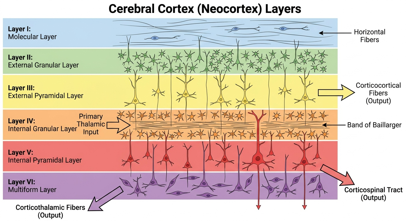
- The most superficial layer
- Contains very few cell bodies
- Primarily composed of dendrites and axons running horizontally (tangentially)
- Contains dendrites of cells from deeper layers and axons from other cortical areas and thalamic nuclei
- Cajal-Retzius cells are found here during development
- Contains small pyramidal cells and numerous stellate (granule) cells
- Densely packed small neurons
- Dendrites extend into Layer I, axons project to deeper layers
- Contains medium-sized pyramidal cells (larger than Layer II)
- This is the primary source of cortico-cortical (association and commissural) fibers
- Axons project to the contralateral hemisphere via the corpus callosum and to ipsilateral association areas
- Contains densely packed stellate (granule) cells
- This is the primary RECEIVING layer for thalamocortical afferents (specific sensory input from thalamus)
- Very prominent in primary sensory cortices — hence called "granular cortex" or "koniocortex"
- Contains a dense band of horizontally running fibers called the external band of Baillarger
- In the primary visual cortex (area 17), this band is so prominent it is visible to the naked eye — the Line (Stria) of Gennari — hence the term "striate cortex"
- Contains the largest pyramidal cells, including the giant cells of Betz in the primary motor cortex
- Primary source of corticospinal, corticobulbar, corticostriatal, and corticopontine fibers
- Contains the internal band of Baillarger
- This is the main OUTPUT layer for subcortical projections
- Deepest layer, blends with subcortical white matter
- Contains fusiform (spindle-shaped) cells and modified pyramidal cells
- Primary source of corticothalamic fibers (feedback projections to the thalamus)
Types of Cortex Based on Layering
- Homotypical Cortex (6 clear layers): Association areas — most of the cortex
- Heterotypical Cortex:
- Granular (Koniocortex): Layer IV is very thick. Found in primary sensory areas (S1, V1, A1). Receives massive thalamic input.
- Agranular: Layer IV is thin or absent; Layers III and V are thick. Found in primary motor cortex (M1) and premotor areas. Heavy output function.
Cell Types in the Cortex
-
Pyramidal Cells:
- Most abundant (~75%)
- Excitatory neurons using glutamate
- Have a triangular cell body with a single large apical dendrite extending toward the surface and multiple basal dendrites
- Long axons that project to distant cortical and subcortical areas
- Found in layers II, III, V, and VI
- The largest are the Betz cells of layer V in the precentral gyrus, measuring up to 120 μm
-
Stellate (Granule) Cells:
- Small, star-shaped neurons
- Spiny stellate cells: Excitatory, use glutamate. Found mainly in Layer IV.
- Smooth stellate cells (aspiny): Inhibitory, use GABA.
- Local circuit interneurons
-
Other Interneurons:
- Basket cells: Inhibitory (GABAergic), synapse on cell bodies of pyramidal cells
- Chandelier cells: Inhibitory, synapse specifically on the axon initial segment of pyramidal cells — very powerful inhibition
- Double bouquet cells: Vertically oriented inhibitory interneurons
- Martinotti cells: Found in deep layers, send axons to layer I
Cortical Columns — The Functional Unit
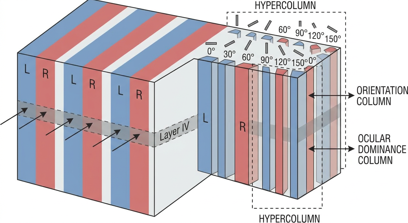
- All neurons within a column respond to the same type of stimulus
- Information flows vertically within columns
- Columns are interconnected horizontally
- Orientation columns — neurons respond to a specific edge orientation
- Ocular dominance columns — neurons preferentially respond to input from one eye
- A hypercolumn contains a complete set of orientation columns and a pair of ocular dominance columns
PART II: FUNCTIONAL AREAS OF THE CEREBRAL CORTEX
Brodmann's Classification
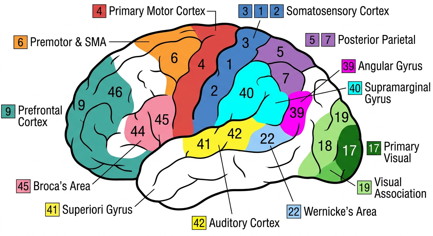
| Brodmann Area | Location | Function |
|---|---|---|
| 4 | Precentral gyrus | Primary motor cortex |
| 6 | Anterior to area 4 | Premotor and supplementary motor area |
| 3, 1, 2 | Postcentral gyrus | Primary somatosensory cortex |
| 5, 7 | Superior parietal lobule | Somatosensory association area |
| 17 | Banks of calcarine sulcus | Primary visual cortex |
| 18, 19 | Surrounding area 17 | Visual association areas |
| 41, 42 | Superior temporal gyrus (Heschl's gyrus) | Primary auditory cortex |
| 22 | Superior temporal gyrus | Auditory association area (Wernicke's area in dominant hemisphere) |
| 44, 45 | Inferior frontal gyrus | Broca's area (dominant hemisphere) |
| 9, 10, 11, 12 | Prefrontal cortex | Higher executive functions |
| 39 | Angular gyrus | Reading, writing, calculation |
| 40 | Supramarginal gyrus | Language integration |
| 28 | Parahippocampal gyrus | Entorhinal cortex (memory gateway) |
PART III: SENSORY AREAS OF THE CORTEX
A. Primary Somatosensory Cortex (S1) — Brodmann Areas 3, 1, 2
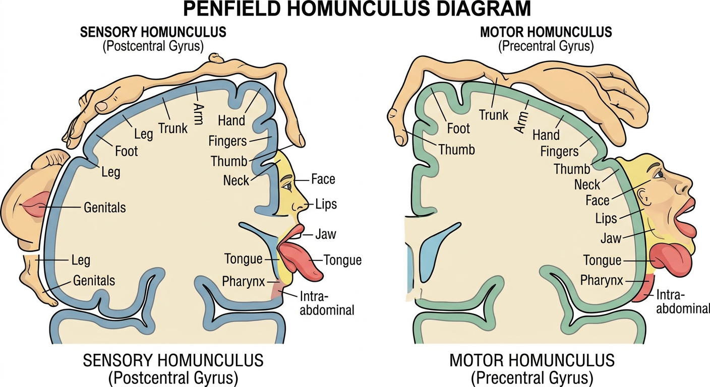
- Somatotopic organization — the famous "Sensory Homunculus" described by Wilder Penfield
- Body is represented upside down: foot and leg on the medial surface, trunk and arm on the lateral convexity, face and tongue at the lateral end
- Contralateral representation — each hemisphere receives sensory information from the opposite side of the body
- The size of cortical representation is proportional to the density of sensory innervation (receptor density), NOT to the physical size of the body part
- The lips, face, thumb, and fingers have disproportionately large representation
- The trunk, back, and proximal limbs have very small representation
- Area 3a: Receives proprioceptive input (from muscle spindles)
- Area 3b: Receives cutaneous tactile input — the TRUE primary somatosensory cortex
- Area 1: Processes texture information
- Area 2: Processes size and shape information (stereognosis)
Clinical Correlates — S1 Lesions:
- Contralateral loss of fine touch, proprioception, vibration, two-point discrimination, and stereognosis
- Pain and temperature are relatively preserved
- Astereognosis (inability to recognize objects by touch)
- Agraphesthesia (inability to recognize letters/numbers traced on skin)
- Positive phenomena: Focal sensory seizures (Jacksonian sensory seizures)
- Combined loss of discriminative sensations with preserved crude touch and pain
- Sensory inattention/extinction on double simultaneous stimulation
B. Somatosensory Association Cortex (S2 and Areas 5, 7)
- Located in the superior wall of the lateral sulcus (parietal operculum)
- Receives bilateral input
- Involved in tactile learning, tactile memory, and attention to somatosensory stimuli
- Area 5: Integrates somatosensory information for spatial awareness of the body and limb position
- Area 7: Integrates visual and somatosensory information for visuospatial processing and hand-eye coordination
Clinical Correlate — Posterior Parietal Lobe Lesions:
- Contralateral hemispatial neglect (hemineglect) — patient ignores the left side of space and their own left body
- Anosognosia — denial of illness, especially denial of left-sided weakness
- Asomatognosia — unawareness or denial of one's own body parts
- Constructional apraxia — inability to draw or copy figures
- Dressing apraxia — difficulty putting on clothes
- Gerstmann syndrome (lesion of angular gyrus, area 39):
- Agraphia (inability to write)
- Acalculia (inability to calculate)
- Finger agnosia (inability to recognize individual fingers)
- Left-right disorientation
- Balint syndrome:
- Simultanagnosia — inability to perceive more than one object at a time
- Optic ataxia — inability to accurately reach for objects under visual guidance
- Oculomotor apraxia — inability to voluntarily direct gaze to objects in the peripheral visual field
C. Primary Visual Cortex (V1) — Brodmann Area 17
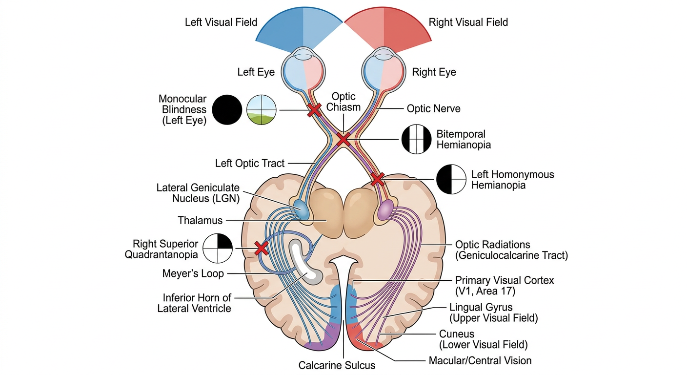
- Classic 6-layered cortex with extremely thick Layer IV (granular/koniocortex)
- Layer IV is subdivided into 4a, 4b, 4cα, and 4cβ
- Layer 4cα receives input from magnocellular layers of LGN (motion, depth)
- Layer 4cβ receives input from parvocellular layers of LGN (color, form)
- Contains the Line of Gennari — a dense band of myelinated fibers in layer 4b, visible to the naked eye
- Contralateral visual field is represented in each hemisphere
- The macula (central vision) has enormous cortical representation — about 50% of V1 is devoted to the central 10 degrees (cortical magnification)
- Upper visual field → represented on the lower bank of the calcarine sulcus (lingual gyrus)
- Lower visual field → represented on the upper bank (cuneus)
- Peripheral visual field → represented anteriorly
- Central (macular) visual field → represented posteriorly at the occipital pole
- Simple cells: Respond to bars of light with a specific orientation at a specific position. Found mainly in Layer IV.
- Complex cells: Respond to bars of light with a specific orientation anywhere within a larger receptive field. Can detect movement direction. Found in Layers II, III, V, VI.
- Hypercomplex (end-stopped) cells: Respond to bars/edges of a specific length, orientation, and position. Detect corners and curvatures.
Clinical Correlates — V1 Lesions:
- Complete bilateral destruction → Cortical blindness — pupillary reflexes intact, but patient cannot see
- Unilateral destruction → Contralateral homonymous hemianopia with macular sparing
- Upper bank lesion (cuneus) → Contralateral inferior quadrantanopia
- Lower bank lesion (lingual gyrus) → Contralateral superior quadrantanopia
D. Visual Association Areas (V2-V5 and beyond)
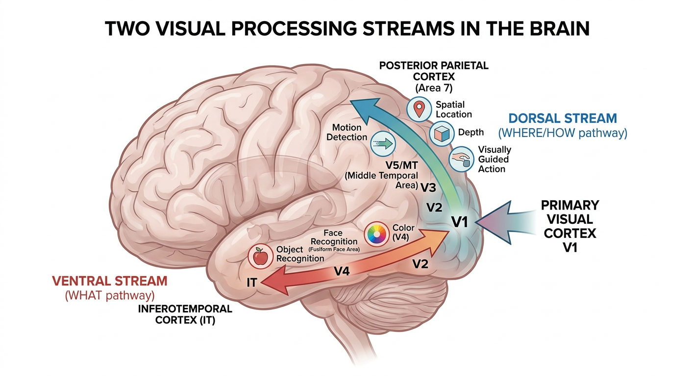
-
Dorsal Stream ("Where/How" pathway):
- V1 → V2 → V3 → V5/MT → Posterior parietal cortex (area 7)
- Processes motion, spatial location, depth, visually guided actions
- Dominated by magnocellular input
- V5/MT: Specialized for motion detection
-
Ventral Stream ("What" pathway):
- V1 → V2 → V4 → Inferotemporal cortex (IT)
- Processes object recognition, face recognition, color
- Dominated by parvocellular input
- V4: Color processing
- Fusiform face area (FFA): Specialized for face recognition (in fusiform gyrus)
- Parahippocampal place area: Scene and place recognition
Clinical Correlates — Visual Association Area Lesions:
- Apperceptive agnosia: Cannot form a complete percept. Cannot copy drawings. (Bilateral parieto-occipital lesions)
- Associative agnosia: Can perceive and copy the object but cannot identify/name it. (Bilateral ventral occipitotemporal lesions)
E. Primary Auditory Cortex (A1) — Brodmann Areas 41, 42
- Tonotopic organization:
- Low frequencies → anterolateral
- High frequencies → posteromedial
- Receives bilateral input (but predominantly contralateral)
Clinical Correlates — A1 Lesions:
- Unilateral lesion → Difficulty in sound localization on the contralateral side, but NO significant deafness
- Bilateral destruction → Cortical deafness (very rare)
F. Auditory Association Cortex — Area 22
G. Gustatory Cortex
- Thalamic input from VPM nucleus
H. Vestibular Cortex
- Receives input from vestibular nuclei via thalamus
I. Olfactory Cortex
- Unique: The ONLY sensory system that reaches the cortex WITHOUT a thalamic relay
- Also projects to entorhinal cortex, amygdala, and orbitofrontal cortex
PART IV: MOTOR AREAS OF THE CORTEX
A. Primary Motor Cortex (M1) — Brodmann Area 4
- Agranular cortex — Layer IV is very thin/absent
- Layer V contains the giant Betz cells (up to 120 μm)
- Betz cells constitute only ~3-5% of corticospinal tract fibers
- Somatotopic organization — Motor Homunculus (Penfield and Rasmussen)
- Body represented upside down (see Homunculus image above)
- The size of cortical representation is proportional to the fineness and complexity of movement (dexterity)
- Hands, fingers, lips, tongue, and larynx have enormous representation
- Contralateral representation — each hemisphere controls the opposite side of the body
- Corticospinal tract → internal capsule → cerebral peduncle → basis pontis → medullary pyramids
- At the pyramidal decussation: 85-90% cross → lateral corticospinal tract (distal limb muscles)
- 10-15% remain uncrossed → anterior corticospinal tract (axial and proximal muscles)
- Corticobulbar fibers → to cranial nerve motor nuclei
Clinical Correlates — M1 Lesions:
- Contralateral spastic paralysis (initially flaccid in acute phase — "cerebral shock")
- Hyperreflexia — exaggerated deep tendon reflexes
- Spasticity — velocity-dependent increase in muscle tone (clasp-knife)
- Clonus — rhythmic involuntary contractions
- Babinski sign positive — extension (dorsiflexion) of the great toe with fanning of other toes
- Loss of fine, skilled movements
- No significant muscle atrophy, no fasciculations
- Absent superficial reflexes (abdominal, cremasteric)
B. Premotor Cortex (PMC) — Brodmann Area 6 (Lateral Part)
- Planning and programming of movements based on external sensory cues
- Controls proximal and trunk muscles for postural stabilization
- Contains mirror neurons — neurons that fire both when performing an action AND when observing someone else perform the same action
- Important for imitation, motor learning, understanding others' actions, empathy, and language evolution
C. Supplementary Motor Area (SMA) — Brodmann Area 6 (Medial Part)
- Planning and initiation of self-generated (internally cued) movements
- Bimanual coordination
- Motor sequencing
- Mental rehearsal of movement — SMA is active even when you THINK about making a movement
- A slowly rising negative electrical potential over the SMA beginning approximately 1-1.5 seconds BEFORE a voluntary self-initiated movement
- Discovered by Kornhuber and Deecke (1965)
- Starts bilaterally over SMA, then lateralizes to contralateral M1 just before movement
Clinical Correlates:
- Transient akinesia (inability to initiate voluntary movements)
- Mutism (if dominant hemisphere SMA is affected)
- Difficulty with bimanual coordination
- Patients recover relatively well over weeks to months
- One hand performs purposeful actions that the patient denies initiating or controlling
- The hand seems to "have a mind of its own"
- Occurs with lesions of medial frontal cortex (SMA) and/or corpus callosum (anterior cerebral artery territory)
D. Frontal Eye Field (FEF) — Brodmann Area 8
E. Broca's Area — Brodmann Areas 44, 45
- Motor programming of speech
- Also involved in grammar and syntax processing
- Connected to Wernicke's area via the arcuate fasciculus
Clinical Correlate — Broca's Aphasia (Motor/Expressive/Non-fluent Aphasia):
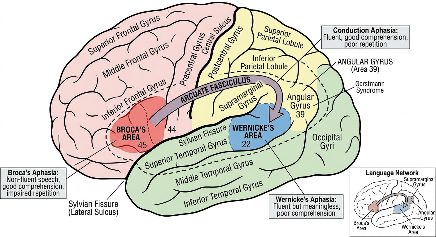
- Non-fluent speech — halting, effortful, telegraphic speech
- Relatively preserved comprehension
- Impaired repetition
- Speech limited to short phrases, lacking prepositions, articles, and conjunctions (agrammatic)
- Patient is aware of deficit and is frustrated
- Often accompanied by right-sided hemiparesis (face and arm)
PART V: ASSOCIATION AREAS OF THE CORTEX
A. Prefrontal Association Cortex (Areas 9, 10, 11, 12, 46, 47)
- Working memory
- Executive functions: planning, strategizing, problem-solving, cognitive flexibility, abstract thinking, judgment, temporal ordering
- Sustained attention and concentration
- Emotional regulation and social behavior
- Impulse control and inhibition of inappropriate behavior
- Personality and social conduct
- Reward-based decision making
- Self-referential processing
- Theory of mind — understanding others' mental states and intentions
- Moral reasoning
- Default mode network (active during rest/daydreaming)
- Language processing (left hemisphere)
- Response inhibition
- Working memory for objects
Clinical Correlates — Prefrontal Lesions:
- Impaired working memory
- Perseveration — inability to shift from one task/response to another; patient repeats the same response even when it's no longer appropriate
- Demonstrated by:
- Wisconsin Card Sorting Test
- Trail Making Test
- Reduced verbal fluency
- Apathy and reduced motivation (abulia)
- Difficulty with abstract thinking
- Disinhibition — inappropriate social behavior, tactlessness
- Personality change — more impulsive, irresponsible
- Emotional lability — inappropriate joking (Witzelsucht)
- Impaired judgment and poor decision-making
- Utilization behavior — compulsive use of objects placed in front of them
- A railway construction foreman who survived an iron tamping rod passing through his left frontal lobe (orbitofrontal cortex)
- Before the accident: responsible, capable, well-liked
- After the accident: "fitful, irreverent, indulging at times in the grossest profanity...impatient of restraint or advice...a child in his intellectual capacity and manifestations, he has the animal passions of a strong man"
- This case was historically crucial in establishing the role of the frontal lobes in personality and social behavior
- Abulia — profound lack of will, motivation, and spontaneous action
- Akinetic mutism (severe cases) — patient is awake but makes no spontaneous movements or speech
- Usually bilateral medial frontal lesions (anterior cerebral artery territory)
- Grasp reflex — stroking the palm causes involuntary grasping
- Rooting reflex — stroking the cheek causes the head to turn toward the stimulus
- Sucking reflex — touching the lips causes sucking movements
- Palmomental reflex — stroking the palm causes contraction of the ipsilateral mentalis (chin) muscle
- Snout reflex — tapping the lips causes lip puckering
- Glabellar tap (Myerson's sign) — repeated tapping on the glabella causes persistent blinking
- Gegenhalten (paratonia) — involuntary resistance to passive movement
- Motor impersistence — inability to sustain a voluntary act
- Echopraxia — involuntary imitation of movements of others
- Incontinence — medial frontal lesions can cause urinary incontinence
B. Parieto-Occipito-Temporal Association Cortex
- Comprehension of spoken language
- Connected to Broca's area via the arcuate fasciculus
- Integration of visual information with language
- Critical for reading
- Phonological processing and language production integration
Clinical Correlates:
- Fluent speech — normal or even excessive speech output
- But the speech is MEANINGLESS — filled with:
- Phonemic (literal) paraphasias: Substituting similar-sounding words/syllables
- Semantic (verbal) paraphasias: Substituting related words
- Neologisms: Made-up words
- Jargon: Unintelligible stream of neologisms and paraphasias
- Severely impaired comprehension
- Impaired repetition
- Patient is unaware of the deficit (anosognosia)
- No hemiparesis (Wernicke's area is distant from the motor cortex)
- Lesion: Arcuate fasciculus
- Fluent speech with frequent phonemic paraphasias
- Good comprehension
- Severely impaired repetition — the hallmark
- Lesion: Large lesion of the dominant hemisphere involving both Broca's and Wernicke's areas
- Non-fluent speech, impaired comprehension, impaired repetition
- Usually accompanied by right hemiplegia, right hemianopia, right hemisensory loss
- Non-fluent speech, good comprehension, INTACT repetition
- Distinguishes it from Broca's aphasia
- Fluent paraphasic speech, impaired comprehension, INTACT repetition
- Distinguishes it from Wernicke's aphasia
- Patient can repeat sentences without understanding them (echolalia)
- Non-fluent speech, impaired comprehension, INTACT repetition (only thing preserved)
| Type | Fluency | Comprehension | Repetition | Lesion |
|---|---|---|---|---|
| Broca's | Non-fluent | Good | Impaired | Inferior frontal (area 44,45) |
| Wernicke's | Fluent | Impaired | Impaired | Superior temporal (area 22) |
| Conduction | Fluent | Good | Impaired | Arcuate fasciculus |
| Global | Non-fluent | Impaired | Impaired | Large perisylvian |
| Transcortical Motor | Non-fluent | Good | Intact | Anterior/superior to Broca's |
| Transcortical Sensory | Fluent | Impaired | Intact | Posterior to Wernicke's |
| Mixed Transcortical | Non-fluent | Impaired | Intact | Watershed zones |
PART VI: CEREBRAL DOMINANCE AND LATERALIZATION
Left Hemisphere (Dominant in Most People)
- Language — speech production, comprehension, reading, writing
- Logical/analytical processing
- Mathematical calculations
- Sequential/temporal processing
- Fine motor control of the dominant hand
Right Hemisphere (Non-dominant in Most People)
- Visuospatial processing — spatial orientation, mental rotation
- Facial recognition
- Music perception and production (especially melody, pitch, timbre)
- Emotional processing:
- Emotional prosody — understanding and expressing emotion through tone of voice
- Attention — especially spatial attention
- Holistic/gestalt processing
- Constructional abilities — drawing, copying complex figures
- Motor aprosodia (right frontal): Cannot express emotion through tone of voice — speech is monotone
- Sensory aprosodia (right temporal): Cannot understand emotional tone of others' speech
Anatomical Asymmetries
- Planum temporale: Larger on the left in 65% of brains. Corresponds to Wernicke's area.
- Sylvian fissure: Longer on the left, angles up more steeply on the right.
- Heschl's gyrus: Often two gyri on the right, one on the left.
- Frontal lobe: Right frontal lobe slightly larger (right frontal petalia)
- Occipital lobe: Left occipital lobe slightly larger (left occipital petalia)
Split-Brain Studies
- Object in left visual field (goes to right hemisphere) → patient cannot name it but can pick it out by touch with the left hand
- Object in right visual field (goes to left hemisphere) → patient can name it normally
- Object placed in left hand → patient cannot name it verbally but can demonstrate its use
- Right hemisphere can understand simple language but cannot produce speech
PART VII: INTELLECTUAL FUNCTIONS OF THE BRAIN
A. Attention
- Alertness/Arousal: Maintained by the reticular activating system (RAS)
- Selective attention: Involves prefrontal cortex, parietal cortex, and thalamus
- Sustained attention (vigilance): Right frontal and parietal cortex
- Divided attention: Prefrontal cortex
- Right parietal cortex — spatial attention (dominant role). Attends to BOTH sides of space, whereas the left parietal cortex primarily attends to the right side. This is why right parietal lesions cause profound left neglect.
- Prefrontal cortex — executive attention, working memory, sustained attention
- Anterior cingulate cortex — conflict monitoring, error detection
- Thalamic reticular nucleus — gates sensory information to the cortex
- Ascending reticular activating system — maintains overall arousal
B. Language
C. Higher Cognitive Functions
PART VIII: LEARNING AND MEMORY
Definitions
- Learning: The process of acquiring new information or modifying existing knowledge, behaviors, skills, values, or preferences
- Memory: The process of encoding, storing, and retrieving information
Classification of Memory
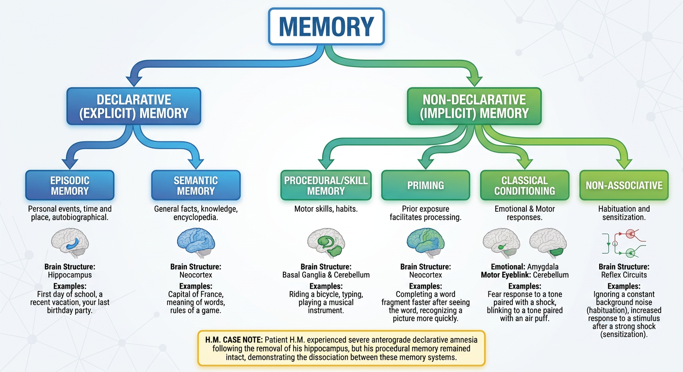
A. By Duration:
- Duration: Milliseconds to seconds
- Types:
- Iconic memory (visual) — lasts ~200-500 milliseconds
- Echoic memory (auditory) — lasts ~3-4 seconds
- Acts as a buffer for incoming sensory information
- Duration: Seconds to minutes (~15-30 seconds without rehearsal)
- Capacity: 7 ± 2 items (Miller's Magic Number, 1956)
- Working Memory (Baddeley's Model):
- Phonological loop: Stores and rehearses verbal/acoustic information
- Phonological store (inner ear) — holds speech-based information for ~2 seconds
- Articulatory rehearsal process (inner voice) — refreshes information by subvocal repetition
- Visuospatial sketchpad: Stores and manipulates visual and spatial information
- Central executive: Attentional control system. Dependent on prefrontal cortex
- Episodic buffer: Integrates information from subsystems and long-term memory
- Phonological loop: Stores and rehearses verbal/acoustic information
- Neural basis: Primarily dorsolateral prefrontal cortex
- Duration: Hours to a lifetime
- Capacity: Virtually unlimited
- Requires consolidation
B. By Content (Types of Long-Term Memory):
-
Memory for facts and events that can be consciously recalled and verbally declared
-
Dependent on medial temporal lobe (hippocampus, entorhinal cortex, perirhinal cortex, parahippocampal cortex) and diencephalic structures (mammillary bodies, anterior and mediodorsal thalamic nuclei)a. Episodic Memory:
- Memory for personal events and experiences — contextual details (time, place, emotions)
- Critically dependent on the hippocampus
b. Semantic Memory:- Memory for general knowledge and facts — independent of personal context
- Less dependent on hippocampus once consolidated; stored in neocortical association areas
- More resistant to hippocampal damage than episodic memory
-
Memory expressed through performance rather than conscious recollection
-
NOT dependent on hippocampus or medial temporal lobea. Procedural (Skill/Habit) Memory:
- Memory for motor skills and cognitive procedures (riding a bicycle, typing, playing piano)
- Neural basis: Basal ganglia (striatum), cerebellum, supplementary motor area
- Intact in amnesic patients — H.M. could learn new motor skills even though he couldn't remember learning them
b. Classical Conditioning:- Emotional conditioning (fear conditioning): Amygdala
- Motor conditioning (eyeblink conditioning): Cerebellum (specifically interpositus nucleus)
c. Priming:- Improved identification/processing of a stimulus based on prior exposure, without conscious awareness
- Neural basis: Neocortex
- Intact in amnesic patients
d. Non-associative Learning:- Habituation: Decreased response to a repeated, innocuous stimulus — the simplest form of learning
- Sensitization: Increased response to a stimulus after exposure to a strong or noxious stimulus
- Studied extensively in Aplysia by Eric Kandel (Nobel Prize, 2000)
Neural Substrates of Memory
The Hippocampus and Medial Temporal Lobe
- Located in the medial temporal lobe, forming the floor of the inferior horn of the lateral ventricle
- Components:
- Hippocampus proper (Cornu Ammonis, CA): CA1, CA2, CA3, CA4
- Dentate gyrus
- Subiculum
- Entorhinal cortex (Brodmann area 28) — the gateway to the hippocampus
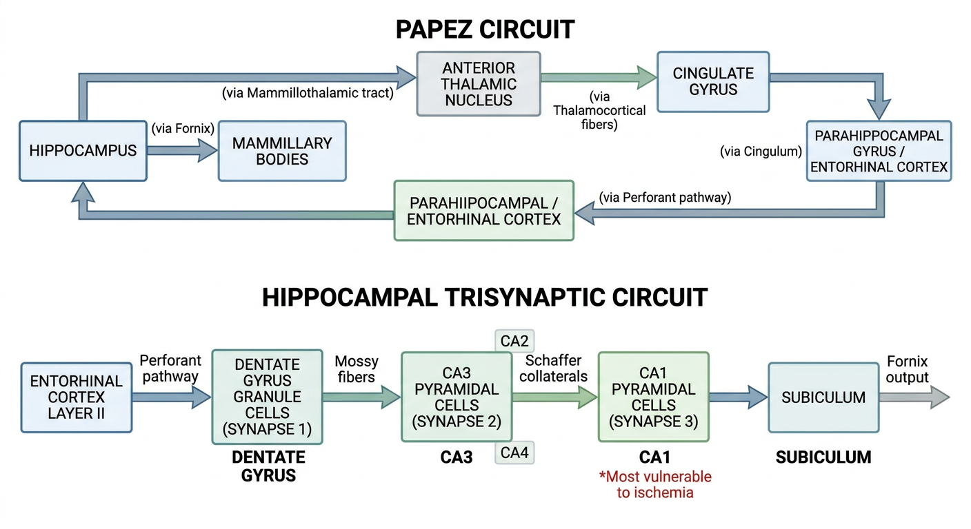
- Entorhinal cortex (Layer II) → via Perforant pathway → Dentate gyrus (Synapse 1)
- Dentate gyrus → via Mossy fibers → CA3 (Synapse 2)
- CA3 → via Schaffer collaterals → CA1 (Synapse 3)
- CA1 → Subiculum → Fornix → Mammillary bodies → Mammillothalamic tract → Anterior thalamic nucleus → Cingulate gyrus → Entorhinal cortex
- NOT the storage site of long-term memories
- Essential for the CONSOLIDATION of new declarative memories
- Acts as a temporary holding station or index that binds together distributed cortical representations during memory formation
- Over time, memories become independent of the hippocampus and are stored in neocortical networks — systems consolidation
- Contains place cells — neurons that fire when the animal is in a specific location. Discovered by John O'Keefe (Nobel Prize, 2014, shared with May-Britt and Edvard Moser who discovered grid cells in the entorhinal cortex)
- Right hippocampus: More involved in spatial memory
- Left hippocampus: More involved in verbal/declarative memory
Clinical Correlate — The Famous Case of H.M. (Henry Molaison):
- In 1953, Dr. William Scoville performed bilateral medial temporal lobe resection (removing hippocampus, amygdala, and surrounding cortex bilaterally) for intractable epilepsy
- Unable to form ANY new declarative (explicit) long-term memories after the surgery
- Could not remember people he met, events that occurred, or things he learned after the operation
- Immediate/working memory was INTACT — could carry on a conversation, but the moment he was distracted, the memory vanished
- Remote memories (from before surgery) were relatively intact
- Procedural/motor learning — H.M. improved on the mirror drawing task with practice each day, even though he had no memory of ever doing the task before
- Priming — showed normal repetition priming
- Classical conditioning
- Immediate memory/working memory
- Intelligence — IQ was normal
- Personality — essentially unchanged
- The hippocampus is essential for formation of new declarative memories
- The hippocampus is NOT needed for immediate/working memory
- The hippocampus is NOT needed for procedural memory
- Memory is NOT a unitary faculty — different types depend on different brain systems
- Short-term and long-term memory are distinct processes
Clinical Correlate — Korsakoff Syndrome:
- Etiology: Chronic alcoholism → thiamine (Vitamin B1) deficiency → damage to mammillary bodies and dorsomedial thalamic nuclei
- Features:
- Severe anterograde amnesia
- Retrograde amnesia — temporal gradient (recent memories more affected than remote)
- Confabulation — fills memory gaps with fabricated, plausible-sounding stories without intention to deceive
- Lack of insight
- Personality and other cognitive functions may be relatively preserved
- Often preceded by Wernicke's encephalopathy (acute phase):
- Classic triad: Confusion, ophthalmoplegia, ataxia
- Together called Wernicke-Korsakoff syndrome
- Wernicke's is a medical emergency requiring immediate IV thiamine
The Amygdala and Emotional Memory
- Modulates memory consolidation based on emotional arousal — emotionally charged events are remembered better
- Critical for fear conditioning
- Essential for recognizing emotional expressions (especially fear) in faces
- Emotional event → activation of amygdala
- Amygdala → activation of hypothalamus and brainstem → release of stress hormones (epinephrine, cortisol)
- These hormones and direct amygdala inputs enhance synaptic plasticity in the hippocampus
- Result: Stronger, more durable memory for emotionally significant events (flashbulb memories)
- Psychic blindness (visual agnosia)
- Hyperorality — tendency to explore objects by mouth
- Hypermetamorphosis — compulsive attention to all visual stimuli
- Emotional blunting — loss of fear and aggression (placidity)
- Hypersexuality
- Dietary changes and hyperphagia
- Rare genetic disorder causing bilateral calcification of the amygdala
- Patient S.M.: Cannot recognize fear in facial expressions, cannot experience fear in normally fear-inducing situations, does not learn fear conditioning, but can form declarative memories normally
- Amygdala hyperactivity → excessive fear conditioning to trauma-related cues
- Hippocampal volume may be reduced
- Prefrontal cortex (which normally inhibits the amygdala) may be hypoactive
Memory Consolidation
- Occurs within minutes to hours after learning
- Requires new protein synthesis
- Can be blocked by protein synthesis inhibitors applied shortly after learning
- Involves the mechanisms of long-term potentiation (LTP)
- Occurs over weeks, months, to years
- Gradual transfer of memory from hippocampal dependence to neocortical storage
- Sleep plays a crucial role:
- Slow-wave sleep (SWS/NREM Stage 3): Important for declarative memory consolidation. Hippocampal sharp-wave ripples during SWS are associated with replay of learned experiences.
- REM sleep: Important for procedural and emotional memory consolidation
- When a consolidated memory is retrieved/reactivated, it temporarily becomes labile (unstable) again and must be re-stabilized
- During this window, the memory can be modified, strengthened, weakened, or even erased
- Therapeutic implications: Potential treatment for PTSD — reactivate traumatic memory, then administer propranolol (a β-blocker) that interferes with reconsolidation → weakened traumatic memory
Synaptic Mechanisms of Learning and Memory
Long-Term Potentiation (LTP)
- Cooperativity: A minimum number of presynaptic fibers must be activated simultaneously to induce LTP
- Associativity: A weak input can be potentiated if activated simultaneously with a strong input — the neural basis of Hebbian learning: "Neurons that fire together wire together"
- Input Specificity: LTP occurs only at the specific synapses that were activated
- Persistence: LTP lasts for a long time (hours to weeks in vivo)
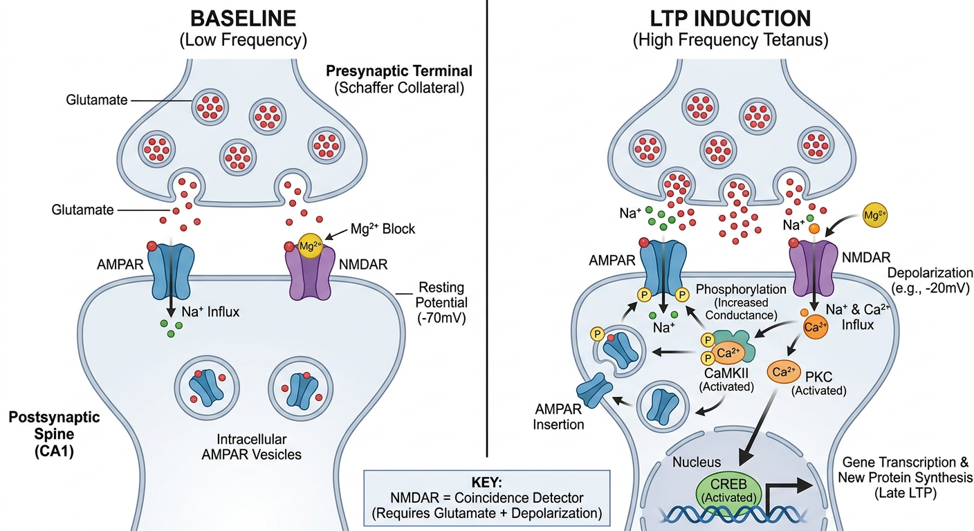
- Presynaptic terminal releases glutamate during normal low-frequency transmission
- Glutamate binds to AMPA receptors → influx of Na⁺ → small depolarization (EPSP)
- Under normal conditions, NMDA receptors are blocked by a Mg²⁺ ion at resting membrane potential
- During high-frequency stimulation:
- Repeated AMPA activation → cumulative depolarization → Mg²⁺ block is relieved
- Now NMDA receptor can pass current → influx of Ca²⁺ into the postsynaptic cell
- NMDA receptor is thus a "coincidence detector" — requires BOTH presynaptic activity (glutamate) AND postsynaptic depolarization (to relieve Mg²⁺ block). This is the molecular basis of Hebbian learning.
- Ca²⁺ influx triggers:
- Activation of CaMKII — most important kinase for LTP
- Activation of PKC
- Activation of tyrosine kinases
- These kinases:
- Phosphorylate existing AMPA receptors → increased conductance
- Insert additional AMPA receptors from intracellular vesicular pools → more AMPA receptors → stronger synaptic response
- Ca²⁺ signal and kinase activity activate:
- Adenylyl cyclase → cAMP → PKA
- MAPK/ERK pathway
- PKA and MAPK translocate to the nucleus
- They phosphorylate CREB (cAMP Response Element Binding protein)
- Activated CREB → transcription of new genes → new protein synthesis, including:
- Structural proteins for new synapse formation
- Growth of new dendritic spines
- BDNF (Brain-Derived Neurotrophic Factor)
- More AMPA receptors and scaffolding proteins
- Result: Structural changes — growth of new synapses, formation of new dendritic spines → long-lasting synaptic strengthening
- This is how short-term memory → long-term memory (at the molecular level)
| Feature | Early LTP | Late LTP |
|---|---|---|
| Duration | 1-3 hours | Hours to weeks |
| Protein synthesis | Not required | Required |
| Gene transcription | Not required | Required |
| Key molecules | CaMKII, PKC | CREB, BDNF, new proteins |
| Mechanism | Phosphorylation, AMPA insertion | New synapse formation, structural changes |
Long-Term Depression (LTD)
- Low-frequency stimulation (e.g., 1-5 Hz for 10-15 minutes)
- Small, sustained Ca²⁺ entry (NOT the large transient Ca²⁺ rise seen in LTP)
- Modest Ca²⁺ rise preferentially activates protein phosphatases (calcineurin/PP2B and PP1) rather than kinases
- Phosphatases dephosphorylate AMPA receptors → AMPA receptors are internalized by endocytosis → weakened synaptic response
- Clearing old/irrelevant memory traces → making room for new learning
- Fine-tuning synaptic connections during development and learning
- Motor learning in the cerebellum (cerebellar LTD at the parallel fiber-Purkinje cell synapse is critical for error correction)
Spike-Timing-Dependent Plasticity (STDP)
- If the presynaptic neuron fires BEFORE the postsynaptic neuron (within ~20 ms) → LTP
- If the presynaptic neuron fires AFTER the postsynaptic neuron → LTD
- Implements Hebb's rule at the millisecond timescale
Kandel's Studies on Aplysia — Molecular Basis of Simple Learning
- Repeated gentle siphon stimulation → progressive decrease in gill withdrawal
- Mechanism:
- Progressive inactivation of presynaptic voltage-gated Ca²⁺ channels
- Less Ca²⁺ entry → less neurotransmitter release → smaller EPSP → weaker reflex
- Presynaptic mechanism
- Short-term habituation: Modification of existing proteins
- Long-term habituation: Reduction in number of active zones and synaptic connections (requires gene expression)
-
A strong tail shock → subsequent gentle siphon touch now produces an EXAGGERATED gill withdrawal
-
Mechanism (Short-term sensitization):
- Tail shock activates facilitatory interneurons releasing serotonin (5-HT) onto sensory neuron presynaptic terminals
- Serotonin → adenylyl cyclase → cAMP → PKA
- PKA phosphorylates and closes K⁺ channels → broader action potential
- Broader AP → Ca²⁺ channels open longer → more Ca²⁺ entry → more neurotransmitter release
- Enhanced EPSP → stronger gill withdrawal
- This is presynaptic facilitation
-
Long-term sensitization (lasting days to weeks):
- Repeated tail shocks → sustained serotonin → persistently elevated cAMP/PKA
- PKA translocates to the nucleus
- PKA phosphorylates CREB-1 (transcription activator) and removes CREB-2 (repressor)
- CREB-1 → gene transcription → new protein synthesis
- Growth of new synaptic connections — sensory neurons may double their synaptic connections
- Long-lasting structural change → long-term memory
- Pairing a gentle siphon touch (CS) with a tail shock (US) → stronger response than sensitization alone
- Activity-dependent presynaptic facilitation: When the sensory neuron fires (CS) just BEFORE the facilitatory interneuron releases serotonin (US), adenylyl cyclase is MORE strongly activated by synergistic action of Ca²⁺ and serotonin → greater presynaptic facilitation → associative learning
Neurogenesis and Memory
- Subventricular zone (SVZ) → new neurons migrate to the olfactory bulb
- Subgranular zone (SGZ) of the dentate gyrus of the hippocampus → new granule cells integrate into hippocampal circuits
- Exercise (running), enriched environment, learning itself, BDNF, antidepressants (SSRIs)
- Stress (cortisol), aging, depression, alcohol, sleep deprivation
- Pattern separation — ability to distinguish between similar memories/stimuli
- Spatial memory
- Mood regulation (may mediate antidepressant effects)
Neurotransmitters and Memory
| Neurotransmitter | Role in Memory |
|---|---|
| Glutamate | Primary excitatory NT; essential for LTP (via NMDA and AMPA receptors) |
| Acetylcholine | Critical for attention and memory encoding; cholinergic neurons from nucleus basalis of Meynert project to cortex and hippocampus; DESTROYED in Alzheimer's disease |
| Dopamine | Reward-based learning; working memory (prefrontal cortex); motivation to learn |
| Norepinephrine | Emotional arousal → enhances memory consolidation (via amygdala); attention |
| Serotonin | Modulates learning (presynaptic facilitation in Aplysia); mood → influences memory |
| GABA | Inhibitory; benzodiazepines (enhance GABA) cause anterograde amnesia |
| BDNF | Neurotrophin; essential for LTP maintenance, synaptic growth, neurogenesis |
PART IX: CLINICAL DISORDERS OF MEMORY
A. Amnesia
- Inability to form new memories after the onset of the condition
- Example: H.M., Korsakoff syndrome
- Bilateral hippocampal/medial temporal lobe damage
- Loss of memories formed BEFORE the onset of the condition
- Temporal gradient (Ribot's law): Recent memories are more vulnerable to loss than remote memories
- Sudden onset of severe anterograde amnesia lasting < 24 hours (typically 4-6 hours)
- Patient is alert, oriented to self, but repeatedly asks the same questions
- No focal neurological deficits
- Complete recovery with no recurrence (usually)
- Cause: Possibly transient ischemia of hippocampal/medial temporal regions
- May be triggered by physical exertion, emotional stress, Valsalva maneuver, sexual intercourse, or cold water immersion
- DWI-MRI may show small lesions in the hippocampus (CA1 region)
- Memory loss due to psychological trauma rather than organic brain damage
- Usually retrograde amnesia for autobiographical information
- May include dissociative fugue — sudden, unexpected travel with inability to recall past identity
- Potentially reversible with psychological treatment or hypnosis
B. Alzheimer's Disease (AD)
-
Amyloid plaques (neuritic plaques/senile plaques):
- Extracellular deposits of β-amyloid protein (Aβ) surrounded by dystrophic neurites
- Aβ derived from abnormal cleavage of Amyloid Precursor Protein (APP) by β-secretase and γ-secretase
- Normal cleavage by α-secretase produces non-amyloidogenic fragments
- Aβ42 is the most toxic and aggregation-prone form
-
Neurofibrillary tangles (NFTs):
- Intracellular twisted fibers of hyperphosphorylated tau protein
- Tau normally stabilizes microtubules; when hyperphosphorylated, tau dissociates → microtubule collapse → disrupted axonal transport → neuronal death
- NFTs correlate better with cognitive decline than amyloid plaques
-
Loss of cholinergic neurons:
- Severe degeneration of the nucleus basalis of Meynert (basal forebrain)
- This nucleus provides the major cholinergic input to the entire cerebral cortex and hippocampus
- Loss of ACh → impaired attention and memory
- Basis for cholinesterase inhibitor therapy (donepezil, rivastigmine, galantamine)
-
Neuronal and synaptic loss:
- Begins in entorhinal cortex and hippocampus → spreads to temporal, parietal, and eventually frontal cortices
- Braak staging: Stage I-II (entorhinal), III-IV (limbic/hippocampal), V-VI (neocortical)
-
Gross pathology:
- Cortical atrophy (especially temporal and parietal)
- Widened sulci, narrowed gyri
- Enlarged ventricles (hydrocephalus ex vacuo)
- Hippocampal atrophy visible on MRI (coronal sections)
- Early stage: Memory loss — particularly recent (episodic) memory; forgetting names, misplacing objects, repeating questions
- Intermediate stage: Word-finding difficulty (anomia), visuospatial disorientation, apraxia, impaired judgment, personality changes
- Late stage: Severe cognitive impairment, inability to perform activities of daily living, incontinence, mutism, death (typically 8-10 years after diagnosis)
-
Early-onset AD (< 65 years, ~5% of cases):
- APP gene (chromosome 21) — Down syndrome patients invariably develop AD pathology by age 40
- Presenilin 1 (PSEN1) (chromosome 14) — most common cause of familial early-onset AD
- Presenilin 2 (PSEN2) (chromosome 1)
-
Late-onset AD (> 65 years, ~95% of cases):
- APOE ε4 allele (chromosome 19) — most important genetic risk factor
- ε4/ε4 homozygotes: ~15× increased risk
- APOE ε2 allele is protective
- APOE ε4 allele (chromosome 19) — most important genetic risk factor
- Cholinesterase inhibitors: Donepezil, rivastigmine, galantamine
- Memantine: NMDA receptor antagonist — reduces glutamate excitotoxicity; used in moderate-severe AD
- Anti-amyloid antibodies: Lecanemab, aducanumab — disease-modifying therapies that clear amyloid plaques (modest clinical benefit, controversial)
C. Other Dementias
- Affects frontal and temporal lobes preferentially
- Prominent behavioral/personality changes and/or language dysfunction early in the course (contrast with AD where memory loss is prominent early)
- Behavioral variant FTD: Personality change, disinhibition, apathy, loss of empathy
- Primary progressive aphasia: Progressive language deterioration
- Pathology: Tau-positive (Pick bodies) or TDP-43 positive inclusions
- Younger onset than AD (typically 50s-60s)
- Memory is relatively preserved early
- Lewy bodies (intracytoplasmic inclusions of α-synuclein) in cortical and brainstem neurons
- Triad:
- Fluctuating cognition with pronounced variations in attention and alertness
- Visual hallucinations — well-formed, detailed, often of people or animals
- Parkinsonism
- Additional features: REM sleep behavior disorder, severe sensitivity to antipsychotics, falls, syncope
- Stepwise deterioration rather than gradual decline
- Executive dysfunction often more prominent than memory impairment
- Associated with hypertension, diabetes, atrial fibrillation
D. Age-Related Memory Changes
- Episodic memory declines
- Working memory capacity decreases
- Processing speed slows
- Semantic memory is well preserved or may even improve
- Procedural memory is well preserved
- Cognitive decline greater than expected for age but NOT severe enough for dementia
- Amnestic MCI: Memory impairment predominant — approximately 10-15% per year convert to Alzheimer's disease
PART X: THE ELECTROENCEPHALOGRAM (EEG) AND CORTICAL ELECTRICAL ACTIVITY
Origin of the EEG
- The EEG records electrical activity of the cerebral cortex from electrodes placed on the scalp
- The signals primarily reflect summated postsynaptic potentials (EPSPs and IPSPs) of cortical pyramidal neurons, NOT action potentials
- The apical dendrites of pyramidal cells (oriented perpendicular to the cortical surface) create extracellular current flow (dipoles)
- EEG requires synchronous activity of large populations of neurons (millions) to generate detectable signals
Normal EEG Rhythms
| Rhythm | Frequency | Amplitude | State/Conditions |
|---|---|---|---|
| Beta (β) | 13-30 Hz | Low (< 20 μV) | Alert, active thinking, eyes open. "Desynchronized" pattern |
| Alpha (α) | 8-13 Hz | Moderate (20-60 μV) | Relaxed, awake, eyes closed. Best seen over occipital region. |
| Theta (θ) | 4-7 Hz | Variable | Drowsiness, light sleep. Normal in children. Abnormal in awake adults. |
| Delta (δ) | 0.5-4 Hz | High (> 75 μV) | Deep sleep. Abnormal in awake adults — indicates serious pathology. |
| Gamma (γ) | 30-100+ Hz | Very low | Higher cognitive functions, perception, consciousness, attention. Associated with "binding" of distributed neural activity. |
- The dominant rhythm of the normal resting EEG
- Frequency: 8-13 Hz
- Best recorded over the occipital region
- Present when the person is relaxed with eyes closed
- Alpha block (desynchronization/Berger effect): When the person opens their eyes or begins mental activity, alpha waves are replaced by faster, lower-amplitude beta waves
- Stage N1: Alpha → theta transition
- Stage N2: Sleep spindles (12-14 Hz bursts, generated by thalamic reticular nucleus) and K-complexes
- Stage N3/Slow-wave sleep: Delta waves dominate. Important for declarative memory consolidation
- REM sleep: Desynchronized, low-voltage fast activity. Important for procedural and emotional memory consolidation
Clinical Correlates — EEG:
- 3 Hz spike-and-wave: Classic for absence seizures (petit mal)
- 4-6 Hz spike-and-wave: Other generalized epilepsies
- Slow spike-and-wave (< 3 Hz): Lennox-Gastaut syndrome
- Focal seizures: Activity begins in one region and may spread
- Generalized seizures: Bilateral synchronous activity from onset
PART XI: CONSCIOUSNESS AND THE RETICULAR ACTIVATING SYSTEM
Consciousness
- Arousal (Wakefulness): Maintained by the ascending reticular activating system (ARAS)
- Awareness (Content): Maintained by the cerebral cortex (especially association areas and thalamocortical circuits)
Ascending Reticular Activating System (ARAS)
- Cholinergic: Pedunculopontine and laterodorsal tegmental nuclei (PPT/LDT) — active during waking and REM sleep
- Noradrenergic: Locus coeruleus — active during waking, silent during REM sleep
- Serotonergic: Raphe nuclei — active during waking, reduced in sleep
- Dopaminergic: Ventral tegmental area (VTA) and substantia nigra
- Histaminergic: Tuberomammillary nucleus (hypothalamus)
- Orexin/Hypocretin: Lateral hypothalamus — promotes wakefulness, stabilizes sleep-wake transitions
- Dorsal pathway: Brainstem → thalamus (intralaminar and midline nuclei) → cortex (diffuse cortical activation)
- Ventral pathway: Brainstem → hypothalamus → basal forebrain → cortex (bypasses thalamus)
Clinical Correlates:
- Bilateral cortical damage (diffuse) — metabolic encephalopathy, global ischemia
- Brainstem/ARAS lesion — small bilateral lesions in the midbrain/upper pons
- Bilateral thalamic lesion
- Note: Unilateral cortical lesion does NOT cause coma
- Arousal preserved (sleep-wake cycles, eyes open) but awareness absent
- Brainstem function intact, cortex severely damaged
- Persistent vegetative state: > 1 month
- Permanent vegetative state: > 3 months (non-traumatic) or > 12 months (traumatic)
- Awareness FULLY INTACT but cannot move or speak (total paralysis except for vertical eye movements and blinking)
- Lesion: Ventral pons (basilar artery thrombosis)
- NOT a disorder of consciousness — it is a motor disorder that mimics unconsciousness
- Disorder of orexin/hypocretin system (loss of orexin-producing neurons in lateral hypothalamus)
- Features:
- Excessive daytime sleepiness with irresistible sleep attacks
- Cataplexy — sudden loss of muscle tone triggered by emotions
- Sleep paralysis — inability to move upon falling asleep or awakening
- Hypnagogic hallucinations — vivid dream-like hallucinations at sleep onset
- Low CSF orexin levels are diagnostic
- Treatment: Modafinil/stimulants for sleepiness; sodium oxybate or antidepressants for cataplexy
PART XII: CORTICAL PLASTICITY
Definition
Types
- Critical periods — windows during development when neural circuits are particularly sensitive to environmental input
- Visual system: If one eye is deprived of visual input during the critical period (birth to ~8 years in humans), the ocular dominance columns serving that eye shrink while those serving the other eye expand → amblyopia (lazy eye)
- Language: Critical period for language acquisition (birth to ~12 years)
- Auditory: Critical period for phoneme discrimination
- Expansion of cortical maps with use:
- Musicians who play string instruments have larger cortical representation of the left hand fingers in S1
- Braille readers have expanded cortical representation of the reading finger
- London taxi drivers have larger posterior hippocampi (spatial navigation demands)
- Contraction of cortical maps with disuse:
- Amputation → the cortical area previously representing the amputated limb is "invaded" by adjacent representations (e.g., after hand amputation, face area expands into former hand area)
- This may contribute to phantom limb pain
- After stroke, surrounding cortex and contralateral cortex may take over functions of damaged area
- Constraint-induced movement therapy (CIMT): Restraining the unaffected arm to force use of the affected arm → promotes cortical reorganization and recovery after stroke
- Young brains are more plastic than adult brains
- If the left hemisphere is damaged very early in life (before age ~5-6), the right hemisphere can take over language function almost completely
- After amputation, patient continues to feel sensation (often pain, tingling, itching) in the missing limb — affects ~80% of amputees
- Mechanisms:
- Cortical reorganization — adjacent cortical areas invade the deafferented limb area
- Peripheral nerve sprouting — neuromas at the stump generate ectopic signals
- Spinal cord reorganization
- Memory of the limb — cortical body representation persists
- Mirror therapy (Ramachandran): Patient views the intact limb in a mirror positioned so it appears to be the missing limb → visual feedback can relieve phantom pain
SUMMARY OF KEY CLINICAL SYNDROMES
| Syndrome | Lesion Location | Key Features |
|---|---|---|
| Broca's aphasia | Left inferior frontal gyrus (44,45) | Non-fluent, good comprehension, impaired repetition |
| Wernicke's aphasia | Left superior temporal (22) | Fluent but meaningless, poor comprehension |
| Conduction aphasia | Arcuate fasciculus | Fluent, good comprehension, poor repetition |
| Global aphasia | Large left perisylvian | Non-fluent, poor comprehension and repetition |
| Gerstmann syndrome | Left angular gyrus (39) | Agraphia, acalculia, finger agnosia, L-R confusion |
| Hemineglect | Right parietal lobe | Ignores left side of space/body |
| Prosopagnosia | Bilateral fusiform gyrus | Cannot recognize faces |
| Akinetopsia | Bilateral V5/MT | Cannot perceive motion |
| Anton syndrome | Bilateral V1 | Cortical blindness + denial of blindness |
| Balint syndrome | Bilateral parietal | Simultanagnosia, optic ataxia, oculomotor apraxia |
| Klüver-Bucy | Bilateral temporal | Visual agnosia, hyperorality, hypersexuality, placidity |
| Korsakoff syndrome | Mammillary bodies/thalamus | Anterograde amnesia, confabulation |
| Alien hand syndrome | Medial frontal/corpus callosum | Involuntary purposeful hand movements |
| Frontal lobe syndrome | Prefrontal cortex | Personality change, disinhibition or apathy |
KEY SUMMARY POINTS
-
The cerebral cortex has 6 layers; Layer IV receives sensory input, Layer V sends motor output, Layer III provides corticocortical connections, Layer VI sends feedback to thalamus.
-
Cortical areas are organized somatotopically (motor and sensory homunculi) with representation proportional to functional importance, not physical size.
-
Language is lateralized to the left hemisphere in most people; Broca's area (production) and Wernicke's area (comprehension) are connected by the arcuate fasciculus.
-
The prefrontal cortex is responsible for executive functions, personality, social behavior, and working memory; its damage produces the frontal lobe syndrome.
-
Memory is not unitary — declarative memory depends on the hippocampus (for consolidation, not storage), while procedural memory depends on the basal ganglia and cerebellum.
-
LTP at the molecular level involves NMDA receptor activation → Ca²⁺ influx → kinase activation → AMPA receptor phosphorylation/insertion (short-term) → CREB-dependent gene expression and new synapse formation (long-term).
-
Kandel's work in Aplysia showed that short-term memory involves modification of existing proteins (cAMP/PKA pathway), while long-term memory requires CREB-mediated gene transcription and structural synaptic changes.
-
The hippocampus is essential for consolidating new declarative memories (H.M. case); damage causes anterograde amnesia with preserved remote memory, procedural memory, and working memory.
-
Alzheimer's disease involves amyloid plaques, neurofibrillary tangles, and cholinergic neuron loss, beginning in the entorhinal cortex/hippocampus and spreading to the neocortex.
-
Cortical plasticity allows reorganization of cortical maps based on experience and after injury, with greater plasticity in younger brains.
| # | Image | Placed After |
|---|---|---|
| 1 | 6 Layers of the Cerebral Cortex — cell types, inputs, outputs of each layer | Histological Layers intro |
| 2 | Sensory & Motor Homunculus — somatotopic map with body parts proportional to cortical representation | Primary Somatosensory Cortex (S1) |
| 3 | Brodmann Areas Map — lateral view, color-coded numbered regions | Brodmann's Classification table |
| 4 | Cortical Hypercolumns — orientation columns and ocular dominance columns in V1 | Cortical Columns section |
| 5 | Visual Pathway — from retina to V1, showing visual field defects at each level | Primary Visual Cortex (V1) |
| 6 | Dorsal vs Ventral Visual Streams — "Where/How" and "What" pathways | Visual Association Areas |
| 7 | Aphasia Brain Map — Broca's area, Wernicke's area, arcuate fasciculus labeled | Broca's Aphasia |
| 8 | Papez Circuit + Hippocampal Trisynaptic Circuit — complete memory circuit diagram | Hippocampal Formation anatomy |
| 9 | LTP Molecular Mechanism — NMDA receptor, Ca²⁺, CaMKII, CREB | LTP section |
| 10 | Memory Classification Tree — declarative vs non-declarative with neural substrates | Memory classification |