
Rima glotadis
Reading File
The rima glottidis is the opening between the vocal folds (true vocal cords) in the larynx. It is the narrowest part of the laryngeal airway.
Searching sources...
Finding Sources
Searching the Web
rima glottidis anatomy larynx vocal cords
Reading File
Reading File
Reading File
Reading File
Loading Image
Finding Sources
Here is a complete overview of the Rima Glottidis from authoritative anatomy textbooks:
Rima Glottidis
Definition and Location
The rima glottidis is the narrow, slit-like opening between the two vocal folds (true vocal cords) within the larynx. It separates the middle chamber of the laryngeal cavity (above) from the infraglottic cavity (below). It is the narrowest part of the laryngeal airway.
The rima glottidis is part of the glottis - the vocal apparatus consisting of the two vocal folds and the structures in their walls.
- Gray's Anatomy for Students, p. 1204
- Color Atlas of Human Anatomy Vol. 2, p. 202
Parts of the Rima Glottidis
The rima glottidis is divided into two parts:
| Part | Location | Description |
|---|---|---|
| Intermembranous part (anterior, longer) | Over the vocal ligament | Between the membranous portions of the vocal folds |
| Intercartilaginous part (posterior, shorter) | Between the arytenoid cartilages | Between the vocal processes and bodies of the arytenoids |
Both portions can be opened to varying degrees depending on functional state.
Shape Changes with Function
The shape and size of the rima glottidis changes dynamically:
| State | Shape / Appearance |
|---|---|
| Quiet respiration | Triangular (arytenoids slightly abducted) |
| Forced inspiration | Wide rhomboid (arytenoids fully abducted by posterior cricoarytenoid muscles) |
| Phonation | First closed, then opened by expiratory airstream, causing the vocal folds to vibrate |
| Whispering | Intermembranous part closed; intercartilaginous part open (triangular) |
| Effort closure | Completely closed (both vocal and vestibular folds adducted) |
| Coughing | Opens explosively after effort closure |
| Swallowing | Closed as part of laryngeal protection |
Muscles Controlling the Rima Glottidis
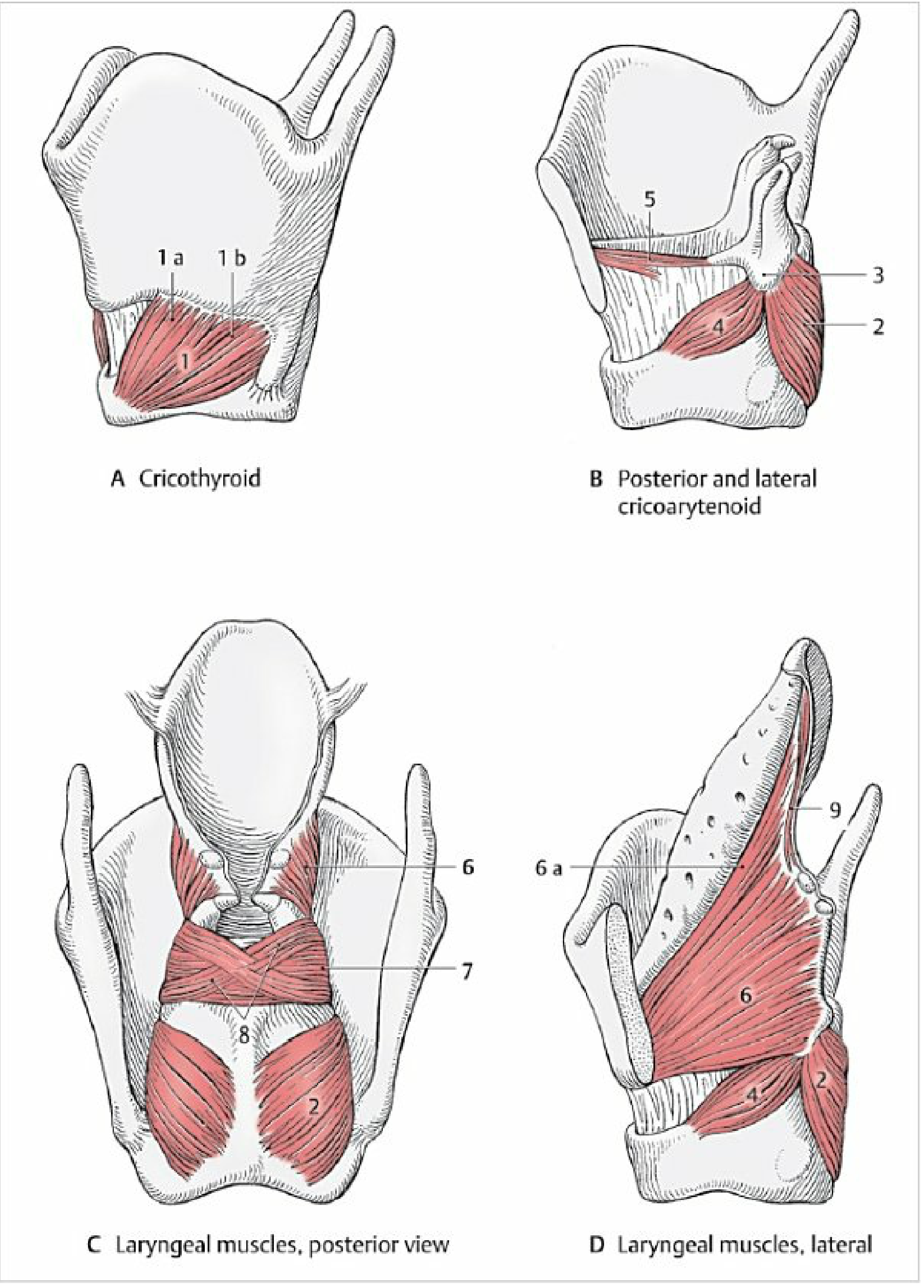
All intrinsic laryngeal muscles are innervated by the recurrent laryngeal nerve (branch of vagus [CN X]), except the cricothyroid (external branch of superior laryngeal nerve).
Openers (Abductors):
- Posterior cricoarytenoid - the ONLY muscle that opens the entire rima glottidis for inspiration. Draws the muscular process posteriorly, moving the vocal process laterally.
Closers (Adductors):
- Lateral cricoarytenoid - draws muscular process anteriorly, closes the rima glottidis
- Transverse arytenoid - closes the intercartilaginous (posterior) part
- Thyroarytenoid / Vocalis - closes the intermembranous (anterior) part; the vocalis completely closes the rima glottidis by becoming thicker as it contracts
Phonation
During phonation, the arytenoid cartilages and vocal folds are adducted (rima glottidis closed), then air is forced through under expiratory pressure, causing the vocal folds to vibrate and produce sound waves. The volume of sound depends on the force of the airstream; the pitch depends on vibration frequency, which varies by length, thickness, and tension of the vocal ligaments.
Rima Glottidis vs. Rima Vestibuli
- Rima vestibuli: the opening between the two vestibular folds (false vocal cords) - it is wider
- Rima glottidis: the opening between the two vocal folds (true vocal cords) - it is narrower, lying just inferior
Clinical Relevance
-
Life-threatening closure (e.g., anaphylaxis, angioedema, foreign body): involuntary closure of the rima glottidis occurs reflexively when a foreign body enters the airway. If this closure is life-threatening, an airway can be re-established below the rima glottidis by incision through the median cricothyroid ligament - this procedure is called cricothyrotomy.
-
Recurrent laryngeal nerve (RLN) injury:
- Unilateral injury: paralysis of intrinsic muscles on one side; that vocal fold lies in an adducted paramedian position - hoarseness results
- Bilateral acute injury: paralyzed vocal folds meet in the rima glottidis, causing stridor and respiratory distress, potentially requiring tracheostomy
-
Laryngoscopy: direct visualization of the rima glottidis is possible. The laryngoscopic image is inverted - anterior structures appear at the top.
Sources: Gray's Anatomy for Students (9780323934237); Color Atlas of Human Anatomy Vol. 2, Internal Organs (9783132424487)
This is a shared conversation. Sign in to Orris to start your own chat.