
Bulbar urethra rupture
Reading File
Now let me search the medical library and images in parallel.
Finding Sources
Finding Sources
Searching Images
bulbar urethra rupture injury anatomy diagram
Good results. Let me read the most relevant sections from Pye's Surgical Handicraft (most focused on bulbar urethral trauma) and Sabiston, in parallel.
Reading File
Reading File
Searching Images
anterior urethral injury straddle perineum extravasation urine
I have a butterfly hematoma image URL. Let me fetch it and also read more content from the Sabiston section for management details.
Loading Image
Reading File
Reading File
I now have comprehensive content. Let me also quickly fetch the retrograde urethrogram image from Sabiston to show the diagnostic study.
Loading Image
Excellent - I now have all the key information and images needed. Here is a comprehensive answer:
Bulbar Urethra Rupture
Anatomy Recap
The urethra is divided into posterior (prostatic + membranous) and anterior (bulbar + penile) segments. The bulbar urethra is the proximal portion of the anterior urethra, lying in the perineum where it curves over the perineal body. It accounts for 85% of all urethral injuries and is the most vulnerable segment of the anterior urethra. - Sabiston Textbook of Surgery, p. 752
Mechanism of Injury
The classic mechanism is a straddle injury - compression of the bulbar urethra against the inferior border of the pubic symphysis by a fall astride a hard object (bicycle crossbar, fence rail, manhole cover, ship's rigging) or a direct perineal blow (assault, sport). The urethra is crushed between the object and the bony symphysis, causing contusion or frank rupture. - Pye's Surgical Handicraft, p. 314
Clinical Features
| Feature | Details |
|---|---|
| Blood at urethral meatus | Present in 37-93% of cases; the single most important sign |
| Inability to void / urinary retention | Due to disruption of urethral continuity |
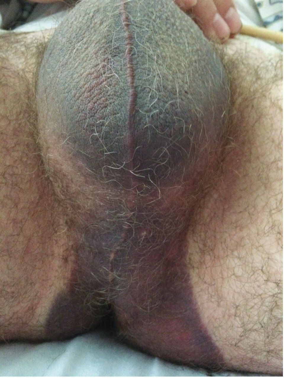
| Butterfly perineal hematoma | Pathognomonic sign (see image below) |
| Perineal pain and swelling | From local crush injury |
Butterfly Hematoma - Explained
When Buck's fascia ruptures, blood and urine spread along the fascial planes. Because Colles' fascia (the perineal extension of Scarpa's fascia) is attached at the:
- Posterior: perineal body
- Lateral: pubic rami and fascia lata
...extravasation is confined to form a "butterfly" pattern spanning the scrotum, perineum, and lower abdominal wall, but NOT the thighs or beyond the inguinal ligament.
Investigations
1. Retrograde Urethrogram (RUG) - Gold Standard
- When: Performed whenever blood is at the meatus after perineal/pelvic trauma
- How: Patient placed obliquely; contrast fills the entire urethra under gentle penile stretch; images taken in the oblique position
- Findings: Loss of urethral contour, extravasation of contrast beyond the normal bulbar cone
Critical rule: Do NOT attempt blind urethral catheterization before RUG - this can convert a partial tear into a complete disruption.
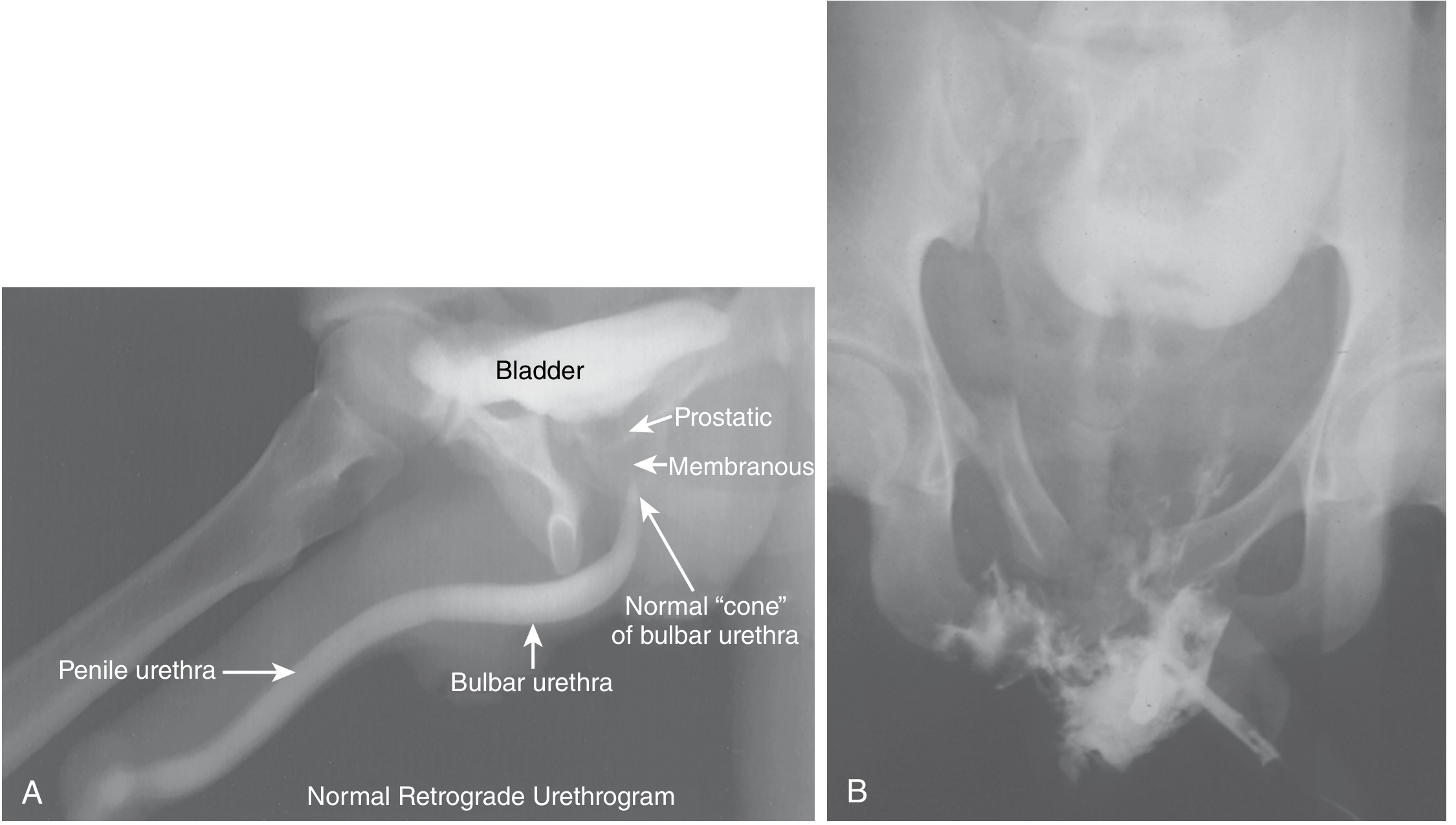
Panel A shows the normal anatomy of the bulbar urethra (the "cone" shape); Panel B shows traumatic urethral disruption with contrast extravasation.
Management
Immediate Priority
Bladder drainage is the first urgent step.
Algorithm
Blood at meatus / retention after straddle injury
↓
Retrograde urethrogram (RUG)
↓
┌────────────────────────────────┐
│ Partial tear │ Complete rupture
│ (contrast reaches bladder) │ (no contrast in bladder)
└──────────┬─────────────────────┘
↓ ↓
Single gentle attempt at Suprapubic catheter (SPC)
urethral catheterization (percutaneous or open)
by experienced urologist
with 12Fr soft catheter
↓
Fails → SPC
- Pye's Surgical Handicraft recommends: one expert attempt at 12Fr soft catheter; if fails, immediate suprapubic catheterization. Urethrography at ~14 days, then urethroscopic assessment with placement of a Silastic catheter across the rupture for a further 10-14 days. - p. 314
- Sabiston states: straddle injuries to the bulbar urethra require prompt urinary drainage; most can be repaired primarily, but if extensive local tissue damage exists, suprapubic drainage + delayed repair is preferred. - p. 752
Delayed Repair / Urethroplasty
Once the acute phase is managed, stricture formation is the major long-term concern:
| Stricture Length | Treatment |
|---|---|
| ≤2 cm | Complete excision + end-to-end primary anastomosis (excise 1 cm beyond each end of stricture) |
| >2 cm | Patch graft urethroplasty - midline urethrotomy + buccal mucosa graft (onlay) |
| Very long / dense fibrosis | Distal penile fasciocutaneous flap ± buccal mucosa; success >80% in single-stage |
- Buccal mucosa grafts in the bulbar region should use an onlay technique (dorsal or ventral) to maximize vascularization from the corpus spongiosum. - Smith & Tanagho's General Urology, p. 667
Complications
| Complication | Notes |
|---|---|
| Urethral stricture | Most common late complication; monitor with uroflowmetry, RUG, cystoscopy |
| Erectile dysfunction | Less common than with posterior urethral injuries but occurs |
| Periurethral abscess | Requires drainage + antibiotics |
| Urethrocutaneous fistula | Usually requires surgical repair |
| Urinary incontinence | Uncommon with anterior urethral injuries |
Key Distinctions: Bulbar vs. Posterior Urethral Rupture
| Feature | Bulbar (Anterior) | Posterior (Membranous) |
|---|---|---|
| Mechanism | Straddle/direct perineal blow | Pelvic fracture (RTA, crush, fall from height) |
| Associated injuries | Usually isolated | Pelvic ring fractures (almost invariable) |
| Hematoma | Butterfly perineal hematoma | Pelvic hematoma, bladder displaced cephalad ("pie-in-sky") |
| Incontinence risk | Low | High |
| ED risk | Moderate | High |
| Initial management | SPC or gentle catheterization | SPC (immediate suprapubic cystotomy) |
Sources: Sabiston Textbook of Surgery (11th ed.), p. 752-753 | Pye's Surgical Handicraft, p. 314-315 | Smith & Tanagho's General Urology (19th ed.), p. 667-668
This is a shared conversation. Sign in to Orris to start your own chat.