
Hello
Hey! How can I help you today?
I want to know about mitochondria
Mitochondria are membrane-bound organelles found in nearly all eukaryotic cells, often called the "powerhouse of the cell" because they generate most of the cell's ATP through oxidative phosphorylation. They have a double membrane structure - an outer membrane and a highly folded inner membrane (cristae) - and contain their own DNA, suggesting they evolved from ancient bacteria (endosymbiotic theory).
Searching sources...
Reading File
Finding Sources
Searching the Web
mitochondria structure function biology overview
Reading File
Reading File
Reading File
Loading Image
Here is a thorough overview of mitochondria, drawn from authoritative medical textbooks:
Mitochondria
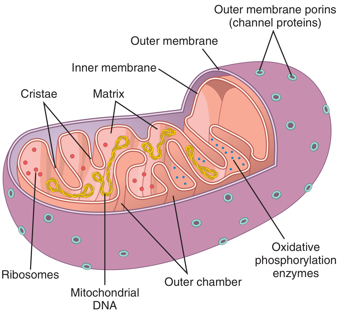
What Are Mitochondria?
Mitochondria (from Greek: mitos = thread, chondros = granule) are membrane-enclosed organelles found in the cytoplasm of nearly all eukaryotic cells. They are best known as the "powerhouses of the cell" - without them, cells cannot extract enough energy from nutrients to survive. - Guyton and Hall Textbook of Medical Physiology
Structure
Mitochondria typically have diameters of 0.5-1 μm and lengths up to 10 μm. They are highly plastic - rapidly changing shape, fusing with one another, and dividing as they move through the cytoplasm along microtubules.
Each mitochondrion has two distinct membranes that create two compartments:
| Component | Description |
|---|---|
| Outer membrane | Contains channel proteins called porins that regulate the flow of ions and small metabolites (up to ~1,000 Da) between the cytoplasm and intermembrane space |
| Intermembrane space | The narrow space between outer and inner membranes |
| Inner membrane | Highly impermeable; forms deep infoldings called cristae - these massively increase surface area and carry the electron transport chain (ETC) and ATP synthase |
| Matrix | The innermost compartment; contains dissolved enzymes for the Krebs/TCA cycle, mitochondrial DNA (mtDNA), ribosomes, and RNA |
- Junqueira's Basic Histology, Basic Medical Biochemistry - A Clinical Approach, 6e
Functions
1. ATP Production (Primary Role)
Mitochondria generate the vast majority of a cell's ATP through oxidative phosphorylation (OXPHOS):
- Cytoplasmic enzymes first convert glucose to pyruvate via glycolysis (anaerobic, produces a little ATP)
- Pyruvate is imported into mitochondria and oxidized via the Krebs/TCA cycle to CO₂ and H₂O
- The ETC on the cristae then uses this to drive ATP synthesis - yielding ~15x more ATP than glycolysis alone
- The liberated energy is captured as ATP, which diffuses out of the mitochondrion to fuel the rest of the cell
2. Heat Production
A small part of the energy released in mitochondria escapes as heat, helping maintain body temperature.
3. Calcium Signaling
Mitochondria store calcium ions and release them for cell signaling events.
4. Apoptosis (Programmed Cell Death)
Mitochondria play a key role in initiating apoptosis by releasing pro-apoptotic factors like cytochrome c.
5. Other Metabolic Roles
They are involved in fatty acid oxidation (beta-oxidation), amino acid metabolism, and steroid hormone synthesis.
Mitochondrial DNA (mtDNA)
-
Mitochondria contain their own DNA, separate from nuclear DNA
-
mtDNA encodes 37 genes essential for mitochondrial function
-
The matrix also contains ribosomes and RNA for protein synthesis
-
mtDNA is primarily maternally inherited (sperm mitochondria are typically lost at fertilization)
-
Mitochondria are self-replicating - they can divide to meet increased energy demands (e.g., in muscle under chronic exercise)
-
Guyton and Hall Textbook of Medical Physiology
Number Per Cell
The number of mitochondria directly reflects a cell's energy needs:
| Cell Type | Mitochondria |
|---|---|
| Cardiac muscle (cardiomyocytes) | Very abundant (high energy demand) |
| Skeletal muscle (trained) | Increased density with exercise |
| Fat cells (adipocytes) | Very few (low metabolic rate) |
| Liver/kidney tubule cells | Abundant |
Endosymbiotic Origin
Mitochondria are thought to have evolved from ancient free-living bacteria that were engulfed by a host cell billions of years ago (the endosymbiotic theory). Evidence includes:
- Their own circular DNA (similar to bacteria)
- Double membrane structure
- Self-replication capability
- Their own ribosomes (similar to bacterial ribosomes)
Clinical Relevance
Mutations in mtDNA can cause primary mitochondrial diseases, which are variable in onset and severity and can affect many organs. An example is:
- MERRF (Myoclonic Epilepsy with Ragged-Red Fibers) - a rare disease where cells inherit mtDNA with a mutated gene for lysine-tRNA, causing defective synthesis of respiratory chain proteins and structural abnormalities in muscle fibers. - Junqueira's Basic Histology
Here's a helpful video if you'd like a visual walkthrough:
This is a shared conversation. Sign in to Orris to start your own chat.