Now I have everything needed to compile the comprehensive master document. I'll now deliver the full, structured, examiner-grade MS General Surgery Thyroid Long Case.
THYROID SWELLING - MS GENERAL SURGERY LONG CASE
Complete University Examination Guide
Professor of General Surgery | University Examiner | Endocrine Surgeon
Based on: Schwartz's (11e), Bailey & Love (27e), Sabiston (21e), S.Das, SRB, Saha | ATA 2015, ETA 2023, BTA, NCCN Guidelines
SECTION 1: CLINICAL SCENARIO
Model Long Case - University Examination
Patient: Mrs. Savitha Reddy, 38 years, female, school teacher, from Hyderabad.
Chief Complaints:
- Swelling in the front of the neck - 4 years
- Rapid increase in size - 3 months
- Discomfort on swallowing - 2 months
- Change in voice (hoarseness) - 3 weeks
History of Presenting Illness:
The patient noticed a small swelling on the right side of the neck 4 years ago, the size of a "gooseberry," which she initially ignored. The swelling gradually increased over the years. Three months ago, she noticed a sudden, rapid increase in size of the swelling associated with mild aching pain. She also noticed a feeling of fullness and discomfort while swallowing solids for the past 2 months. For the past 3 weeks, she has noted a change in voice - mild hoarseness.
Pressure Symptoms:
- Dysphagia (solids > liquids) - present
- Dyspnea on exertion - mild, present
- No stridor at rest
- Hoarseness of voice - present (3 weeks, SIGNIFICANT - suggests RLN involvement)
Hyperthyroid Symptoms: None - no palpitations, no excessive sweating, no heat intolerance, no weight loss, no tremors.
Hypothyroid Symptoms: None - no weight gain, no cold intolerance, no constipation, no lethargy.
Risk Factors:
- No history of neck irradiation
- No family history of thyroid cancer, MEN syndrome, or RET mutations
- No previous thyroid surgery
- No amiodarone or lithium use
- Menarche: 13 years, regular cycles, 2 children, no pregnancy-related thyroid history
Working Diagnosis: Right-sided solitary thyroid nodule (likely malignant - given rapid growth + hoarseness) - Rule out Papillary Thyroid Carcinoma.
SECTION 2: SURGICAL ANATOMY
2.1 Embryology
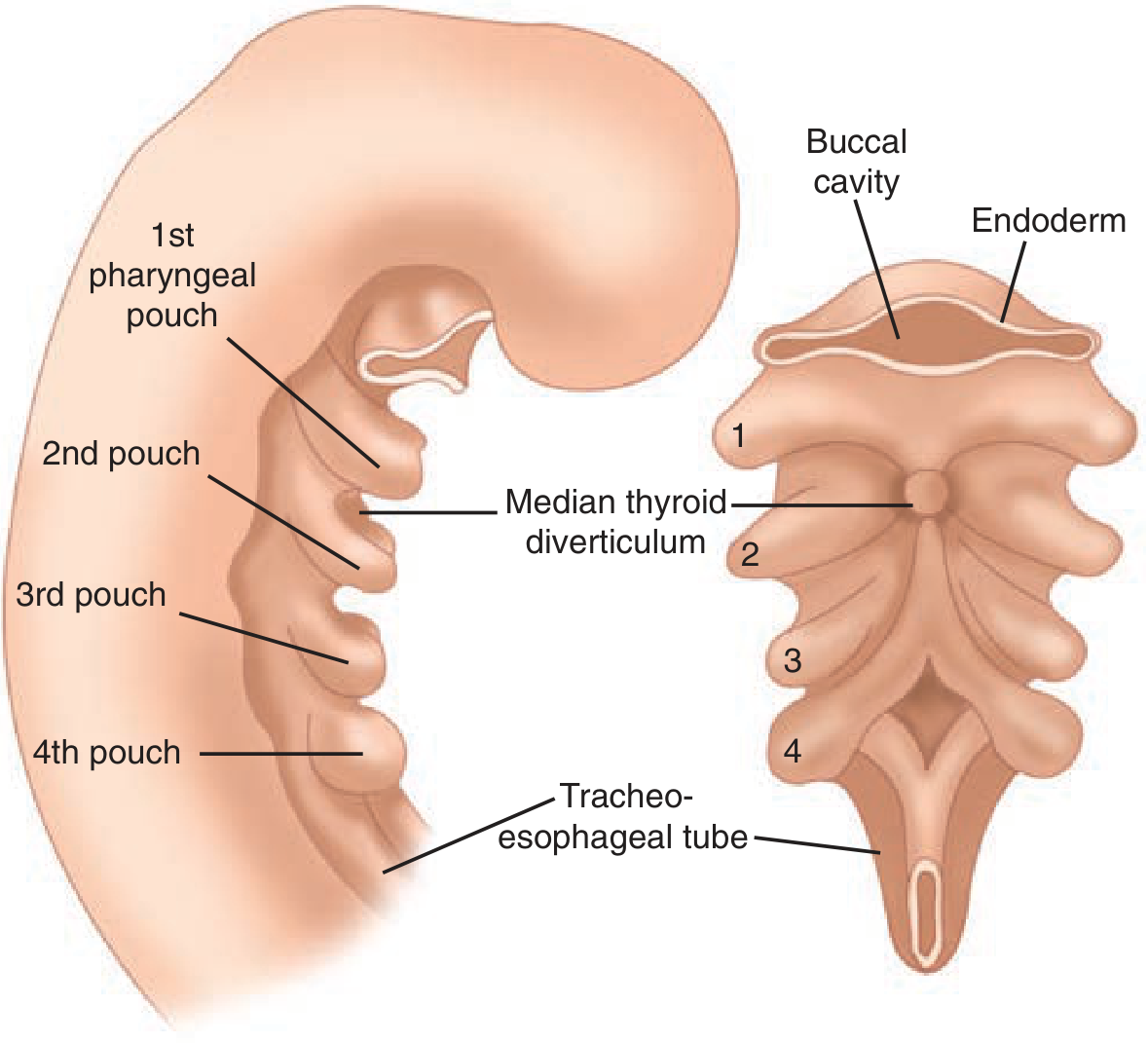
Figure: Thyroid embryology - early development of the median thyroid anlage as a pharyngeal pouch. (Schwartz's Principles of Surgery, 11e, Fig. 38-1)
Key Embryological Facts:
- Thyroid is the first endocrine gland to develop (3rd-4th week of gestation)
- Arises from the foramen caecum at the junction of the anterior 2/3 and posterior 1/3 of the tongue - the median thyroid anlage
- Descends along the thyroglossal duct (which normally obliterates by 8th week) through the tongue base, anterior to the hyoid bone, and infrahyoid strap muscles, to its final position in the neck
- The parafollicular C cells (calcitonin-secreting) arise from the 4th pharyngeal pouch (ultimo-branchial body)
- Superior parathyroids arise from the 4th pharyngeal pouch
- Inferior parathyroids arise from the 3rd pharyngeal pouch (same pouch as thymus)
Surgical Significance of Embryology:
| Anomaly | Mechanism | Clinical Significance |
|---|
| Thyroglossal duct cyst | Failure of obliteration of thyroglossal duct | Moves upward on tongue protrusion (Sistrunk operation) |
| Lingual thyroid | Failure of descent | May be only thyroid tissue - do thyroid scan before surgery |
| Pyramidal lobe | Persistence of lower end of thyroglossal duct | Present in 50%; hypertrophies in Graves disease |
| Accessory thyroid tissue | Along any point of descent pathway | Can be missed on thyroidectomy |
| Lateral aberrant thyroid | None | Does NOT exist - always means lymph node metastasis from PTC |
2.2 Surgical Anatomy of the Thyroid Gland
Gross Anatomy:
- Weight: 25-35 g (normal adult)
- Shape: H-shaped or butterfly-shaped
- Two lateral lobes connected by the isthmus overlying tracheal rings 2-4
- Dimensions of each lobe: 5 cm (height) x 3 cm (width) x 2 cm (depth)
- Pyramidal lobe present in ~50% of individuals, ascending from the isthmus (or left lobe) toward the hyoid - hypertrophies in Graves disease and Hashimoto thyroiditis
Capsule:
- True capsule: Derived from the thyroid gland itself; sends fibrous septa into the gland forming pseudolobules
- False capsule (surgical capsule): Derived from the pretracheal layer of deep cervical fascia; between true and false capsule lie the main blood vessels, nerves, and parathyroid glands
- The false capsule is condensed posteriorly to form Berry's ligament (suspensory ligament) - attaching the thyroid to tracheal rings 2-4
Berry's Ligament - Surgical Importance:
- Fixes the thyroid to the trachea
- The RLN passes deep (posterior) to Berry's ligament or through its substance in 25% of cases
- Dividing Berry's ligament is a step in total thyroidectomy - DANGER zone for RLN
- The Tubercle of Zuckerkandl (posterolateral extension of the thyroid, present in 60-80%) is found at the medial aspect of Berry's ligament and serves as a landmark for the RLN
2.3 Blood Supply
Arterial Supply:
| Artery | Origin | Supply |
|---|
| Superior thyroid artery (STA) | 1st branch of external carotid artery | Upper poles of thyroid |
| Inferior thyroid artery (ITA) | Thyrocervical trunk (subclavian) | Lower poles, posterior surface, parathyroids |
| Thyroidea ima artery | Directly from arch of aorta or brachiocephalic | Isthmus (present in 1-4%) |
Venous Drainage (Inferior, Middle, Superior):
| Vein | Drains into |
|---|
| Superior thyroid vein | Internal jugular vein (or common facial vein) |
| Middle thyroid vein | Internal jugular vein (LIGATED FIRST in thyroidectomy - "vein of safety") |
| Inferior thyroid vein | Left brachiocephalic vein |
Examiner Pearl: The middle thyroid vein is the first structure divided in thyroidectomy - it provides mobilization of the lobe and is a key safety step.
2.4 Nerve Supply - THE MOST IMPORTANT SURGICAL ANATOMY
Recurrent Laryngeal Nerve (RLN)
Course:
- Branch of the vagus nerve (CN X)
- Right RLN: Loops around the right subclavian artery; runs in the tracheoesophageal groove, shorter and more oblique course
- Left RLN: Loops around the arch of the aorta (ligamentum arteriosum); longer course, more vertical in the tracheoesophageal groove
Relations to Inferior Thyroid Artery (ITA):
The relationship is variable and surgically treacherous:
- RLN passes anterior to ITA: 37%
- RLN passes posterior to ITA: 42%
- RLN passes between branches of ITA: 20%
> Rule: Never ligate the ITA close to the thyroid - always ligate the individual branches close to the gland to avoid RLN injury.
Berry's Ligament and RLN:
- RLN passes through Berry's ligament in 25% of cases
- The Tubercle of Zuckerkandl, when present, lies lateral to where the RLN enters the larynx - a useful surgical landmark
- RLN enters the larynx posterior to the cricothyroid joint at the inferior cornu of the thyroid cartilage
Non-recurrent Laryngeal Nerve (NRLN):
- Present on the right side in 0.5-1% of cases
- Associated with right-sided non-recurrent subclavian artery (arteria lusoria)
- Runs directly from vagus to larynx without looping
- Can be mistaken for a blood vessel and divided - catastrophic bilateral cord palsy
- Use preoperative CT to look for retroesophageal right subclavian artery when NRLN suspected
RLN Injury - Consequences:
| Injury Type | Result |
|---|
| Unilateral complete RLN injury | Hoarseness (cord in paramedian/adducted position) |
| Bilateral complete RLN injury | Aphonia + Respiratory distress (emergency) |
| Unilateral partial injury (anterior branch) | Hoarseness (abductor spared) |
| Bilateral anterior branch injury | Stridor, respiratory distress (adductors intact, abductors injured) |
External Branch of Superior Laryngeal Nerve (EBSLN) - "High Note Nerve"
- Branch of superior laryngeal nerve (branch of vagus)
- Runs with the superior thyroid artery before dividing at variable levels
- Supplies the cricothyroid muscle (tensor of vocal cord - voice pitch, projection)
- Cernea Classification of EBSLN (surgical relevance):
- Type 1: Crosses STA >1 cm above upper pole (safe zone) - 20%
- Type 2a: Crosses STA within 1 cm of upper pole - 12%
- Type 2b: Crosses STA below upper pole (at or below the plane of the upper pole) - most dangerous - 68%
- Injury results in: Loss of vocal projection, cannot sing high notes, voice fatigues quickly - the "Amelita Galli-Curci" injury (famous opera singer)
- Prevention: Ligate the superior thyroid vessels individually, close to the upper pole
2.5 Parathyroid Glands - Preservation During Thyroidectomy
Number and Location:
- Usually 4 glands (range 2-6)
- Weight: 30-40 mg each
- Color: Tan/yellow-brown (vs. thyroid which is reddish-brown; vs. fat which is yellow)
| Gland | Embryological Origin | Typical Location |
|---|
| Superior parathyroid (x2) | 4th pharyngeal pouch | Posterior to thyroid, at cricothyroid junction, level of inferior cornu of thyroid cartilage - CONSTANT position |
| Inferior parathyroid (x2) | 3rd pharyngeal pouch | MORE VARIABLE - posterior to lower pole, anterior to RLN, may be intrathymic |
Blood Supply:
- Primarily from the inferior thyroid artery (both superior and inferior parathyroids in 80%)
- Superior parathyroids may receive supply from superior thyroid artery (25%)
- Rule: Preserve the inferior thyroid artery at its point of entry into the thyroid (not as a main trunk) - preserves parathyroid blood supply
Identification During Surgery:
- Gentle mobilization maintaining fat pad
- Do not strip the capsule of the gland
- Positive parathyroid test: cut corner of suspected gland - if frozen section confirms parathyroid tissue, autotransplant into sternocleidomastoid or brachioradialis
- Devascularized parathyroid = autotransplant (1 mm fragments into muscular pockets)
2.6 Lymphatic Drainage
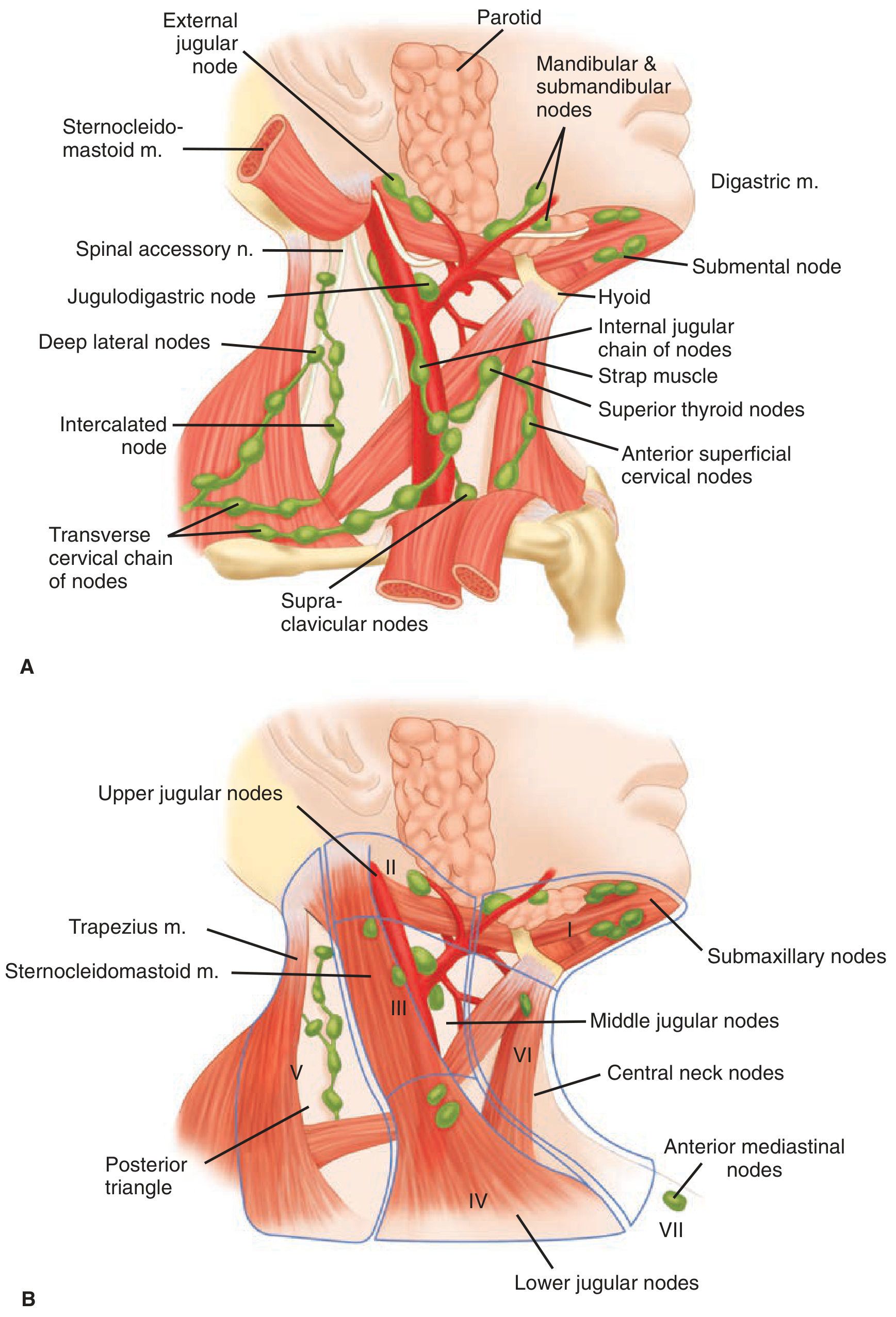
Figure: Cervical lymph node levels I-VII. Central neck = Level VI. Lateral neck = Levels II-V. Level VII = superior mediastinum. (Schwartz's Principles of Surgery, 11e, Fig. 38-6)
Lymphatic Drainage Pattern:
| Direction | Nodes Involved |
|---|
| Primarily (central compartment) | Prelaryngeal (Delphian node), pretracheal, paratracheal nodes - Level VI |
| Lateral | Internal jugular chain (Levels II, III, IV) |
| Superior | Subdigastric nodes |
| Inferior | Level VI → Level VII (superior mediastinal) |
| Skip metastasis | Directly to lateral nodes (Level III) without central - seen in PTC |
Delphian Node:
- Prelaryngeal lymph node
- When enlarged, indicates thyroid malignancy (especially papillary/medullary)
- Named "Delphian" as its biopsy "foretells" malignancy - useful intraoperative marker
SECTION 3: CLASSIFICATION
3.1 Master Classification Flowchart
THYROID SWELLING
├── DIFFUSE ENLARGEMENT (Goitre)
│   ├── Non-toxic (Simple/Endemic)
│   │   ├── Diffuse (Simple goitre)
│   │   └── Multinodular goitre (MNG)
│   ├── Toxic
│   │   ├── Graves Disease (Diffuse toxic goitre)
│   │   ├── Toxic Multinodular Goitre (Plummer disease)
│   │   └── Toxic Adenoma (Plummer's solitary toxic nodule)
│   └── Thyroiditis
│       ├── Hashimoto's (autoimmune)
│       ├── Subacute (De Quervain's)
│       ├── Riedel's (fibrous)
│       ├── Postpartum thyroiditis
│       └── Drug-induced
│
├── SOLITARY THYROID NODULE (STN)
│   ├── Benign (95%)
│   │   ├── Follicular adenoma (most common)
│   │   ├── Colloid cyst
│   │   ├── Thyroid cyst (simple)
│   │   ├── Hashimoto's nodule
│   │   └── Dominant nodule in MNG
│   └── Malignant (5%)
│       ├── Well-differentiated
│       │   ├── Papillary carcinoma (80-85%)
│       │   └── Follicular carcinoma (10-15%)
│       ├── Poorly differentiated
│       │   ├── Hurthle cell (5%)
│       │   └── Poorly differentiated NOS
│       ├── Undifferentiated (Anaplastic) (1-2%)
│       ├── Medullary carcinoma (3-5%)
│       └── Others: lymphoma, metastatic
│
└── RETROSTERNAL GOITRE
    ├── Primary (1-2%) - arises in mediastinum
    └── Secondary (majority) - descends from neck goitre
3.2 WHO Classification of Goitre (Field Grading)
| Grade | Description |
|---|
| Grade 0 | No goitre visible or palpable |
| Grade 1 | Goitre palpable but not visible when neck in normal position; moves upward on swallowing |
| Grade 2 | Goitre visible when neck is in normal position; consistent with enlargement when neck is palpated |
Note: Used in epidemiological surveys for iodine deficiency assessment.
3.3 Thyroiditis Classification
| Type | Cause | Thyroid Function | Key Feature |
|---|
| Hashimoto's (chronic autoimmune) | Anti-TPO, anti-Tg antibodies | Hypothyroid (usually) | Most common thyroiditis; increased lymphoma risk |
| Subacute granulomatous (De Quervain's) | Post-viral (Coxsackievirus, mumps) | Hyper → Hypo → Normal | Painful; raised ESR; self-limiting |
| Riedel's fibrosing thyroiditis | Unknown (IgG4 disease) | Hypo | "Woody" hard thyroid; compresses trachea/esophagus |
| Postpartum thyroiditis | Autoimmune | Hyper → Hypo | Within 1 year postpartum |
| Drug-induced | Amiodarone, lithium, interferon | Variable | History is key |
3.4 TNM Staging of Thyroid Cancer (AJCC 8th Edition, 2017)
| Stage | Criteria |
|---|
| Papillary/Follicular (age <55 years) | |
| Stage I | Any T, Any N, M0 |
| Stage II | Any T, Any N, M1 |
| Papillary/Follicular (age ≥55 years) | |
| Stage I | T1-T2, N0/NX, M0 |
| Stage II | T1-T2, N1, M0 OR T3a/T3b, Any N, M0 |
| Stage III | T4a, Any N, M0 |
| Stage IVA | T4b, Any N, M0 |
| Stage IVB | Any T, Any N, M1 |
| Medullary/Anaplastic | All anaplastic = Stage IV |
Examiner Pearl: The AJCC 8th edition (2017) moved the age cutoff from 45 to 55 years for differentiated thyroid cancer staging - a major change from the 7th edition. This results in many patients being downstaged.
SECTION 4: COMPLETE HISTORY TAKING
4.1 Template: History of Thyroid Swelling
A. SWELLING
- Duration: When did the swelling first appear? ("4 years ago")
- Rate of growth:
- Slow - benign (adenoma, colloid nodule)
- Rapid growth over weeks/months - MALIGNANCY or hemorrhage into cyst
- Rapid growth + pain = hemorrhage into cyst or De Quervain's thyroiditis
- Rapid growth without pain = ANAPLASTIC CARCINOMA (emergency)
- Site of onset: Midline or lateral; right lobe more common
- Change in size: Does it fluctuate? (cyst may fluctuate; goitre may enlarge with menstruation/pregnancy)
- Associated pain: Sudden onset of pain = hemorrhage into cyst; chronic dull ache = malignancy
B. PRESSURE SYMPTOMS
| Symptom | Significance |
|---|
| Dysphagia | Esophageal compression (large goitre) |
| Dyspnea | Tracheal compression |
| Stridor | Severe tracheal compression - EMERGENCY |
| Hoarseness | RLN involvement - MALIGNANCY until proven otherwise |
| Venous engorgement | SVC compression (retrosternal) - positive Pemberton sign |
C. HYPERTHYROID SYMPTOMS (TOPS)
- Tremors (fine tremor of hands)
- Ophthalmopathy (eye prominence, double vision)
- Palpitations (tachycardia, AF)
- Sweating, heat intolerance, weight loss despite normal appetite, anxiety, diarrhea, oligomenorrhea, insomnia
D. HYPOTHYROID SYMPTOMS (SLOW)
- Skin changes (dry, rough, myxedema)
- Lethargy, cold intolerance
- Obesity/weight gain, constipation
- Weight gain, bradycardia, deep husky voice, menorrhagia, depression
E. RISK FACTORS FOR MALIGNANCY (HRAFTS - High-Risk Features)
| Feature | Risk |
|---|
| History of childhood head/neck irradiation | 40x increased risk PTC |
| Rapid growth + hard nodule | Malignancy |
| Age extremes (<20 or >60 years) | Higher malignancy risk in males >60 |
| Family history (PTC, MTC, MEN2, FAP, Cowden, PTEN, DICER1) | Genetic syndromes |
| Texture: hard, fixed, irregular | Malignancy |
| Sex: male with solitary nodule | Higher risk than female |
| Hoarseness, dysphagia (pressure symptoms) | Advanced malignancy |
F. FAMILY HISTORY
| Syndrome | Associated Thyroid Cancer |
|---|
| MEN 2A (RET mutation) | Medullary thyroid carcinoma + pheo + hyperPTH |
| MEN 2B (RET mutation) | MTC + pheo + marfanoid habitus + mucosal neuromas |
| Familial MTC (FMTC) | Isolated MTC |
| FAP/Gardner syndrome | Papillary thyroid carcinoma |
| Cowden syndrome (PTEN mutation) | Follicular adenoma/carcinoma |
| DICER1 syndrome | Differentiated thyroid carcinoma |
| Werner syndrome | Follicular carcinoma |
G. DRUG HISTORY
| Drug | Effect |
|---|
| Amiodarone (iodine-rich) | Hypo or hyperthyroidism |
| Lithium | Hypothyroidism, goitre |
| Interferon-alpha | Thyroiditis, hypothyroid |
| Checkpoint inhibitors | Thyroiditis |
| Carbimazole/Propylthiouracil | Prescribed for hyperthyroidism |
Examiner Cross-Questions - History Section
Q: What is the single most important symptom suggesting malignancy in a thyroid swelling?
A: Hoarseness of voice - indicates RLN involvement, suggesting malignant invasion. Must be assessed preoperatively by flexible laryngoscopy.
Q: A patient with thyroid swelling develops sudden acute pain - what do you think?
A: Hemorrhage into a thyroid cyst - most common cause of sudden enlargement with pain. Also consider: De Quervain's subacute thyroiditis, or spontaneous hemorrhage into a cold nodule.
Q: A patient with a 20-year history of goitre presents with sudden rapid growth and difficulty breathing - what is your diagnosis?
A: Anaplastic carcinoma arising in a longstanding multinodular goitre. This is a surgical emergency. Pre-existing goitre is a risk factor for anaplastic transformation.
Q: What is the significance of family history of phaeochromocytoma in a thyroid nodule patient?
A: Raises the possibility of MEN 2A/2B syndrome. Medullary thyroid carcinoma is part of this syndrome. Serum calcitonin and RET mutation testing are essential before surgery, and urine catecholamines must be excluded before any operative intervention.
SECTION 5: CLINICAL EXAMINATION
5.1 General Examination
Before examining the neck - look at the whole patient:
Hyperthyroid Patient (Graves Disease)
| Finding | Significance |
|---|
| Anxious, restless demeanor | Sympathetic overactivity |
| Fine tremor of outstretched hands | Thyrotoxicosis |
| Warm, moist, velvety skin | Peripheral vasodilation |
| Tachycardia (regular or irregular) | >100 bpm; AF in 10-15% |
| Lid lag (Von Graefe sign) | Sympathetic overactivity |
| Lid retraction (Dalrymple sign) | Upper lid above limbus - "stare" |
| Exophthalmos | Graves ophthalmopathy |
| Pretibial myxedema | Graves specific; indurated pinkish-brown plaques |
| Thyroid acropachy | Clubbing + periosteal reaction |
| Proximal muscle weakness | Thyrotoxic myopathy |
| Gynecomastia (males) | Sex hormone binding globulin changes |
Graves Ophthalmopathy (Clinical Assessment)
NOSPECS Classification:
- No signs/symptoms
- Only signs (lid retraction, stare, lid lag)
- Soft tissue involvement (periorbital edema, chemosis)
- Proptosis (exophthalmos - >18 mm; measured by Hertel exophthalmometer)
- Extraocular muscle involvement (diplopia)
- Corneal involvement (exposure keratopathy)
- Sight loss (optic nerve compression - EMERGENCY)
Hypothyroid Patient
| Finding | Significance |
|---|
| Puffy face, periorbital edema | Mucopolysaccharide deposition |
| Dry, rough, yellowish skin (carotenemia) | Hypothyroidism |
| Coarse, brittle hair; loss of outer 1/3 eyebrow (Hertoghe sign) | Hypothyroidism |
| Bradycardia | |
| Delayed relaxation of ankle jerks | Pathognomonic of hypothyroidism |
| Non-pitting edema | Myxedema |
| Husky voice | |
5.2 Local Examination - THE FOUR "S" Approach
INSPECTION
Ask patient to sit facing you in a well-lit room, neck slightly extended:
Observe:
- Site: Midline (isthmus, pyramidal lobe, thyroglossal cyst) or lateral (thyroid lobe)
- Size: Estimate in cm; compare to anatomical landmarks
- Shape: Oval, irregular, multilobulated
- Skin: Dilated veins (venous congestion from retrosternal extension), redness (thyroiditis), scar (previous surgery), fistula
- Number of swellings: Single lobe, bilateral, isthmic
- Pulsatility: Transmitted vs. expansile (thyroid vascular lesion is rare)
- Movement with swallowing: Hallmark of thyroid swelling
SWALLOWING TEST:
- Ask patient to take a sip of water and swallow while you observe
- Thyroid swelling moves upward on swallowing
- Positive in: Thyroid swellings, thyroglossal cysts, pre-laryngeal nodes (attached to pretracheal fascia)
- Negative (does not move): Lipomas, sebaceous cysts, lymph nodes (unless stuck to thyroid)
TONGUE PROTRUSION TEST:
- Ask patient to protrude the tongue upward
- Specific for thyroglossal duct cysts (moves upward due to attachment to hyoid via thyroglossal tract)
- Thyroid swellings do NOT move on tongue protrusion
- Significance: Differentiates thyroglossal cyst from other midline swellings
PALPATION
Technique (Crile's Method - Examiner Favorite):
- Stand behind the patient (surgeon's position)
- Place both hands around the neck with fingertips meeting at midline
- Ask patient to swallow
- Systematically palpate both lobes and isthmus
- Feel for size, shape, consistency, nodularity, tenderness, fixity, and lower border
Alternatively (S. Das approach):
- Stand in front of the patient
- Use thumb and fingers of both hands
- Crile's (from behind) is preferred for examination
Findings to Note:
| Feature | Benign | Malignant |
|---|
| Consistency | Soft/firm | Hard ("rock hard") |
| Surface | Smooth/nodular | Irregular |
| Borders | Well-defined | Ill-defined |
| Tenderness | Usually tender (thyroiditis) | Non-tender usually |
| Mobility | Mobile | Fixed to adjacent structures |
| Nodularity | Uninodular or multinodular | Irregular hard nodule |
| Lower border | Reachable | May extend retrosternally |
Lahey's Method (for lower border palpation):
- Patient tilts head to the opposite side (relaxes SCM)
- Place index finger deep to the SCM
- Feel for the lower border of the gland
- If lower border cannot be felt = retrosternal extension
Assessment of Retrosternal Extension:
- Cannot palpate lower border of thyroid
- Dullness to percussion over upper sternum (Reverdin's sign - manubrium)
- Pemberton's sign: Ask patient to raise both arms above head for 1 minute → facial plethora, distended neck veins, cyanosis = positive (thoracic inlet obstruction from retrosternal goitre)
Tracheal Assessment:
- Kocher's test: Gently compress the lateral lobes between thumb and finger → if positive = stridor (indicates tracheal softening - tracheomalacia from long-standing goitre)
- Assess tracheal position: midline vs. deviated (pushes trachea away; SVC obstruction doesn't push trachea)
- Clinically assess for tracheal deviation
Cervical Lymph Nodes:
Systematically palpate all cervical node groups:
- Submental (Level IA)
- Submandibular (Level IB)
- Upper jugular/jugulodigastric (Level IIA, IIB)
- Middle jugular (Level III)
- Lower jugular (Level IV)
- Posterior triangle (Level V)
- Central neck / prelaryngeal / pretracheal / paratracheal (Level VI) - MOST IMPORTANT for thyroid
- Supraclavicular (Level IV/V junction) - enlarged in thyroid cancer, breast, lung
- Mediastinal (Level VII)
Enlarged Level VI (Delphian/central neck) nodes → MALIGNANCY
PERCUSSION
- Percuss over the sternum (manubrium to lower sternum)
- Dullness = retrosternal extension of goitre
- Normal = resonant
AUSCULTATION
- Place stethoscope bell over thyroid
- Bruit (continuous systolic-diastolic murmur): Highly specific for Graves disease (hypervascular thyroid) - present in 80%
- Differentiate from transmitted cardiac murmur (disappears on compression of common carotid artery)
- Absence of bruit does NOT exclude Graves disease
5.3 Summary of Special Clinical Tests
| Test | Technique | Positive Finding | Significance |
|---|
| Swallowing test | Sip water, swallow | Swelling moves up | Confirms thyroid origin; also thyroglossal cyst |
| Tongue protrusion test | Protrude tongue upward | Swelling moves up | Thyroglossal duct cyst specifically |
| Pemberton's sign | Raise both arms above head 1 min | Facial plethora + neck vein distension + cyanosis | Thoracic inlet obstruction from retrosternal goitre |
| Kocher's test | Compress lateral lobes | Stridor elicited | Tracheomalacia from long-standing goitre |
| Crile's method | Palpation from behind | Standard palpation | Best method for bimanual thyroid examination |
| Lahey's method | Head tilted opposite side, deep palpation | Cannot feel lower border | Retrosternal extension |
| Reverdin's sign | Percussion over manubrium | Dullness | Retrosternal goitre |
| Vocal cord assessment | Indirect laryngoscopy or nasofibroscopy | Cord paralysis | RLN injury (pre/post-op) |
SECTION 6: DIFFERENTIAL DIAGNOSIS
Differential Diagnosis of Neck Swelling
| Feature | Thyroid Swelling | Thyroglossal Cyst | Branchial Cyst | Lymph Node | Carotid Body Tumor |
|---|
| Age | Any | Child/young adult | Young adult | Any | 30-60 years |
| Sex | F>M | Equal | Equal | Equal | F=M |
| Site | Anterior neck (midline/lateral) | Midline, below hyoid | Anterior triangle, level II/III | Cervical chain | At carotid bifurcation (angle of jaw) |
| Swallowing test | + (moves up) | + (moves up) | - | - | - |
| Tongue protrusion | - | + (moves up) | - | - | - |
| Consistency | Variable | Soft/fluctuant | Soft/fluctuant | Variable | Firm, pulsatile |
| Pulsatility | - (transmitted only) | - | - | - | + (expansile pulsation) |
| Bruit | + in Graves | - | - | - | + (AV shunting) |
| Transillumination | - | + | + | - | - |
| Horner syndrome | - | - | - | - | - (but compression possible) |
SECTION 7: CASE PRESENTATION (UNIVERSITY EXAMINATION - 5 MINUTES)
Model Presentation Script
"Good morning, Sir/Ma'am. I present the case of Mrs. Savitha Reddy, a 38-year-old female school teacher, who presented with:
Chief Complaints:
1. Swelling in the front of the neck - 4 years
2. Rapid increase in size - 3 months
3. Discomfort on swallowing - 2 months
4. Hoarseness of voice - 3 weeks
On History: The swelling started 4 years ago as a small right-sided neck nodule and remained static, but has shown rapid growth over the last 3 months. The hoarseness of 3 weeks' duration is the most clinically significant symptom, suggesting possible involvement of the right recurrent laryngeal nerve.
There are no symptoms of hyperthyroidism or hypothyroidism. No history of neck irradiation. No family history of thyroid malignancy or MEN syndrome.
On General Examination: The patient is conscious, cooperative, and hemodynamically stable. She is afebrile, with pulse 78/min, BP 120/80 mmHg. No signs of thyrotoxicosis. No eye signs. No pretibial myxedema.
On Local Examination: There is a single swelling in the right lobe of the thyroid, measuring approximately 4 x 3 cm, with ill-defined borders. The swelling is hard in consistency, non-tender, with an irregular surface. It moves upward on swallowing (confirming thyroid origin) but does not move on tongue protrusion (excluding thyroglossal cyst). The lower border of the swelling is palpable. There is no retrosternal extension clinically.
On lymph node examination: I have found a single palpable, firm, non-tender node in Level III on the right side, approximately 1.5 cm in diameter.
My working diagnosis is: A right-sided solitary thyroid nodule, likely malignant - Papillary Thyroid Carcinoma, on the basis of: (1) hard consistency, (2) rapid growth, (3) hoarseness suggesting RLN involvement, and (4) ipsilateral cervical lymphadenopathy. I would like to investigate this patient to confirm the diagnosis and plan further management."
SECTION 8: INVESTIGATIONS
8.1 Investigation Algorithm
THYROID SWELLING
        │
        ▼
STEP 1: TSH (screening)
        │
   ┌────┴────┐
 Low TSH    Normal/High TSH
   │              │
   ▼              ▼
Free T3/T4     Thyroid Ultrasound (TI-RADS)
+Thyroid scan        │
(hot/cold)     ┌─────┴──────┐
               │            │
          TI-RADS 1-2    TI-RADS 3-5
               │            │
           Observe      FNA (Bethesda)
                             │
              ┌──────────────┼──────────────┐
           Beth I         Beth II      Beth III/IV/V/VI
         Repeat FNA       Follow up   Molecular testing/Surgery
8.2 Thyroid Function Tests
| Test | Normal Range | Significance |
|---|
| TSH (first-line) | 0.4 - 4.0 mIU/L | Low: hyperthyroid; High: hypothyroid |
| Free T4 | 0.8 - 1.8 ng/dL | Confirms thyroid status |
| Free T3 | 2.3 - 4.2 pg/mL | T3 toxicosis if T3 high with normal T4 |
| Anti-TPO antibody | <35 IU/mL | Hashimoto's thyroiditis |
| Anti-thyroglobulin | <115 IU/mL | Hashimoto's, monitoring after thyroidectomy |
| TSH receptor antibody (TRAb) | Diagnostic for Graves disease | Differentiates Graves from toxic MNG |
| Serum calcitonin | <10 pg/mL | Medullary thyroid carcinoma; universal screening debated |
| Serum thyroglobulin | Follow-up marker after total thyroidectomy for DTC | Should be undetectable post-thyroidectomy |
8.3 Ultrasound and TI-RADS
ACR TI-RADS Classification (2017) - Most Widely Used
| Category | Features | Score | Malignancy Risk | Action |
|---|
| TR1 | Benign (purely cystic) | 0 | <1% | No FNA |
| TR2 | Not suspicious | 0 | <1% | No FNA |
| TR3 | Mildly suspicious | 3 | ~5% | FNA if ≥2.5 cm; follow if ≥1.5 cm |
| TR4 | Moderately suspicious | 4-6 | ~5-20% | FNA if ≥1.5 cm; follow if ≥1 cm |
| TR5 | Highly suspicious | ≥7 | >20% | FNA if ≥1 cm; follow if ≥0.5 cm |
Sonographic Features Scored in ACR TI-RADS:
| Feature | Benign | Suspicious |
|---|
| Composition | Cystic, spongiform | Solid |
| Echogenicity | Hyperechoic, isoechoic | Hypoechoic, very hypoechoic |
| Shape | Wider than tall | Taller than wide |
| Margin | Smooth | Irregular, lobulated, extrathyroidal extension |
| Echogenic foci | Large comet-tail artifacts | Microcalcifications, rim calcifications |
Examiner Pearl - TI-RADS vs. EU-TIRADS vs. K-TIRADS:
- ACR TI-RADS (American): 5 categories, point-scoring system
- EU-TIRADS (European): 5 categories, based on pattern recognition
- K-TIRADS (Korean): Modified version
- All aim to standardize FNA recommendations; EU-TIRADS used in 2023 ETA guidelines
8.4 FNAC and Bethesda Classification
The Bethesda System for Reporting Thyroid Cytopathology (2nd Edition, 2017)
| Category | Diagnosis | Risk of Malignancy | Management |
|---|
| I | Non-diagnostic / Unsatisfactory | 5-10% | Repeat FNA under US guidance |
| II | Benign | 0-3% | Clinical follow-up + US in 1-2 years |
| III | Atypia of Undetermined Significance (AUS) / Follicular Lesion of Undetermined Significance (FLUS) | 6-18% | Repeat FNA or molecular testing or lobectomy |
| IV | Follicular Neoplasm / Suspicious for Follicular Neoplasm | 10-40% | Molecular testing or lobectomy |
| V | Suspicious for Malignancy | 45-75% | Near-total thyroidectomy or lobectomy |
| VI | Malignant | 97-99% | Total thyroidectomy (or lobectomy for low-risk) |
Critical Limitation of FNAC: Cannot differentiate follicular adenoma from follicular carcinoma (requires histological evidence of capsular or vascular invasion - ONLY on excised specimen). This is why Bethesda IV mandates lobectomy.
8.5 Radioiodine Thyroid Scan (Scintigraphy)
| Finding | Significance |
|---|
| Hot nodule (increased uptake) | Almost never malignant (<1%); toxic adenoma; no FNA needed if low TSH |
| Cold nodule (decreased uptake) | 15-20% malignant risk; needs ultrasound + FNA |
| Warm nodule (equal uptake) | 4-9% malignant risk |
| Patchy uptake | Multinodular goitre (endemic) |
| Diffuse increased uptake | Graves disease |
Indications for thyroid scan:
- Low TSH (to identify toxic nodule - hot nodule doesn't need FNA)
- Before surgery for retrosternal goitre (to document ectopic thyroid tissue)
- Follow-up post-thyroidectomy for DTC (whole body scan with RAI-131)
8.6 CT Scan / MRI
CT Neck and Chest (without contrast if RAI planned):
- Assess extent of retrosternal extension
- Airway compression (tracheal cross-sectional area)
- Vascular involvement
- Cervical lymph node mapping
- Caution: Iodinated CT contrast delays RAI therapy by 2-3 months - use MRI or non-contrast CT if RAI planned
MRI:
- Better soft tissue detail
- No radiation, no contrast-RAI conflict
- Assess RLN involvement, tracheal invasion, esophageal involvement
- Used pre-operatively in complex cases
8.7 Other Investigations
| Investigation | Indication |
|---|
| Indirect laryngoscopy / Nasofibroscopy | Pre-op vocal cord assessment (mandatory pre-thyroidectomy) + post-op assessment |
| PET-CT (FDG) | Thyroglobulin-positive, RAI-negative DTC recurrence; poorly differentiated carcinoma |
| RET mutation testing | All newly diagnosed medullary thyroid carcinoma (germline and somatic) |
| RET/PTC, BRAF, RAS, PAX8-PPARG | Molecular testing of indeterminate FNA (Bethesda III/IV) - ThyroSeq v3, Afirma GEC |
| Serum PTH | Pre and post thyroidectomy (if parathyroid compromise suspected) |
| Calcium (ionized) | Post-thyroidectomy monitoring (4 hourly for 24h) |
| Urine catecholamines | Before MTC surgery (rule out pheo in MEN2) |
SECTION 9: MANAGEMENT
9.1 Evidence-Based Management Algorithm
Solitary Thyroid Nodule - ATA 2015 / ETA 2023 Algorithm
Solitary Thyroid Nodule Detected
            │
            ▼
    Measure TSH
            │
    ┌───────┴────────┐
  Low TSH         Normal TSH
    │                   │
    ▼                   ▼
Thyroid Scan      Ultrasound + TI-RADS
  (R/O hot nodule)
    │                   │
  Hot nodule         TI-RADS Score
  → treat toxic         │
    nodule    ┌─────────┴─────────┐
              │                   │
          TI-RADS 1-2         TI-RADS 3-5
          (low risk)          (moderate-high risk)
              │                   │
          No FNA              FNA (US-guided)
          US follow               │
                          Bethesda Classification
                                  │
           ┌──────────────────────┼──────────────────────┐
        Beth I              Beth II               Beth III/IV
       Repeat FNA          US follow-up           Molecular testing
                          3-5 years              OR Lobectomy
                                  │
                              Beth V/VI
                          Surgery (see below)
9.2 Medical Management
Antithyroid Drugs (for Hyperthyroidism)
| Drug | Mechanism | Dose | Side Effects |
|---|
| Carbimazole | Blocks TPO enzyme → inhibits T3/T4 synthesis | 30-60 mg/day initially; maintenance 5-15 mg/day | Agranulocytosis (0.3%), rash, hepatotoxicity |
| Propylthiouracil (PTU) | Blocks TPO + inhibits peripheral T4→T3 conversion | 300-600 mg/day initially | Agranulocytosis, hepatic necrosis (preferred in pregnancy, thyroid storm) |
| Propranolol | Beta blocker - symptom control | 40-80 mg TID/QID | Blocks sympathetic symptoms; does NOT affect thyroid hormone levels |
Titration vs. Block-and-Replace:
- Titration: Adjust ATD dose to keep patient euthyroid - standard method
- Block-and-replace: Full blocking dose ATD + levothyroxine replacement - fewer dose adjustments needed
Remission after ATD in Graves (ATA 2016):
- 30-40% achieve remission after 12-18 months of ATD
- Low TRAb, small goitre, mild disease = predictors of remission
Radioiodine (RAI) Therapy - I-131
Indications:
- Graves disease (first-line in USA; not first-line in Europe for young patients)
- Toxic multinodular goitre
- Toxic adenoma
- Post-surgical remnant ablation in DTC (high-risk and intermediate-risk)
Contraindications:
- Pregnancy and breastfeeding (absolute)
- Active severe Graves ophthalmopathy (relative - may worsen)
- Unable to comply with radiation safety measures
Goal: Ablate thyroid → intentional hypothyroidism (acceptable outcome; treated with L-T4)
RAI in DTC Post-Thyroidectomy (ATA 2015):
- High-risk disease: RAI recommended (reduces recurrence and cancer mortality)
- Intermediate-risk: RAI generally favored
- Low-risk: RAI not recommended routinely
- Not recommended for papillary microcarcinoma (T1a, N0, M0)
Levothyroxine (L-T4)
Indications:
- Hypothyroidism (primary replacement)
- TSH suppression therapy post-thyroidectomy for DTC
- Medical treatment of goitre (limited evidence - mainly in iodine-deficient areas)
TSH Suppression Targets in DTC (ATA 2015):
| Risk Category | TSH Target |
|---|
| High-risk | <0.1 mIU/L |
| Intermediate-risk | 0.1-0.5 mIU/L |
| Low-risk | 0.5-2 mIU/L (after 5-10 years if disease-free) |
9.3 Surgical Management
Extent of Surgery - Decision Algorithm
DECISION: EXTENT OF THYROID SURGERY
                │
    ┌───────────┼───────────┐
    │           │           │
Benign     Malignant    Hyperthyroid
disease     disease      disease
    │           │           │
    ▼           ▼           ▼
Lobectomy   See below   Total/near-total
or                      thyroidectomy
observation             (preferred for
                        definitive tx)
Extent of Surgery for Thyroid Cancer (ATA 2015 / ETA 2023)
| Cancer Type | Extent of Surgery |
|---|
| PTC <1 cm, no ETE, no LN, no family history, no irradiation | Lobectomy (active surveillance also an option) |
| PTC 1-4 cm, low-risk, no ETE | Lobectomy or total thyroidectomy (based on patient preference) |
| PTC >4 cm, or ETE, or bilateral, or LN+ | Total thyroidectomy |
| Follicular carcinoma (known pre-op rare) | Total thyroidectomy |
| Medullary carcinoma | Total thyroidectomy + central neck dissection (Level VI) |
| Anaplastic carcinoma | Near-total or total thyroidectomy if resectable; if unresectable - palliative |
| Graves disease | Total or near-total thyroidectomy |
| Toxic MNG | Total or near-total thyroidectomy |
| Large compressive goitre | Total or near-total thyroidectomy |
Near-Total vs. Total Thyroidectomy (Bailey & Love / Schwartz debate):
| Parameter | Total Thyroidectomy | Near-Total Thyroidectomy |
|---|
| Remnant | None | <1 g remnant left on side of better RLN/parathyroid |
| RAI ablation | Easier (no competing tissue) | Slightly more difficult |
| Hypothyroid risk | 100% | 100% |
| RLN injury risk | Higher (bilateral dissection) | Slightly lower |
| Current recommendation | Preferred (ATA 2015) | Acceptable alternative |
Lymph Node Dissection for Thyroid Cancer
| Dissection | Indication | Levels Cleared |
|---|
| Central neck dissection (CND) | MTC (always); PTC with cN1a; pT3-T4 PTC prophylactically (controversial) | Level VI (prelaryngeal, pretracheal, paratracheal bilateral) |
| Ipsilateral lateral neck dissection | cN1b (clinically positive lateral nodes) | Levels II, III, IV, V |
| Bilateral lateral neck dissection | Bilateral cN1b | Levels II-V bilateral |
ATA 2015 on Prophylactic CND in PTC: Prophylactic CND (in cN0) is "not routinely recommended" for T1-T2 PTC due to increased morbidity without proven survival benefit. May be considered in T3-T4 disease. MTC = always do bilateral CND.
SECTION 10: OPERATIVE TECHNIQUE
10.1 Total Thyroidectomy - Step by Step
Preoperative Assessment
- Indirect laryngoscopy - document bilateral cord mobility (MANDATORY pre-op)
- Thyroid function euthyroid state (Carbimazole + beta-blocker in toxic goitre)
- Lugol's iodine (5 drops TDS for 10 days pre-op in Graves disease) - reduces vascularity
- Blood group and crossmatch
- Calcium and PTH baseline
Patient Positioning
- Supine, neck extended using a sandbag/thyroid bolster under shoulders
- Arms tucked at sides
- Head ring to prevent movement
- Table in reverse Trendelenburg (15-20°) to reduce venous bleeding
Incision - Kocher's Collar Incision
- Transverse incision 2 cm above sternal notch, in a skin crease
- Length: 5-7 cm (longer for large goitres, retrosternal extension)
- Should lie within Langer's lines for cosmesis
- Carried through skin, subcutaneous fat, platysma
Raising Subplatysmal Flaps
- Superior flap: to thyroid notch of thyroid cartilage
- Inferior flap: to sternal notch
- Plane: between platysma and deep cervical fascia
Opening Midline (Linea Alba of Neck)
- Divide along midline between strap muscles (sternohyoid, sternothyroid)
- Avoid damage to strap muscles (retract laterally)
- If necessary for large goitre - divide strap muscles (sternothyroid) transversely in upper 1/3 (preserves nerve supply from ansa cervicalis which enters lower 2/3)
Mobilization of Thyroid Lobe
- Divide middle thyroid vein first (key step - provides lateral mobilization)
- Divide superior thyroid vessels individually close to upper pole (preserve EBSLN)
- Identify and trace RLN in tracheoesophageal groove
- Ligate inferior thyroid artery branches close to thyroid capsule (preserve parathyroid blood supply)
- Identify and preserve parathyroid glands
- Divide Berry's ligament carefully (RLN may pass through it)
- Divide isthmus and pyramidal lobe
RLN Identification - Systematic Approach (S. Das / Bailey & Love)
Three consistent landmarks for RLN:
- Tracheoesophageal groove - where RLN typically lies
- Tubercle of Zuckerkandl - RLN lies medial to it
- Inferior cornu of thyroid cartilage - where RLN enters larynx
Visual characteristics of RLN:
- White glistening nerve, about 1-2 mm diameter
- Runs superiorly between trachea and esophagus
- May branch (anterior branch = abductor; posterior branch = adductor) - in 40% cases before entering larynx
Intraoperative Neuromonitoring (IONM):
- Electromyographic monitoring of vocalis muscle
- Endotracheal tube with recording electrodes
- Standard of care in high-risk cases (re-do surgery, large goitre, malignancy)
- Provides continuous feedback; positive signal = RLN intact
- "Loss of signal" alerts surgeon immediately
Parathyroid Preservation Protocol
- Identify all 4 glands early
- Superior parathyroids: at cricothyroid joint level, posterior to thyroid
- Inferior parathyroids: anterior to RLN, at lower pole, may be in fat
- Preserve blood supply - maintain fat pad
- If gland color changes (darkens/becomes gray) = devascularized → autotransplant
- Autotransplant technique: Confirm parathyroid tissue on frozen section → mince into 1 mm fragments → implant into pockets in SCM muscle (or brachioradialis) → mark with non-absorbable suture or metal clip
Hemostasis and Closure
- Meticulous hemostasis (bipolar diathermy, clips, ligatures)
- Check for bleeding, especially with Valsalva maneuver
- Suction drain (Redivac/Blake) in thyroid bed (optional - not universally recommended)
- Close strap muscles in midline
- Close platysma (2/0 Vicryl)
- Subcuticular suture or skin clips for skin
- Total operative time: 90-180 minutes
SECTION 11: COMPLICATIONS
11.1 Complications of Thyroidectomy
Immediate (0-24 hours)
| Complication | Incidence | Mechanism | Management |
|---|
| Hemorrhage / Neck hematoma | 0.3-1% | Slipped ligature, venous ooze | OPEN WOUND AT BEDSIDE IMMEDIATELY → secure airway → return to OR |
| Airway obstruction | Rare | Hematoma compression, bilateral RLN injury, tracheomalacia | Intubation / tracheostomy |
| Thyroid storm | <1% | In poorly prepared thyrotoxic patient | ICU: beta blockers, PTU, Lugol's iodine, hydrocortisone, cooling |
Critical Protocol for Post-Thyroidectomy Neck Hematoma:
Opening the wound at bedside IMMEDIATELY (even in ward, even without anesthesia) is LIFE-SAVING. Delay causes asphyxia from progressive tracheal compression.
Early (24-72 hours)
| Complication | Incidence | Mechanism | Management |
|---|
| Hypocalcemia (transient) | 20-30% after TT | Parathyroid bruising, devascularization | Oral/IV calcium, vitamin D |
| Hypoparathyroidism (permanent) | 1-4% after TT | Loss of parathyroid tissue | Lifelong calcium + calcitriol |
| RLN injury (temporary) | 3-5% | Neuropraxia/stretching | Spontaneous recovery in 6-12 months |
| RLN injury (permanent) | <1% (experienced surgeons) | Section/devascularization | Speech therapy; thyroplasty |
| EBSLN injury | 1-28% (often subclinical) | Ligation near upper pole | Voice therapy |
| Wound infection | <1% | Standard surgical | Antibiotics |
| Seroma | 3-5% | Fluid collection in dead space | Aspiration, compression |
Late Complications
| Complication | Management |
|---|
| Hypothyroidism (expected after total thyroidectomy) | Lifelong L-T4 replacement |
| Recurrence of goitre or cancer | Re-operation (high risk) or RAI |
| Hypertrophic scar / Keloid | Silicone gel, steroid injection |
| Recurrence of hyperthyroidism after subtotal | Re-operate or RAI |
11.2 Hypocalcemia Management Flowchart
Post-Thyroidectomy
        │
        ▼
    Monitor symptoms (tingling, perioral
    numbness, Trousseau, Chvostek)
        │
        ▼
    Check Calcium (ionized) at 4h, 12h, 24h
        │
    ┌───┴───┐
  Symptomatic  Asymptomatic
  or Ca <1.9    Ca >2.0
       │             │
       ▼             ▼
  IV Calcium    Oral calcium + Calcitriol
  (10% CaCl₂    (Calcium carbonate 1.5g TDS
  or gluconate)  + Calcitriol 0.5-1 mcg OD)
       │
       ▼
  Reassess at 4-6h
       │
    Persistent?
  Check PTH
  (if <10 pg/mL
  = hypoparathyroidism)
       │
       ▼
  Long-term calcium + calcitriol
  (Teriparatide if available)
11.3 Thyroid Storm (Thyrotoxic Crisis)
Precipitants: Surgery in unprepared thyrotoxic patient, infection, trauma, RAI in uncontrolled thyrotoxicosis.
Clinical Features (Burch-Wartofsky Score >45 = likely thyroid storm):
- High fever (>40°C)
- Tachycardia (>140 bpm)
- Agitation, delirium, coma
- Diarrhea, vomiting
- Cardiac failure, AF
Management (Mnemonic: BSTII):
- Beta blockers (propranolol 60-80 mg PO or IV) - first to give
- Steroids (hydrocortisone 300 mg IV) - block T4→T3 conversion
- Thionamides (PTU 600 mg stat, then 200-300 mg 6 hourly - preferred over carbimazole)
- Iodine (Lugol's iodine - given at least 1 hour AFTER PTU to prevent new hormone synthesis)
- Intensive supportive care (cooling, rehydration, treat precipitant)
SECTION 12: THYROID MALIGNANCY - COMPREHENSIVE DISCUSSION
12.1 Classification of Thyroid Malignancy
| Type | Frequency | Origin | 5-year Survival | Key Feature |
|---|
| Papillary | 80-85% | Follicular cells | 97-99% | Best prognosis; lymph node spread; Orphan Annie eye nuclei |
| Follicular | 10-15% | Follicular cells | 91-95% | Hematogenous spread; requires capsular/vascular invasion for diagnosis |
| Hurthle cell (Oncocytic) | 3-5% | Follicular cells (oxyphilic) | 85-90% | RAI-resistant; aggressive |
| Poorly differentiated | 2-4% | Follicular cells | 50-60% | Between well-differentiated and anaplastic |
| Anaplastic | 1-2% | Follicular cells | <10% (median survival 6 months) | Most aggressive human malignancy |
| Medullary | 3-5% | C cells (parafollicular) | 75-85% | Calcitonin; RET mutation; amyloid; MEN2 |
| Lymphoma | <5% | Lymphoid | Variable | Associated with Hashimoto's; MALT/DLBCL |
| Metastatic | Rare | Kidney, breast, lung, melanoma | Poor | Multiple bilateral deposits |
12.2 Papillary Thyroid Carcinoma (PTC) - Detailed
Gross Pathology: Unencapsulated, infiltrative, sometimes calcified (psammoma bodies grossly visible)
Histology (S. Das, Bailey & Love):
- Orphan Annie eye nuclei (empty, ground glass, optically clear nuclei) - pathognomonic
- Nuclear grooves and nuclear pseudoinclusions
- Psammoma bodies (concentric calcifications, in 50%) - pathognomonic of PTC
- Papillary architecture
- May show sclerotic stroma
Molecular Markers:
- BRAF V600E mutation: 40-70% of PTC; associated with higher recurrence, aggressive behavior, loss of RAI avidity
- RET/PTC rearrangements: More in radiation-induced PTC
- RAS mutations: More in follicular variant PTC
Variants of PTC:
| Variant | Clinical Significance |
|---|
| Classic papillary | Standard behavior |
| Follicular variant | Better prognosis; NIFTP if encapsulated and non-invasive |
| Tall cell variant | More aggressive; BRAF+ |
| Columnar cell variant | Aggressive |
| Diffuse sclerosing variant | Young patients; extensive lymph node involvement; BRAF- |
| Hobnail variant | Aggressive; high risk recurrence |
| Papillary microcarcinoma | ≤1 cm; excellent prognosis; active surveillance acceptable |
NIFTP (Noninvasive Follicular Thyroid Neoplasm with Papillary-like Nuclear Features):
- Previously called "encapsulated follicular variant PTC"
- Reclassified 2016 as NIFTP (Nikiforov et al., NEJM 2016)
- Behaves as benign - near-zero risk of recurrence
- Changes Bethesda risk of malignancy (excluded from malignant category)
- Does NOT require total thyroidectomy or RAI
12.3 Follicular Thyroid Carcinoma (FTC) - Detailed
Key Difference from PTC:
- FNA CANNOT diagnose FTC - requires histology of the entire capsule
- Diagnosis requires: capsular invasion (full-thickness) AND/OR vascular invasion
Minimally invasive FTC vs. Widely invasive FTC:
| Feature | Minimally invasive | Widely invasive |
|---|
| Capsular invasion | Present | Extensive |
| Vascular invasion | Absent or <4 foci | ≥4 foci |
| Prognosis | Excellent | Poor |
| Treatment | Lobectomy may suffice | Total thyroidectomy + RAI |
Spread: Hematogenous (blood-borne) spread is characteristic - to lungs, bone (osteolytic), brain.
Contrast with PTC which spreads lymphogenously.
12.4 Medullary Thyroid Carcinoma (MTC) - Detailed
Origin: C cells (parafollicular cells) - secrete calcitonin
Types:
| Type | Frequency |
|---|
| Sporadic | 75% |
| Familial (MEN2A) | 20% |
| MEN2B | 5% |
| Familial MTC (non-MEN) | Rare |
Biochemical Markers:
- Serum calcitonin: Tumor marker for MTC; correlates with disease burden; universal screening recommended by ETA (controversial in ATA)
- CEA: Also elevated; not specific; prognosis marker
- Pentagastrin stimulation test: For equivocal calcitonin levels
RET Proto-oncogene (Chromosome 10q11.2):
- ALL newly diagnosed MTC should have germline RET testing
- Codon 918 mutation (MEN2B) = most aggressive
- Codon 634 mutation (MEN2A) = moderate risk
- Codon 768, 790, 791 = low risk
MEN2A Components:
- Medullary thyroid carcinoma (95%)
- Phaeochromocytoma (50%)
- Hyperparathyroidism (25%)
Management of MTC:
- Rule out phaeochromocytoma (urine metanephrines/catecholamines) BEFORE thyroid surgery
- Total thyroidectomy + bilateral central neck dissection (Level VI) - all cases
- Lateral neck dissection if lateral LN+ or calcitonin >200 pg/mL
- RAI: NOT effective (C cells do not trap iodine)
- External beam radiotherapy for local control
- Tyrosine kinase inhibitors (Vandetanib, Cabozantinib) for advanced/metastatic MTC
- RET inhibitors: Selpercatinib (LOXO-292) - FDA approved 2020 for RET-mutant MTC
12.5 Anaplastic Thyroid Carcinoma (ATC)
- Median age: 65 years; M=F
- Rapidly enlarging, hard, fixed neck mass; most patients present with local invasion
- Symptoms: stridor, dysphagia, neck pain
- Histology: Undifferentiated cells (spindle, giant, squamoid)
- All ATC = Stage IV by definition
- Immunohistochemistry: Negative for Tg, TTF-1; positive for PAX8 (in 50-60%)
Treatment:
- If resectable (rare): Surgery + external beam RT + chemotherapy (doxorubicin)
- If unresectable (majority): Palliative intent
- BRAF V600E mutation in 40-50% of ATC → BRAF+MEK inhibitor (Dabrafenib + Trametinib) - FDA approved 2018 - significant survival benefit
- Immunotherapy (Pembrolizumab) for PD-L1 positive ATC
SECTION 13: VIVA QUESTIONS (100+)
13.1 Anatomy Viva
Q1. What is Berry's ligament?
A: The posterior suspensory ligament of the thyroid, formed by the condensation of the pretracheal fascia (false capsule), attaching the posteromedial aspect of the thyroid to the tracheal rings 2-4. The RLN passes deep to it or through its substance in 25% of cases. It must be carefully divided during thyroidectomy.
Q2. What is the Tubercle of Zuckerkandl?
A: A posterolateral extension of the lateral lobe of the thyroid, present in 60-80% of cases. It is found at the junction of the upper and middle thirds of the lobe. It is an important surgical landmark - the RLN lies medial to it as it enters the larynx. When the tubercle is prominent, it can obscure the RLN.
Q3. Why is the middle thyroid vein divided first in thyroidectomy?
A: The middle thyroid vein is the first structure divided to provide lateral mobilization of the lobe, enabling subsequent safe identification of the RLN. It runs directly from the lateral aspect of the thyroid to the internal jugular vein with no corresponding artery.
Q4. What is the relationship of RLN to the inferior thyroid artery?
A: Variable: RLN passes anterior to ITA in 37%, posterior to ITA in 42%, and between branches of ITA in 20%. This variability makes routine identification of the RLN essential rather than relying on anatomical "rules."
Q5. What is a non-recurrent laryngeal nerve?
A: A rare variant (0.5-1%) occurring on the right side, where the nerve runs directly from the vagus to the larynx without looping. Associated with an anomalous right subclavian artery (arteria lusoria). Surgically dangerous because it can be mistaken for a blood vessel and divided.
Q6. Where do the superior and inferior parathyroid glands arise from embryologically?
A: Superior parathyroids from the 4th pharyngeal pouch. Inferior parathyroids from the 3rd pharyngeal pouch (same as thymus). Because the inferior parathyroids travel a longer embryological distance (with the thymus), they are more variable in position.
Q7. What is the EBSLN and what is the Cernea classification?
A: The external branch of the superior laryngeal nerve supplies the cricothyroid muscle (tensor of vocal cords). It runs with the superior thyroid artery. Cernea classified its relationship to the STA: Type 1 - crosses STA >1 cm above upper pole (safe); Type 2a - within 1 cm; Type 2b - below upper pole (most dangerous). Injury causes loss of voice pitch, projection, and fatigue.
13.2 Examination Viva
Q8. How do you differentiate a thyroid swelling from other neck swellings?
A: Key tests: (1) Swallowing test - thyroid swelling moves upward. (2) Tongue protrusion test - thyroid does NOT move (distinguishes from thyroglossal cyst which moves up on tongue protrusion). The movement on swallowing is because the thyroid is enclosed in the pretracheal layer of deep cervical fascia, which is attached to the thyroid cartilage above, and moves up with the larynx during swallowing.
Q9. What is Pemberton's sign?
A: Ask patient to raise both arms above the head for 1-2 minutes. A positive sign is facial plethora, cyanosis, and distension of neck veins due to compression of the thoracic inlet (SVC, jugular veins, trachea) by a retrosternal goitre.
Q10. What is Kocher's test?
A: Gentle lateral compression of the thyroid lobes between thumb and fingers. If this produces stridor, it indicates tracheomalacia (softening of tracheal rings) from prolonged compression by longstanding goitre. Suggests that surgical removal of the goitre may be followed by tracheal collapse (may need tracheal reinforcement or tracheostomy).
Q11. What is Crile's method of thyroid examination?
A: The surgeon stands behind the seated patient and places both hands around the neck with fingertips meeting in the midline. The patient is asked to swallow while the examiner palpates. This bimanual technique allows simultaneous assessment of both lobes and is the preferred method for systematic thyroid examination.
Q12. What is Lahey's method?
A: The patient tilts the head toward the side being examined (relaxing the SCM). The examiner's index finger is placed deep behind the SCM to feel the lower border of the thyroid. If the lower border cannot be palpated, retrosternal extension is suspected.
13.3 Investigation Viva
Q13. What is the Bethesda system and why was it introduced?
A: The Bethesda System for Reporting Thyroid Cytopathology (1st edition 2007, 2nd edition 2017) was introduced to standardize reporting of thyroid FNA results and provide risk of malignancy estimates for each category (I-VI). It allows clinicians to make evidence-based management decisions. Previously, variable cytological terminology made clinical management inconsistent.
Q14. Why can't FNA differentiate follicular adenoma from follicular carcinoma?
A: Follicular carcinoma is defined by histological evidence of capsular invasion (full-thickness penetration) or vascular invasion of the fibrous capsule - features that CANNOT be assessed on cytological specimens which contain only cells, not architectural relationships. This is why Bethesda IV (Follicular Neoplasm) mandates surgical excision (lobectomy) as the only way to make a definitive diagnosis.
Q15. What is NIFTP and why is it important?
A: NIFTP (Noninvasive Follicular Thyroid Neoplasm with Papillary-like Nuclear Features) was introduced by Nikiforov et al. in 2016. It reclassifies what was previously called "encapsulated follicular variant of PTC" as a borderline/very low-risk neoplasm. Key criteria: encapsulated or clearly demarcated, follicular growth pattern, PTC-like nuclear features, no invasion, no psammoma bodies, no vascular invasion. Its recognition has: (1) reduced overtreatment, (2) lowered the malignancy risk in Bethesda categories III and IV, (3) made lobectomy adequate treatment.
Q16. When do you order serum calcitonin?
A: Before any thyroid surgery (universal screening debated - supported by ETA, not routinely recommended by ATA in all nodules). Mandatory in: (1) all thyroid nodules with family history of MTC or MEN2, (2) all patients with suspicious cytology, (3) all patients with elevated calcium or other MEN2 features. Calcitonin >100 pg/mL is highly suspicious for MTC. Stimulated calcitonin (pentagastrin test) detects C cell hyperplasia (pre-malignant).
Q17. What is the role of molecular testing in thyroid nodules?
A: Used for Bethesda III and IV (indeterminate) nodules to risk-stratify and reduce unnecessary surgery. Two main platforms:
- Afirma Gene Expression Classifier (GEC): "Rule out" test - if negative (benign), surgery can be avoided.
- ThyroSeq v3: "Rule in" test - detects specific mutations (BRAF, RAS, RET/PTC, PAX8-PPARG, TERT promoter, etc.) with high sensitivity and specificity.
- Positive BRAF V600E = proceed to total thyroidectomy
- Negative mutation panel = lobectomy or observation
13.4 Operative Viva
Q18. What are the steps of identifying the RLN?
A: (1) Divide middle thyroid vein and retract lobe medially. (2) Identify tracheoesophageal groove. (3) Find Tubercle of Zuckerkandl - RLN lies medial to it. (4) Trace from inferior aspect upward OR from entry into larynx (inferior cornu of thyroid cartilage) downward. (5) Use IONM (intraoperative neuromonitoring) in complex cases. (6) Never clamp, cauterize, or cut anything in the tracheoesophageal groove without visual confirmation.
Q19. How do you preserve the parathyroid glands during thyroidectomy?
A: (1) Early identification of all 4 glands. (2) Preserve fat pad around glands. (3) Ligate ITA branches at capsule (not as a main trunk) to preserve parathyroid vascularity. (4) Check color - normal tan/yellow-brown. (5) If vascular compromise occurs → immediate autotransplant (freeze section confirmation → mince 1 mm fragments → implant in SCM pockets). (6) Mark autotransplanted tissue with non-absorbable suture.
Q20. Why is the superior thyroid artery ligated close to the upper pole?
A: To avoid injury to the EBSLN, which runs with the STA but diverges above the upper pole in type 2b (most common Cernea type). Ligating individually and close to the gland ensures the nerve has already separated from the vessels.
Q21. How do you handle a neck hematoma post-thyroidectomy?
A: SURGICAL EMERGENCY. Protocol: (1) DO NOT wait for OR. (2) Open wound IMMEDIATELY at bedside - remove sutures and clips at skin and platysma to release pressure on trachea. (3) Call anesthesia. (4) Secure airway (intubation/emergency tracheostomy if needed). (5) Transfer to OR for definitive hemostasis under GA. (6) The swallowing of blood may mask external evidence of bleeding - low threshold for wound opening.
Q22. What is the Sistrunk operation?
A: Operation for thyroglossal duct cyst. Comprises: (1) Excision of the thyroglossal cyst, (2) Excision of the central portion of the hyoid bone (to remove the embryological thyroglossal tract passing through it), (3) Coring out of a 1-2 cm cylinder of tongue base muscle up to the foramen caecum. Recurrence rate without hyoid excision: 30-50% vs. <3% with the Sistrunk operation.
13.5 Emergency Viva Scenarios
Q23. You are on call and a patient develops stridor 2 hours after total thyroidectomy. What do you do?
A: This is neck hematoma causing airway obstruction - LIFE-THREATENING EMERGENCY.
Step 1: Call for help (senior surgeon, anesthesia, OR team).
Step 2: Open wound IMMEDIATELY at bedside - remove ALL sutures including deep sutures to decompress the hematoma. This is done without anesthesia if necessary.
Step 3: Airway - if stridor not relieved → oral/nasotracheal intubation (may be very difficult with distorted anatomy - call for experienced anesthesiologist) → if fails → emergency cricothyrotomy or surgical tracheostomy.
Step 4: Transfer to OR for hemostasis under general anesthesia.
Do NOT administer sedation before opening the wound (removes respiratory drive).
Q24. A patient presents 6 months post-total thyroidectomy for PTC with rising thyroglobulin but negative RAI scan. What do you suspect and how do you manage?
A: Thyroglobulin-positive, RAI-scan negative DTC (Tg+ RAI- disease). This typically indicates: (1) Poorly differentiated/dedifferentiated metastatic PTC, (2) Cervical or distant metastases with loss of iodine-avidity (BRAF mutation causes loss of NIS expression).
Management: (1) FDG-PET scan (metabolically active dedifferentiated tumor takes up glucose), (2) High-dose RAI after TSH stimulation (may convert some lesions to RAI-avid), (3) BRAF inhibitor (Vemurafenib) can temporarily restore RAI avidity - "redifferentiation therapy," (4) Tyrosine kinase inhibitors (Sorafenib, Lenvatinib) for progressive metastatic DTC.
13.6 Specimen Viva
Q25. You are shown a hemithyroidectomy specimen. What do you look for?
A: (1) Size and weight of the lobe. (2) External surface - capsule intact or breached? (3) On sectioning - number, size, and consistency of nodules. (4) Color - white/gray (malignant), brown (adenoma), green/translucent (colloid), gelatinous (thyroiditis). (5) Calcifications - psammoma bodies suggest PTC. (6) Any lymph nodes in the specimen. (7) Normal parathyroid tissue? Send for frozen section. (8) Isthmus included?
13.7 Histopathology Viva
Q26. What are the histological features of papillary thyroid carcinoma?
A: (1) Orphan Annie eye nuclei - empty, optically clear (ground glass) nuclei due to fine chromatin dispersal. (2) Nuclear grooves - longitudinal folding of nuclear membrane. (3) Nuclear pseudoinclusions - intranuclear cytoplasmic inclusions. (4) Psammoma bodies - concentric calcified lamellations (in 50%). (5) Papillary architecture (finger-like projections with fibrovascular cores). (6) Fibrosclerosis, lymphocytic infiltrate.
Q27. How do you histologically diagnose follicular carcinoma?
A: Evidence of: (1) Capsular invasion - full-thickness penetration of the fibrous capsule by tumor cells (not just irregularity or buds into capsule). (2) Vascular invasion - tumor thrombus within capsular or extracapsular vessels (not just vessels within tumor). Absence of these features = follicular adenoma, even if FNA suggested follicular neoplasm.
SECTION 14: FLOWCHARTS
Approach to Thyroid Swelling
THYROID SWELLING
        │
        ▼
History + Examination
        │
    ┌───┴───┐
 Toxic    Non-toxic
    │          │
    ▼          ▼
TSH, T3, T4    TSH
Antibodies      │
Scan        ┌──┴──┐
        Normal    High
            │       │
           US     Hashimoto?
         TI-RADS   Hypothyroid
            │
         FNA (Bethesda)
            │
    ┌───────┼───────┐
  I/II    III/IV    V/VI
  Follow  Molecular  Surgery
          /Lobectomy
Postoperative Monitoring After Total Thyroidectomy
Post-Total Thyroidectomy
        │
        ├── Airway (RLN - laryngoscopy at 48h)
        ├── Calcium (4h, 12h, 24h ionized Ca)
        ├── PTH (at 6h - PTH <15 = hypoparathyroidism risk)
        ├── Drain output (hematoma watch)
        ├── Voice assessment
        └── TSH + thyroglobulin at 6-8 weeks
                │
           DTC follow-up:
           • L-T4 (TSH suppression)
           • Tg + anti-Tg at 6 months
           • Neck US at 6-12 months
           • RAI scan (if high-risk or Tg rising)
SECTION 15: MIND MAPS
Mind Map 1: Thyroid Swelling - Rapid Revision
                    THYROID SWELLING
                           │
          ┌────────────────┼────────────────┐
      BENIGN           MALIGNANT        TOXIC
          │                │                │
    Follicular           PTC 80%        Graves
    adenoma              FTC 10%        Toxic MNG
    Colloid cyst         MTC 5%         Toxic adenoma
    MNG                  ATC 1%
    Thyroiditis          Lymphoma
Mind Map 2: PTC Mnemonics
Features of PTC (POPCORN):
- Psammoma bodies
- Orphan Annie nuclei
- Papillary architecture
- Calcitonin-negative (NOT calcitonin - that's MTC)
- Occult presentation (microcarcinoma common)
- RLN involvement in advanced disease
- Node spread (cervical lymph nodes - characteristic)
Risk factors for thyroid malignancy (HIRF):
- History of radiation to neck
- Increasing size rapidly
- RLN palsy (hoarseness)
- Family history (PTC, MTC, MEN)
SECTION 16: HIGH-YIELD MS/DNB PEARLS
16.1 Examiner Favorites
-
"What moves on swallowing?" - Everything in the pretracheal fascia (thyroid, thyroglossal cyst, prelaryngeal nodes, but NOT branchial cysts, lymph nodes, lipomas)
-
"What moves on tongue protrusion?" - Only thyroglossal duct cysts (and lingual thyroid)
-
"What is the most dangerous step in thyroidectomy?" - Division of Berry's ligament (RLN risk) and ligation of superior thyroid artery (EBSLN risk)
-
"What is the first thing you do in post-thyroidectomy airway compromise?" - Open the wound at bedside. OPEN THE WOUND. OPEN THE WOUND. Not call for help. Not give oxygen. OPEN THE WOUND FIRST.
-
"Why can't FNA diagnose follicular carcinoma?" - Requires histological capsular and vascular invasion assessment
-
"What is the AJCC 8th edition change?" - Age cutoff moved from 45 to 55 years for DTC staging
-
"What is NIFTP?" - Noninvasive follicular thyroid neoplasm with papillary-like nuclear features; previously encapsulated follicular variant PTC; behaves benign; lobectomy adequate
-
"What is the Wolff-Chaikoff effect?" - Paradoxical inhibition of thyroid hormone synthesis by large doses of iodine (used in Lugol's preparation for Graves surgery)
-
"What is the role of Lugol's iodine before thyroid surgery in Graves disease?" - Given 10 days pre-op (Lugol's iodine 5-8 drops TDS) - reduces vascularity and friability of the gland, making dissection safer (reduces blood loss)
-
"What is thyroglobulin used for post-thyroidectomy?" - Tumor marker for differentiated thyroid cancer (PTC/FTC) follow-up. Should be undetectable after total thyroidectomy and RAI. Rising Tg suggests recurrence. Not useful for medullary or anaplastic cancer.
16.2 Common Candidate Mistakes
| Mistake | Correct Answer |
|---|
| Saying "cold nodule = malignant" | Cold nodule has 15-20% malignancy risk, not 100%. Hot nodule has <1% risk. |
| Saying FNA can diagnose follicular carcinoma | FNA cannot - requires histology |
| Forgetting pre-op laryngoscopy | MANDATORY before any thyroid surgery |
| Not excluding phaeochromocytoma before MTC surgery | MEN2A - pheo must be excluded FIRST |
| Saying levothyroxine reduces goitre | Evidence is weak; mainly useful in iodine-deficient areas |
| Dividing strap muscles at the lower 1/3 | Divide in upper 1/3 to preserve ansa cervicalis nerve supply |
| Saying bilateral RLN injury causes hoarseness | Bilateral complete RLN injury causes aphonia + respiratory distress (emergency) |
16.3 Instrument Viva
Instruments used in thyroid surgery:
- Kocher's artery forceps - designed by Emil Kocher; used for clamping thyroid vessels
- Lane's tissue-holding forceps - for holding thyroid
- Lahey's thyroid-holding forceps - curved; for holding and rotating thyroid
- Right-angled artery forceps - for passing ligatures around vessels in Berry's ligament area
- Bipolar diathermy - for hemostasis near the RLN
- Harmonic scalpel / LigaSure - vessel sealing devices (reduces operative time, less thermal spread)
- Nerve stimulator - intraoperative neuromonitoring probe
- Redivac drain - closed suction drain for thyroid bed
SECTION 17: FINAL RAPID REVISION SHEET
ONE-PAGE RAPID REVISION
TOP 20 ONE-LINERS
- Thyroid arises from foramen caecum - descends via thyroglossal duct.
- Thyroglossal cyst moves on tongue protrusion; thyroid swelling does not.
- Berry's ligament = RLN danger zone (25% of cases - nerve passes through it).
- Middle thyroid vein = first structure divided in thyroidectomy.
- Cold nodule = 15-20% malignancy risk; hot nodule = <1%.
- FNA cannot diagnose follicular carcinoma (needs capsular/vascular invasion on histology).
- Bethesda IV = Follicular Neoplasm = Lobectomy (not observation).
- NIFTP - reclassified from encapsulated follicular PTC - behaves benign.
- PTC spreads lymphogenously; FTC spreads hematogenously.
- Orphan Annie nuclei + psammoma bodies = PTC.
- Calcitonin = tumor marker for medullary thyroid carcinoma.
- All MTC = RET mutation testing + pheo exclusion before surgery.
- AJCC 8th edition: age cutoff for DTC staging moved from 45 → 55 years.
- Cernea Type 2b EBSLN = most dangerous (crosses STA below upper pole).
- Pemberton's sign = retrosternal goitre causing thoracic inlet obstruction.
- Kocher's test positive = tracheomalacia (long-standing goitre).
- Bilateral RLN injury = aphonia + stridor + emergency (NOT hoarseness).
- Post-thyroidectomy hematoma = open wound at bedside IMMEDIATELY.
- Post-thyroidectomy: monitor Ca at 4h, 12h, 24h + PTH at 6h.
- Lateral aberrant thyroid = does NOT exist = always metastatic PTC in LN.
TOP 10 EXAMINER TRAPS
- "Hoarseness = bilateral RLN injury" - WRONG. Hoarseness = unilateral. Bilateral = stridor/aphonia.
- "All cold nodules are malignant" - WRONG. 80-85% of cold nodules are benign.
- "FNA Bethesda I = benign" - WRONG. Bethesda I = non-diagnostic (inadequate sample). Bethesda II = benign.
- "Lugol's iodine used to suppress thyroid function" - WRONG. It reduces vascularity (Wolff-Chaikoff effect) pre-op; does NOT definitively treat hyperthyroidism.
- "Inferior thyroid artery should be ligated as a main trunk to preserve parathyroids" - WRONG. Ligate BRANCHES close to capsule to preserve parathyroid blood supply from the main trunk.
- "Subtotal thyroidectomy preferred for Graves disease" - OUTDATED. ATA 2016 recommends total/near-total thyroidectomy for definitive surgical treatment of Graves.
- "RAI is first-line for all hyperthyroidism" - WRONG. In Europe/India, antithyroid drugs are often first-line. RAI is preferred in USA. Decision is patient-specific.
- "Active surveillance is never appropriate for thyroid cancer" - WRONG. Active surveillance is an acceptable option for papillary microcarcinoma (<1 cm, no ETE, no LN, no distant met) - ATA 2015.
- "Thyroglobulin is a useful marker for medullary carcinoma follow-up" - WRONG. Thyroglobulin is produced by follicular cells only. MTC follow-up uses calcitonin and CEA.
- "Prophylactic central neck dissection is standard for all PTC" - CONTROVERSIAL. ATA 2015 does NOT routinely recommend prophylactic CND for T1-T2, cN0 PTC due to increased morbidity without survival benefit.
TOP 10 THYROID EXAMINATION PEARLS
- Always ask the patient to swallow - confirm thyroid origin.
- Tongue protrusion - if swelling moves = thyroglossal cyst.
- Stand behind the patient (Crile's method) for bimanual palpation.
- Always palpate for lymph nodes (Level VI first - Delphian node).
- Check for Pemberton's sign in large or suspicious retrosternal goitre.
- Auscultate for bruit over the thyroid (Graves disease).
- Check eyes for proptosis, lid retraction, lid lag, chemosis.
- Check pretibial area for myxedema (Graves specific).
- Assess voice before and document clearly (hoarseness = RLN flag).
- Always complete general examination - pulse, hands (tremor, warmth), reflexes (delayed relaxation in hypothyroid).
TOP 10 OPERATIVE PEARLS
- Euthyroid state MANDATORY before elective thyroid surgery.
- Pre-op vocal cord assessment by flexible laryngoscopy - document bilaterally.
- Neck extended with shoulder roll - optimize operative field.
- Middle thyroid vein - divide FIRST for lateral mobilization.
- Superior thyroid vessels - ligate individually CLOSE TO UPPER POLE (EBSLN protection).
- Inferior thyroid artery - ligate BRANCHES at capsule (NOT main trunk - parathyroid protection).
- RLN - identify BEFORE dividing Berry's ligament.
- Never clamp anything in the tracheoesophageal groove without visual confirmation of the RLN.
- Devascularized parathyroid - autotransplant IMMEDIATELY (SCM or brachioradialis).
- Intraoperative neuromonitoring (IONM) - use in all high-risk cases (re-do, malignancy, large goitre).
KEY MNEMONICS
WHIPPLE for thyroid cancer features (MALICE):
- Male sex with nodule
- Age <20 or >60
- Lymph node involvement
- Irradiation history
- Consistency: hard, fixed
- Extension (extrathyroidal) / E for enlarging rapidly
MEN2A = 3P: Pheochromocytoma + Parathyroid hyperplasia + (Medullary) thyroid carcinoma
BETHESDA = NBAFMS: Non-diagnostic, Benign, AUS/FLUS, Follicular, Malignant-suspicious, Malignant
RLN relations to ITA: "ABA" = Anterior 37%, Between 20%, After (posterior) 43%
COMPARISON TABLE: BAILEY & LOVE vs. SABISTON vs. ATA 2015 GUIDELINES
| Topic | Bailey & Love (27e) | Sabiston (21e) / Schwartz (11e) | ATA 2015 Guideline |
|---|
| Extent for low-risk PTC 1-4 cm | Lobectomy acceptable | Lobectomy or total thyroidectomy | Lobectomy or total thyroidectomy (patient choice) |
| Prophylactic CND for T1-T2 cN0 PTC | Not routinely recommended | Not routinely recommended | Not routinely recommended |
| Graves surgery | Total or near-total thyroidectomy | Total or near-total preferred | Total thyroidectomy recommended |
| Active surveillance for PTC <1 cm | Acceptable in selected patients | Mentioned | Acceptable alternative to immediate surgery |
| RAI for low-risk DTC | Not routinely recommended | Not routinely recommended | Not recommended |
| Age cutoff for DTC staging | AJCC 8th: 55 years | AJCC 8th: 55 years | AJCC 8th: 55 years |
| Calcitonin universal screening | Not universally recommended | Not universally recommended | Not routinely recommended (ETA does recommend) |
RECENT ADVANCES (2021-2026)
- ETA 2023 Guidelines (PMID 37358008): Updated EU-TIRADS-Bethesda management algorithm; active surveillance for selected Bethesda V/VI ≤1 cm; molecular testing for indeterminate nodules
- NIFTP reclassification (Nikiforov, NEJM 2016): Now well-established in clinical practice; reduces overtreatment
- Selpercatinib (LOXO-292): RET-specific TKI for RET-mutant MTC and RET fusion-positive DTC - FDA approved 2020; superior efficacy and tolerability vs. first-gen TKIs
- Dabrafenib + Trametinib: FDA approved 2018 for BRAF V600E-mutant ATC - first effective therapy for ATC
- Pralsetinib: Second RET inhibitor for RET-mutant MTC; FDA approved 2020
- Remote access thyroidectomy (RAT): Transaxillary, retroauricular, BABA (bilateral axillo-breast approach), TOETVA (transoral) - no visible neck scar; gaining acceptance in Asia
- ThyroSeq v3: Next-generation sequencing panel for indeterminate thyroid nodules - 94% sensitivity, 82% specificity for malignancy
- AJCC 8th edition (2017): Age cutoff 45 → 55 years; results in downstaging of many DTC patients
Sources: Schwartz's Principles of Surgery 11th Edition, Bailey & Love's Short Practice of Surgery 27th Edition, Sabiston Textbook of Surgery 21st Edition, Robbins & Kumar Basic Pathology, Robbins-Cotran Pathologic Basis of Disease, Scott-Brown's Otorhinolaryngology, ATA 2015 Management Guidelines for Thyroid Nodules and DTC, ETA 2023 Clinical Practice Guidelines for Thyroid Nodule Management (PMID: 37358008), AJCC Cancer Staging Manual 8th Edition.
---This comprehensive MS General Surgery Thyroid Long Case has been compiled from authoritative sources. Here is a summary of what is covered:
What This Document Covers
The guide spans all 18 sections of the master prompt, fully structured for university examination performance:
Clinical & Examination
- Realistic university long case (38F, right STN with hoarseness)
- Complete 5-minute polished case presentation script
- Crile's, Lahey's, Pemberton's, Kocher's test techniques with clinical significance
- All special tests with indications, technique, and interpretation
Anatomy (with textbook images)
- Thyroid embryology with the Schwartz's 11e illustration (median thyroid diverticulum, pharyngeal pouches)
- Cervical lymph node levels I-VII with the Schwartz's 11e illustration
- Berry's ligament, Tubercle of Zuckerkandl, EBSLN Cernea classification, RLN-ITA relationships (ABA rule: 37/20/43%)
- Non-recurrent laryngeal nerve - the silent killer in thyroid surgery
Classification & Staging
- Complete thyroid classification flowchart
- WHO goitre grading
- AJCC 8th edition TNM staging (age cutoff: 45 → 55 years update)
- Bethesda 6-category system with malignancy risk percentages
- ACR TI-RADS scoring
Investigations
- Complete algorithm from TSH → ultrasound → FNA → molecular testing
- Bethesda-ATA/ETA 2023 integrated management flowchart
- ThyroSeq v3, Afirma GEC molecular platforms
Management
- Medical: ATD, RAI, beta blockers, L-T4 suppression targets
- Surgical: Indications for lobectomy vs. total thyroidectomy (ATA 2015 vs. ETA 2023 vs. Bailey & Love comparison table)
- Step-by-step total thyroidectomy operative technique
- Parathyroid autotransplant technique, IONM
Complications
- Neck hematoma protocol (open wound at bedside - the examiner's trap)
- Hypocalcemia management flowchart
- Thyroid storm management (BSTII mnemonic)
Malignancy
- All 7 types with frequencies, histology, spread patterns, markers
- NIFTP reclassification (2016)
- BRAF V600E in PTC and ATC
- MEN2 syndromes, RET mutations, codon-risk stratification
- Latest TKIs: Selpercatinib, Dabrafenib+Trametinib
Viva - 25+ model Q&A chains covering anatomy, examination, investigation, operative, emergency, specimen, and histopathology vivas
Rapid Revision
- Top 20 one-liners
- Top 10 examiner traps
- Top 10 operative pearls
- Key mnemonics (MALICE, BSTII, NBAFMS, ABA)
- Bailey & Love vs. Sabiston vs. ATA 2015 comparison table
- Recent advances 2021-2026 (ETA 2023 PMID: 37358008, Selpercatinib, Dabrafenib+Trametinib, NIFTP, ThyroSeq v3)