
What is apoptosis? Explain the alterations of apoptotic mechanisms in cancer?
Reading File
Finding Sources
Finding Sources
Searching PubMed
"Apoptosis"[MeSH Terms] AND "Neoplasms"[MeSH Terms]
Searching the Web
NCI apoptosis cancer p53 BCL2 evasion cell death
Reading File
Reading File
Reading File
Searching Images
diagram intrinsic extrinsic apoptosis pathways BCL2 BAX cytochrome c caspases
Reading File
Finding Sources
Finding Sources
Apoptosis
Apoptosis is an energy-dependent, genetically regulated form of programmed cell death in which a cell activates an orderly self-destruction process. The cell is fragmented into membrane-bound apoptotic bodies, which are rapidly phagocytosed, so there is usually no surrounding inflammation.
Main morphological features
- Cell shrinkage with dense eosinophilic cytoplasm
- Chromatin condensation and nuclear fragmentation
- Membrane blebbing
- Fragmentation into apoptotic bodies
- Rapid phagocytic removal without leakage of intracellular contents or inflammation
Caspases, a family of cysteine proteases, execute this process by cleaving structural, nuclear, and DNA-repair proteins.
Major apoptotic pathways
1. Intrinsic or mitochondrial pathway
Triggered by internal cellular stress, such as:
- DNA damage
- Growth-factor deprivation
- Hypoxia
- Oxidative or metabolic stress
- Cytotoxic chemotherapy and radiation
The balance between BCL-2 family proteins controls mitochondrial outer-membrane permeability:
- Proapoptotic: BAX, BAK, and BH3-only proteins such as BAD, BID, BIM, PUMA
- Antiapoptotic: BCL-2, BCL-XL, MCL-1
When BAX/BAK predominate, mitochondrial cytochrome c enters the cytosol, binds APAF-1, and forms the apoptosome. This activates caspase-9, followed by executioner caspases, particularly caspase-3.
2. Extrinsic or death-receptor pathway
External death signals, especially Fas ligand, TNF, or TRAIL, bind death receptors such as Fas (CD95). This recruits adaptor proteins such as FADD and activates caspase-8, which then activates executioner caspases.
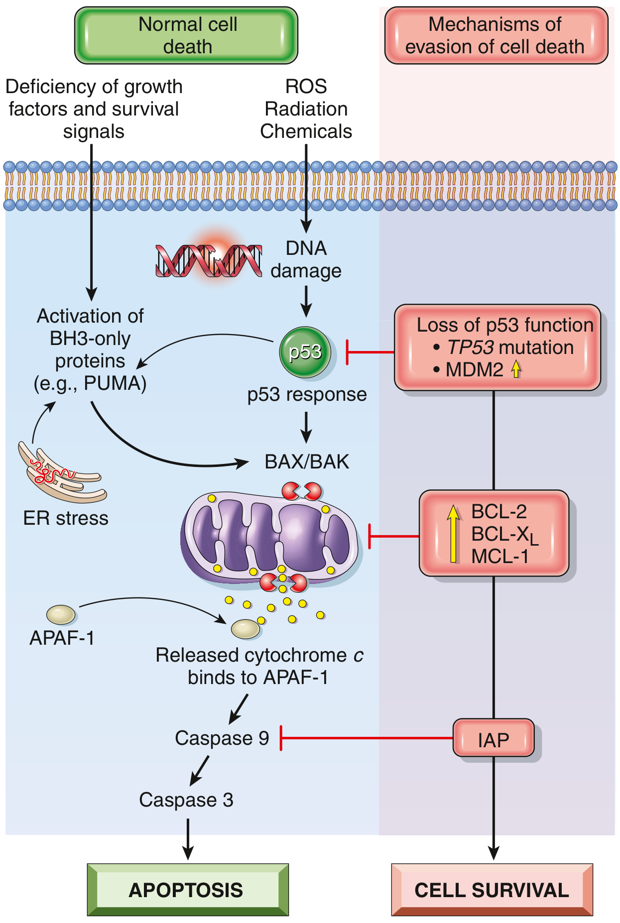
Intrinsic apoptotic pathway and major mechanisms by which tumor cells evade cell death.
Alterations of apoptosis in cancer
Evasion of apoptosis is a hallmark of cancer. Normally, a cell with severe DNA damage or oncogenic stress undergoes apoptosis, preventing survival of a potentially malignant clone. Cancer cells acquire mutations and expression changes that disable this safeguard. The intrinsic pathway is affected most often.
1. Loss of TP53 function
p53 is the key tumor-suppressor response to DNA damage and oncogenic stress. It can:
- Stop the cell cycle to allow DNA repair
- Induce proapoptotic proteins such as BAX, PUMA, and NOXA
- Promote mitochondrial apoptosis if damage is irreparable
Inactivation or mutation of TP53 prevents this response. Damaged cells therefore survive, continue proliferating, acquire further mutations, and become more resistant to chemotherapy and radiotherapy.
Overexpression of MDM2, which promotes p53 degradation, can produce a similar functional loss of p53.
2. Increased antiapoptotic BCL-2 family proteins
Tumors can overexpress proteins that prevent mitochondrial membrane permeabilization:
- BCL-2
- BCL-XL
- MCL-1
These proteins inhibit BAX and BAK, prevent cytochrome-c release, and block activation of caspase-9 and caspase-3.
Classic example: Follicular lymphoma often has a t(14;18) translocation. This places BCL2 under immunoglobulin heavy-chain regulatory control, causing high BCL-2 expression and prolonged survival of B lymphocytes. The key result is impaired cell death, not necessarily increased cell division.
3. Loss or reduced activity of proapoptotic proteins
Cancer cells may lose or suppress:
- BAX or BAK
- BH3-only proteins such as PUMA, BIM, BAD, or BID
- Components of the apoptosome, such as APAF-1
This prevents mitochondrial outer-membrane permeabilization or interrupts downstream caspase activation.
4. Increased inhibitors of apoptosis proteins
IAPs inhibit caspases, particularly caspase-9 and executioner caspases. Their overexpression allows tumor cells to remain alive even after an apoptotic signal has been initiated.
5. Impaired death-receptor signaling
Some cancers reduce sensitivity to immune-cell-mediated killing by:
- Decreasing Fas/death-receptor expression
- Altering downstream FADD or caspase-8 signaling
- Increasing FLIP, which inhibits caspase-8 activation
This makes tumor cells resistant to apoptosis induced by cytotoxic T cells and natural killer cells.
6. Resistance to anoikis
Anoikis is apoptosis induced when epithelial cells lose attachment to the extracellular matrix. Metastatic cancer cells acquire resistance to anoikis through altered integrin signaling, oncogene activation, growth-factor signaling, and increased antiapoptotic proteins. This permits detached cells to survive in blood, lymphatics, or distant tissues and helps metastasis.
Consequences in cancer
Defective apoptosis allows cancer cells to:
- Survive despite DNA damage and hypoxia
- Accumulate mutations and genomic instability
- Avoid immune destruction
- Resist chemotherapy and radiotherapy
- Survive detachment and metastasize
- Persist after therapy and contribute to relapse
Therapeutic relevance
Several therapies try to restore apoptosis:
- BCL-2 inhibitors, such as venetoclax, promote apoptosis in selected B-cell malignancies.
- MDM2 inhibitors aim to reactivate p53 in cancers retaining functional TP53.
- Cytotoxic chemotherapy and radiotherapy often act by inducing DNA damage and activating the mitochondrial apoptotic pathway.
The central cancer alterations are therefore loss of p53-mediated apoptotic signaling, excess antiapoptotic BCL-2-family activity, loss of proapoptotic mediators, and inhibition of caspases.
Robbins & Kumar Basic Pathology, p. 240-241.
Robbins & Kumar Basic Pathology, p. 240-241.
This is a shared conversation. Sign in to Orris to start your own chat.