

3 question answer
vermiform appendix anatomy positions blood supply diagram
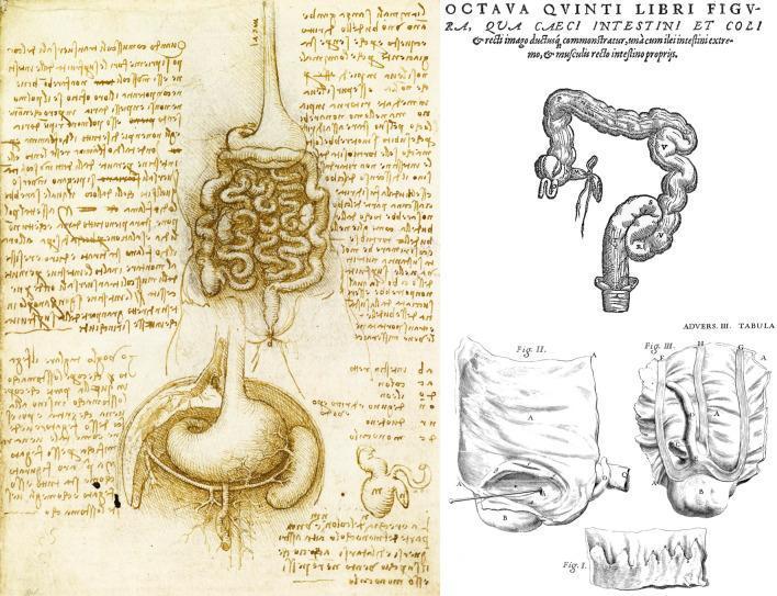
This composite image presents a historical comparison of anatomical drawings focusing on the vermiform appendix and gastrointestinal structures. On the left is a pen-and-ink anatomical diagram by Leonardo da Vinci (c. 1508), depicting the stomach with internal rugae, the esophagus, and the densely coiled small intestines. Below the stomach, a schematic representation of the cecum and appendix is visible, accompanied by mirror-writing notes. The upper right section features a woodcut by Andreas Vesalius showing a more morphologically accurate colon, highlighting the haustra (pouches) and taeniae coli (longitudinal bands), with a clear depiction of the cecum and the vermiform appendix. The lower right contains precise etchings by Giovanni Battista Morgagni, illustrating detailed observational studies of the cecal-appendiceal junction, including variations in appendiceal position and luminal characteristics. These illustrations represent the evolution of anatomical understanding in gastroenterology, moving from schematic representations to detailed structural observations relevant to the history of appendicitis diagnosis and surgical anatomy.
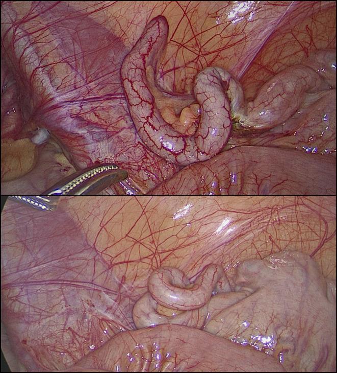
This clinical photograph consists of two laparoscopic views showing a macroscopically normal vermiform appendix during a diagnostic laparoscopy. The appendix appears as an elongated, tubular, and slightly coiled structure with a smooth, pale pink serosal surface. Fine, branching subserosal blood vessels are clearly visible across its length, indicating normal vascularity without the congestion or purulent exudate typical of acute appendicitis. In the upper frame, a laparoscopic grasper is visible, gently manipulating the appendix to facilitate inspection. The surrounding anatomy includes the mesoappendix containing adipose tissue and the glistening peritoneal lining of the right iliac fossa. There are no visual signs of inflammation, perforation, or abscess formation. This image serves as a reference for a 'grossly negative' appendix in the context of clinical suspicion of appendicitis, where symptoms may actually stem from non-inflammatory conditions such as follicular lymphoid hyperplasia or obstructive fecaliths.
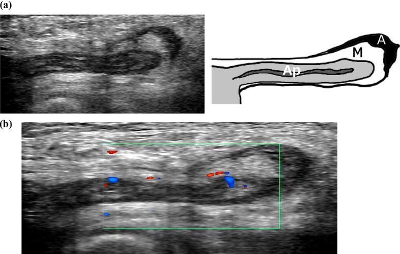
This composite diagnostic image features ultrasonography findings of a de Garengeot hernia, where the vermiform appendix is incarcerated within a femoral hernia sac. Panel (a) presents a B-mode ultrasound and a corresponding schematic diagram. It shows a longitudinal view of a blind-ended, isoechoic tubular structure (labeled 'Ap' for appendix) approximately 5 mm in diameter, which is continuous with the cecum. Surrounding the appendix is a reticular hyperechoic area representing the mesoappendix ('M'), and a localized anechoic fluid collection indicating ascites ('A') medial to the femoral vein. Panel (b) displays a Color Doppler ultrasound of the same anatomical region. Within the green sampling box, distinct red and blue pulsatile blood flow signals are visible along the appendiceal wall. These findings demonstrate preserved vascularity and the absence of significant inflammatory changes such as wall thickening or hyperemia, supporting a clinical diagnosis of an incarcerated but non-inflamed appendix within a femoral hernia.
Question 3 - Vermiform Appendix (Appendicitis Case)
(a) Positions, Blood Supply & Nerve Supply
Positions
| Position | Frequency | Description |
|---|---|---|
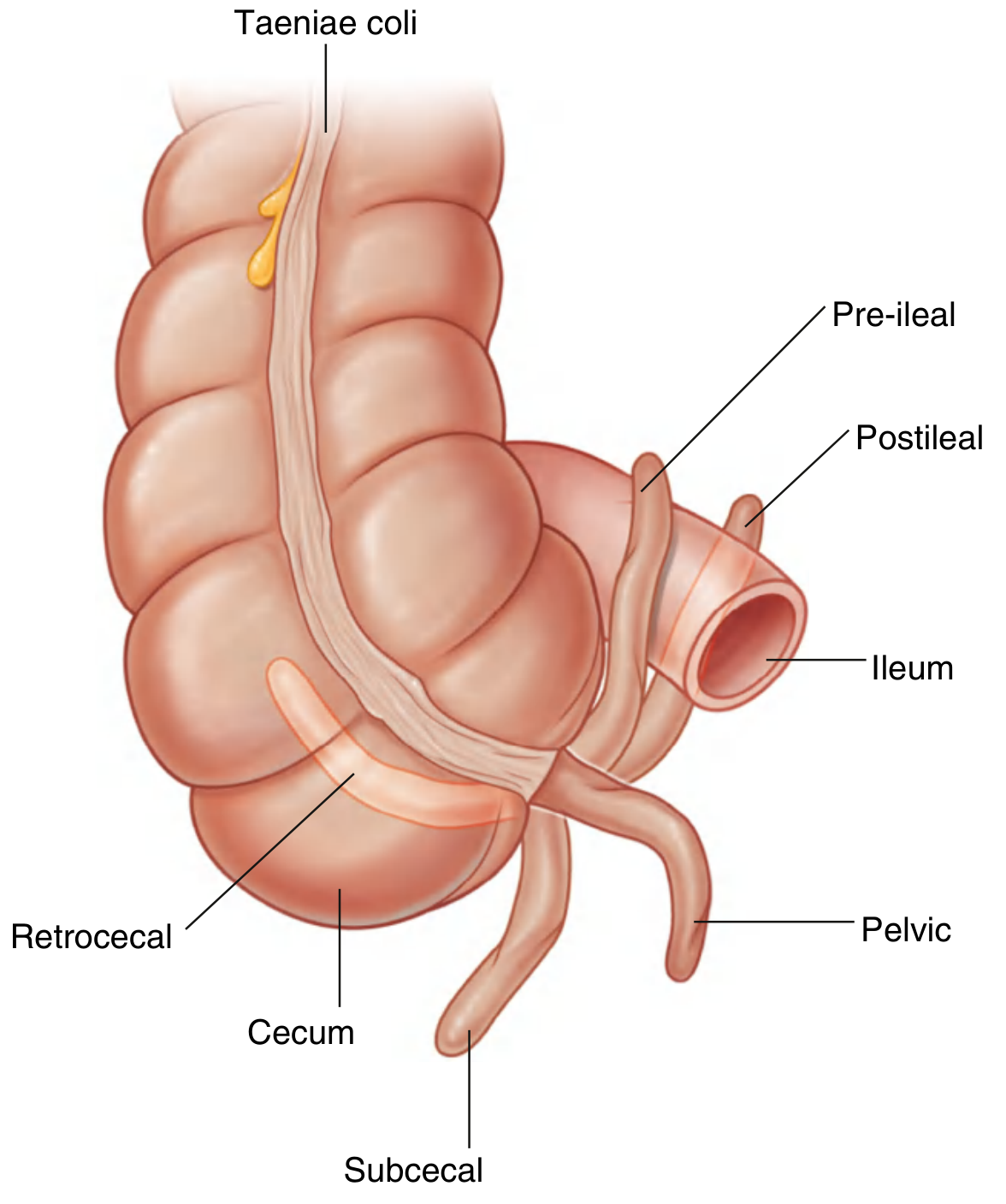
| Retrocecal (ascending) | ~65% | Behind the cecum - MOST COMMON |
| Pelvic / Subcecal (descending) | ~31% | Hangs into the lesser pelvis |
| Retrocecal (transverse) | >2% | Lies transversely behind cecum |
| Paracecal pre-ileal (preileal) | ~1% | Anterior to the terminal ileum |
| Paracecal post-ileal (retroileal) | ~0.5% | Posterior to the terminal ileum |
Blood Supply
- Artery: The appendicular artery, a branch of the lower division of the ileocolic artery (itself the last branch of the superior mesenteric artery). It passes behind the terminal ileum, enters the mesoappendix a short distance from the base, and runs in the free border of the mesoappendix to reach the appendix. It is an end-artery - thrombosis of this artery results in necrosis of the appendix (gangrenous appendicitis).
- An accessory appendicular artery may occasionally be present.
- Vein: The appendicular vein drains into the ileocolic vein, then into the superior mesenteric vein and finally the portal vein.
- Lymphatics: 4-6 or more lymphatic channels pass through the mesoappendix to drain into ileocolic (ileocecal) lymph nodes, then into the mesenteric nodes and intestinal trunks.
Nerve Supply
- Sympathetic: T10 fibers via the lesser splanchnic nerve and superior mesenteric plexus. Pain from appendicitis travels via these fibers - referred initially to the periumbilical region (T10 dermatome), which is the classic early symptom.
- Parasympathetic: Vagus nerve (CN X) via the superior mesenteric plexus.
- As inflammation progresses to involve the parietal peritoneum, somatic pain localizes to the right iliac fossa (McBurney's point).
(b) Clinical Anatomy
-
Acute Appendicitis: The most important clinical condition. Occurs when the appendix is obstructed (by a fecalith, foreign body, or lymphoid hyperplasia). Bacteria proliferate and invade the wall, leading to ischemia, necrosis, and possible perforation causing localized or generalized peritonitis.
-
McBurney's Point: The surface marking of the appendix base. Tenderness here (McBurney's sign) + rebound tenderness (Blumberg's sign) are cardinal signs of appendicitis.
-
Referred Pain Pathway: Early pain is periumbilical (referred via T10 sympathetic fibers). As inflammation involves the parietal peritoneum, pain shifts to the right iliac fossa - the classical "shifting pain" pattern seen in this patient.
-
Retrocecal Position (clinical relevance): In ~65% of cases, the appendix lies retrocecally. In this position, psoas irritation produces a positive Psoas sign (pain on passive hip extension). The appendix may be protected from palpation, making diagnosis harder.
-
Surgeon's Clue: At operation, the convergence of the three taeniae coli at the base guides the surgeon to locate the appendix even when it is hidden. The appendicular artery must be ligated in the mesoappendix before removal.
-
Meckel's Diverticulum vs. Appendicitis: Both produce right lower quadrant pain; Meckel's is located ~2 feet from the ileocecal junction.
-
Portal Pyemia: If appendicular vein thrombosis occurs and infected thrombus travels via the portal vein to the liver, it can cause pylephlebitis and hepatic abscesses - a serious complication.
(c) Microanatomy (Histology)
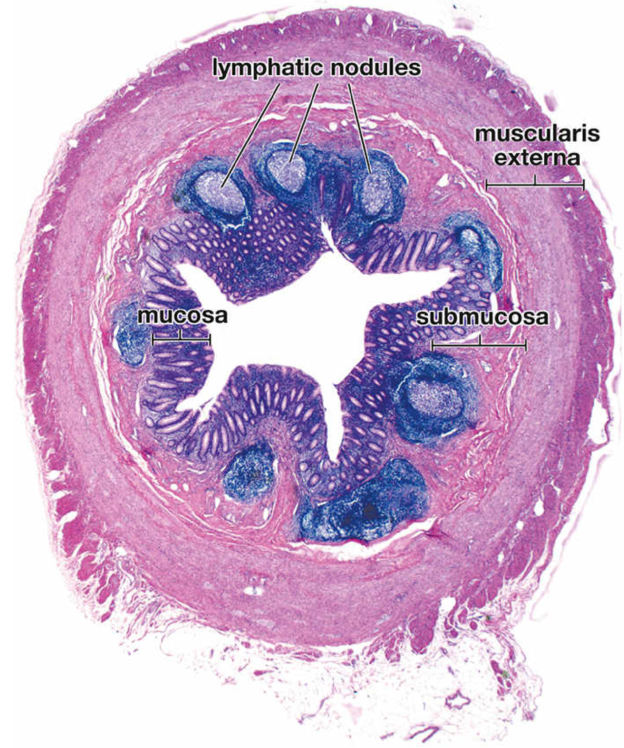
Layer by Layer:
- Lined by simple columnar epithelium (enterocytes + goblet cells), identical to large intestine
- Contains crypts of Lieberkühn (intestinal glands), but they are shallow and irregular (fewer and less developed than the rest of the colon)
- No villi are present
- At the base of the crypts: Argentaffin cells (Kulchitsky cells / enterochromaffin cells) - these are neuroendocrine cells that may give rise to carcinoid tumors (NETs) of the appendix - the most common tumor of the appendix
- The hallmark feature: contains an abundance of aggregated lymphoid nodules (Peyer's patches equivalent) with distinct germinal centers
- These lymphoid follicles extend from the lamina propria through the muscularis mucosae into the submucosa
- This massive lymphoid tissue makes the appendix part of the gut-associated lymphoid tissue (GALT) - it is an important immune organ, particularly in young adults
- The prominence of lymphoid tissue in young adults is important in the pathogenesis of appendicitis (lymphoid hyperplasia can obstruct the lumen)
- Inner circular muscle layer (relatively thick) + outer longitudinal layer (thinner)
- IMPORTANT: unlike the rest of the large intestine, the longitudinal muscle forms a complete continuous coat around the appendix - there are no taeniae coli in the wall of the appendix (the three taeniae of the cecum converge at the base and merge into this complete coat)
- The appendix is entirely surrounded by peritoneum (intraperitoneal) with its own mesentery - the mesoappendix
| Feature | Detail |
|---|---|
| Most common position | Retrocecal (65%) |
| Arterial supply | Appendicular artery (branch of ileocolic artery from SMA) |
| Type of artery | End-artery (thrombosis → gangrene) |
| Venous drainage | Ileocolic vein → SMV → Portal vein |
| Lymphatics | Ileocolic nodes → mesenteric nodes |
| Nerve supply | Sympathetic T10, Parasympathetic Vagus |
| Referred pain site | Periumbilicus (T10) → shifts to RIF |
| Hallmark histology | Massive submucosal lymphoid follicles; no taeniae; shallow crypts; Kulchitsky cells |
| Common tumor | Carcinoid / NET (from Kulchitsky cells) |