
MUSA criteria Adenomyosis
MUSA criteria adenomyosis morphological uterus sonographic assessment 2022 2023
MUSA criteria adenomyosis ultrasound
https://pmc.ncbi.nlm.nih.gov/articles/PMC10886873
MUSA criteria adenomyosis ultrasound features diagram
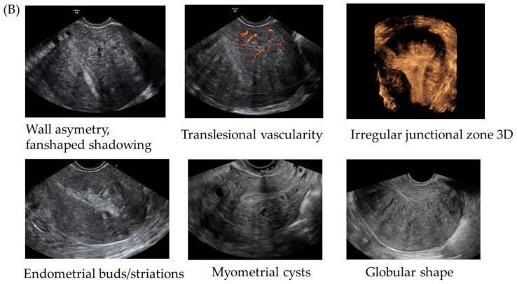
This composite educational graphic displays six ultrasound panels illustrating the classic sonographic MUSA (Morphological Uterine Sonographic Assessment) features of uterine adenomyosis. The images utilize transvaginal sonography (TVS) in grayscale, color Doppler, and 3D reconstruction. Key diagnostic signs depicted include: 1) Wall asymmetry with characteristic fan-shaped shadowing (rainshower appearance); 2) Translesional vascularity demonstrated via color Doppler, showing vessels crossing the myometrium perpendicularly; 3) An irregular junctional zone (JZ) visualized in a 3D reconstructed coronal plane; 4) Endometrial buds or striations appearing as echogenic linear structures extending from the endometrium into the myometrium; 5) Myometrial cysts, which are small anechoic fluid-filled spaces within the uterine wall; and 6) A globular uterine shape, where the uterus appears enlarged and rounded. These diagnostic criteria are essential for gynecological imaging to differentiate adenomyosis from other conditions like uterine fibroids (leiomyomas). The content is tailored for intermediate to advanced medical imaging education in obstetrics and gynecology.
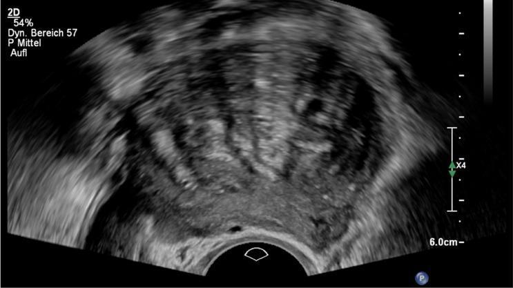
This diagnostic transvaginal ultrasound image displays a sagittal view of a uterus demonstrating classic sonographic features of adenomyosis, classified here under the #Enzian(u)FA category. Key visible findings include a significant global enlargement of the uterus with a marked asymmetry between the anterior and posterior myometrial walls. The myometrial texture is heterogeneous, exhibiting mixed echogenicity with several scattered small anechoic cystic areas (intramyometrial cysts) and bright hyperechogenic islands. A pathognomonic 'fan-shaped' or 'rain-shower' shadowing pattern is evident, where vertical stripes of acoustic shadowing originate from the heterogeneous tissue, obscuring deeper structures. The endo-myometrial junction appears blurred or ill-defined. These features are essential for the non-invasive diagnosis of uterine adenomyosis and are used to differentiate it from other pathologies like leiomyomas. The image serves as an educational tool for identifying Morphological Uterine Sonographic Assessment (MUSA) criteria in gynecological imaging.
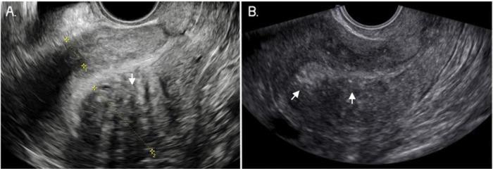
This diagnostic image consists of two grayscale panels (A and B) showing two-dimensional transvaginal ultrasound (TVUS) scans of a uterus in longitudinal section, demonstrating classic sonographic features of adenomyosis. Panel A illustrates marked myometrial asymmetry, where the posterior uterine wall is significantly thicker than the anterior wall. The myometrium exhibits a heterogeneous echo texture, characterized by prominent hypoechoic linear striae (fan-shaped shadowing) marked with arrows, representing subendometrial glandular invasion. Panel B focuses on the internal architecture of the uterine wall, highlighting a poorly defined junctional zone (JZ). White arrows point to the blurred, irregular interface between the endometrium and the underlying myometrium, which lacks a distinct, measurable boundary. These findings are key diagnostic criteria established by the Morphological Uterus Sonographic Assessment (MUSA) group for identifying adenomyosis. The images serve as an educational resource for gynecological radiology and ultrasound technicians to recognize signs of diffuse myometrial disease and uterine wall disproportion.
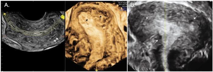
This diagnostic image set showcases 2D and 3D transvaginal ultrasound (TVUS) findings of diffuse adenomyosis in a human uterus. Panel A displays a heterogeneous myometrium with an irregular and interrupted junctional zone (JZ). Key markers include small, anechoic myometrial cysts (indicated by arrows), which represent ectopic endometrial glands. The 3D reconstruction in the center emphasizes the structural distortion of the uterine cavity. Panel B demonstrates severe diffuse adenomyosis, characterized by a globular, enlarged, and asymmetrical uterus. The image reveals an ill-defined, thickened JZ and extensive infiltration of the myometrium, highlighted by further myometrial cysts and echogenic islands (indicated by asterisks and arrows). These sonographic features are classic indicators for the Morphological Uterus Sonographic Assessment (MUSA) criteria used to diagnose adenomyosis and differentiate it from leiomyomas. The content is suitable for gynecological and radiological education at an intermediate to advanced level, focusing on identifying uterine pathologies through imaging modalities.
MUSA Criteria for Adenomyosis
Key Evolution: 2022 Delphi Revision
| Category | Meaning |
|---|---|
| Direct features | Evidence of ectopic endometrial glands/stroma within the myometrium |
| Indirect features | Myometrial reaction/hypertrophy secondary to ectopic endometrial tissue |
Direct Features (3 features)
| Feature | Description |
|---|---|
| Myometrial cysts | Small anechoic or hyperechoic round/oval structures within the myometrium; represent dilated ectopic endometrial glands. Specificity 86-98%, sensitivity 47-55% |
| Hyperechoic islands | Bright echogenic foci within the myometrium; most common direct sign. Specificity 83-95.5%, sensitivity 12-54% |
| Echogenic subendometrial lines and buds | Echogenic linear striations or bud-like projections extending from the endometrium into the myometrium. Specificity ~78%, sensitivity ~51% |
Indirect Features (6 features)
| Feature | Description |
|---|---|
| Globular uterus | Rounded uterine shape; corpus:cervix ratio >1; AP diameter approaches transverse diameter |
| Asymmetrical myometrial thickening | Disproportionate wall thickness between anterior and posterior myometrium (ratio >1.2) |
| Fan-shaped shadowing | "Venetian blind" / rain-shower vertical acoustic shadowing emanating from altered myometrium |
| Translesional vascularity | Color Doppler shows vessels crossing the lesion perpendicularly (distinguishes from fibroids where flow is peripheral) |
| Irregular junctional zone (JZ) | JZ appears uneven/undulating but still visible on 3D ultrasound |
| Interrupted junctional zone | JZ is discontinuous with focal breaks - visualized best on 3D coronal reconstruction |
The Eight Morphological Descriptors (Classification Framework)
- Presence - adenomyosis present/absent
- Location - anterior, posterior, lateral wall; fundal; diffuse
- Differentiation - diffuse vs. focal (adenomyoma)
- Cystic vs. non-cystic
- Myometrial layer involved - inner, outer, or full thickness
- Extent of disease - proportion of myometrium affected
- Size of lesion - measured in three planes
- Vascularity - color Doppler pattern
Scoring and Diagnostic Thresholds
| Threshold | Sensitivity | Specificity |
|---|---|---|
| Total feature score >4 | 27.5% | 90% |
| Direct feature score >1 | 12.5% | 95% |
| Indirect feature score >4 | 17.5% | 93% |
| 2 direct + ≥3 indirect | Higher PPV | 98.7% |
Visual Examples
Clinical Notes
- 3D ultrasound is particularly valuable for visualizing the junctional zone in the coronal plane, improving assessment of irregular and interrupted JZ.
- Adenomyosis often coexists with endometriosis - look for both.
- Translesional vascularity on Doppler is the key feature distinguishing adenomyosis from uterine fibroids (fibroids show peripheral flow).
- The "question mark" sign (uterine flexion with posterior DIE - deep infiltrating endometriosis) was added as an associated sign in 2020 by Chapron et al.
- MRI remains useful when ultrasound is inconclusive or to differentiate adenomyoma from leiomyoma.
- Bailey and Love's Short Practice of Surgery, 28th Ed., p. 2643-2648