
What is bulla
bulla skin lesion definition types dermatology
bulla blister skin lesion dermatology
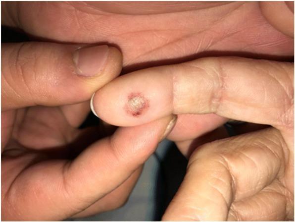
This clinical photograph displays a solitary skin lesion on the distal phalanx of a human ring finger. The lesion is a well-circumscribed, circular bulla (blister) approximately 5-8 mm in diameter. It features a centrally opaque, whitish roof, indicating potential epidermal necrosis or serous fluid accumulation. The blister is bordered by a distinct erythematous halo, suggesting localized inflammation. The surrounding skin appears normal with typical age-related wrinkling, particularly at the interphalangeal joints. In a clinical context, this lesion illustrates a phototoxic skin burn resulting from photodynamic therapy (PDT) and subsequent light exposure from a pulse oximeter probe. It serves as an educational example of iatrogenic skin injury and photosensitivity reactions related to talaporfin sodium. The educational focus is on perioperative dermatology and the importance of shielding photosensitized patients from specific light wavelengths used in routine monitoring equipment.
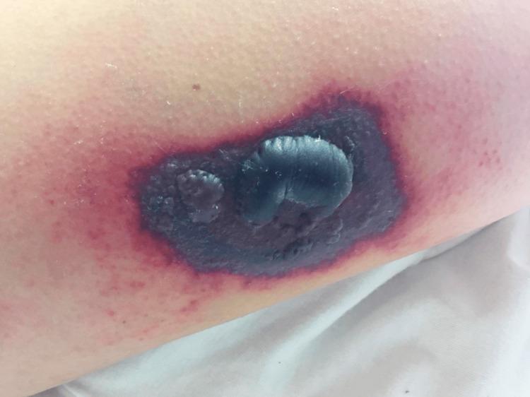
This clinical photograph displays a focal, necrotic skin lesion on an extremity. The lesion is characterized by a central, tense, dark-purple-to-black bulla (blister) approximately 2–3 cm in diameter, indicating epidermal detachment and underlying tissue necrosis or hemorrhage. Surrounding this central bulla is a well-demarcated area of deep violaceous or purpuric plaque. A prominent outer erythematous halo is visible, suggesting secondary acute inflammation. The morphology is consistent with ecthyma gangrenosum, vasculitis, or a progressive cutaneous manifestation of systemic illness such as T-cell lymphoma or leukemia cutis. This visual provides high-yield educational value for identifying necrotic bullae and understanding the progression from livedo reticularis to necrotic plaque and ulceration in a clinical dermatology or oncology context.
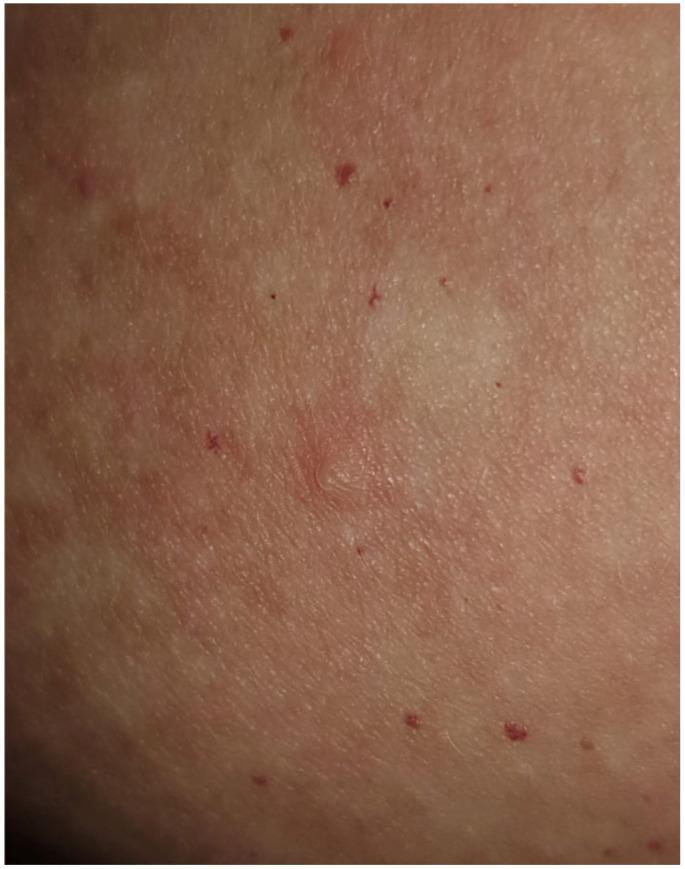
This clinical photograph shows a close-up view of the skin, likely from the trunk (seborrheic area), demonstrating characteristic features of an autoimmune bullous disease, specifically Pemphigus foliaceus. The primary finding is a small, superficial, flaccid blister (vesicle/bulla) located centrally. The lesion is pale and translucent compared to the surrounding skin, appearing thin-roofed and easily collapsible. The background skin is notably erythematous, with diffuse redness and scattered, tiny, punctate red papules or petechiae. The skin texture is irregular, indicating subtle edema and early epidermal involvement. This visual presentation illustrates acantholysis within the granular layer, a hallmark of superficial blistering disorders where bullae are transient and often rupture quickly to leave behind erosions and crusts. The image is a valuable educational resource for dermatology, focusing on the morphology of superficial bullous eruptions and their clinical distinction from deeper blistering conditions like pemphigus vulgaris or bullous pemphigoid.
Bulla - Medical Definition
1. Dermatology - Skin Bulla (most common usage)
Structure
- Has a thin overlying roof (epidermis or part of it)
- Contains clear serous, serosanguinous, or purulent fluid
- Can be flaccid (thin wall, ruptures easily) or tense (thick wall, does not rupture easily)
Location within skin
| Type | Location | Clinical feature |
|---|---|---|
| Intraepidermal (subcorneal) | Within the epidermis | Flaccid, fragile, ruptures easily |
| Subepidermal | Below the epidermis, at dermal-epidermal junction | Tense, more durable |
Common causes
- Burns and physical injury
- Autoimmune blistering diseases:
- Pemphigus vulgaris - flaccid bullae in oral mucosa and skin; positive Nikolsky sign
- Pemphigus foliaceus - superficial flaccid bullae on scalp, chest, back
- Bullous pemphigoid - tense bullae on normal or erythematous skin in elderly; linear immunofluorescence at dermo-epidermal junction
- Infections: bullous impetigo, herpes zoster
- Drug reactions: Stevens-Johnson syndrome, toxic epidermal necrolysis
- Genetic/inherited: epidermolysis bullosa
- Contact dermatitis (irritant or allergic)
2. Pulmonary - Lung Bulla
-
A giant bulla occupies more than 30% of a hemithorax
-
Visible on chest X-ray as radiolucent areas with fine hairline shadows
-
Bullae can compress adjacent lung tissue, and very large ones may warrant bullectomy (surgical removal)
-
Natural history is unpredictable - some resolve, some grow, some rupture causing spontaneous pneumothorax
-
Goldman-Cecil Medicine, Bullectomy section
3. ENT / Anatomy - Ethmoid Bulla
- KJ Lee's Essential Otolaryngology
Summary Table
| Context | What it is | Size criterion |
|---|---|---|
| Skin (dermatology) | Fluid-filled blister | > 1 cm diameter |
| Lung (pulmonology) | Air-filled emphysematous space | > 1 cm; giant if > 30% hemithorax |
| Nose/sinus (ENT) | Ethmoid air cell (anatomical landmark) | Variable |
Types of cellities
types of cellulitis classification dermatology
cellulitis skin infection erythema swelling leg
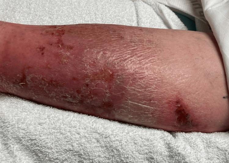
This clinical photograph displays the lower extremity of a patient, demonstrating classic physical examination findings of cellulitis. The skin exhibits diffuse erythema and swelling extending across the anterior and lateral aspects of the leg. The texture is notably compromised, showing signs of desquamation (peeling), xerosis (dryness), and lichenification, particularly in the mid-to-lower portion. Multiple discrete skin lesions are visible, including areas of darker, crusted scabbing and potential ulceration that may serve as a portal of entry for infection. Small petechiae or ecchymotic spots are scattered within the erythematous region. The presentation is highly characteristic of an acute bacterial skin and soft tissue infection (SSTI), emphasizing the visual transition from healthy proximal skin to distal inflamed, edematous, and scaling tissue. This image is an educational resource for identifying inflammatory dermatological conditions and assessing secondary skin changes associated with chronic or severe cellulitis.
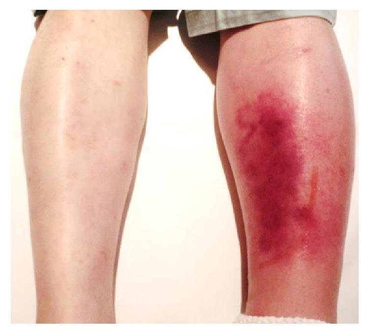
This clinical photograph shows a posterior view of a patient's lower legs, providing a side-by-side comparison between a healthy limb and a symptomatic one. The left lower leg (right side of the image) exhibits classic clinical signs of cellulitis, characterized by significant unilateral edema and diffuse erythema. The affected skin shows a spectrum of inflammatory discoloration, ranging from bright red to deep purplish hues, particularly over the calf region. The skin texture appears tense and glossy due to underlying swelling. A thin, vertical line of serous fluid exudate is visible on the posterior-lateral aspect of the left calf. In contrast, the right lower leg (left side of the image) appears normal with healthy skin tone and no evidence of swelling. This visual demonstrates the acute presentation of a soft tissue infection, useful for teaching the differential diagnosis of unilateral leg swelling and redness, which may include cellulitis, deep vein thrombosis (DVT), or localized vasculitis.
Types of Cellulitis
A. By Location / Anatomical Site
1. Limb Cellulitis (most common)
- Usually affects the lower leg
- Most common organisms: Streptococcus (Group A beta-hemolytic) and Staphylococcus aureus
- Risk factors: lymphedema, chronic venous insufficiency, tinea pedis, diabetes, obesity
- Rarely bilateral - bilateral presentation should prompt reconsideration of the diagnosis
2. Preseptal (Periorbital) Cellulitis
- Infection of eyelids and periocular tissues anterior to the orbital septum
- Generally benign; can be treated as an outpatient
- Primarily a disease of childhood (most patients < 10 years)
- Common organisms: S. aureus, S. epidermidis, Streptococcus spp., anaerobes
- Causes: sinusitis, hordeolum, chalazion, insect bite, trauma
- Tintinalli's Emergency Medicine
3. Postseptal (Orbital) Cellulitis
- Infection of orbital soft tissues posterior to the orbital septum
- Vision and life-threatening - requires inpatient IV antibiotics and possible surgical drainage
- May complicate paranasal sinusitis (most common cause)
- Features distinguishing from preseptal: proptosis, restricted/painful eye movement (ophthalmoplegia), chemosis, decreased visual acuity
- Complications if untreated: subperiosteal abscess, orbital abscess, cavernous sinus thrombosis, meningitis, blindness
- Tintinalli's Emergency Medicine
4. Facial Cellulitis
- More common in adults > 50 years; pneumococcal facial cellulitis in young children
- May be of odontogenic (dental) origin
- Older patients with diabetes are particularly vulnerable
5. Perianal Cellulitis
- Usually caused by Group A beta-hemolytic Streptococcus (GABHS)
- Occurs mainly in children < 3 years
6. Orbital / Ear Cellulitis
- Elderly diabetic patients are more prone to Pseudomonas cellulitis of the ear
- Orbital cellulitis in elderly is typically polymicrobial (aerobic + anaerobic organisms)
- Fitzpatrick's Dermatology
B. By Purulence (Clinical Distinction)
| Type | Features | Main Organism | First-line Treatment |
|---|---|---|---|
| Nonpurulent cellulitis | No abscess/pus; diffuse spreading infection | Streptococcus spp. | Beta-lactam antibiotics (e.g. penicillin/cephalexin) |
| Purulent cellulitis | Associated with furuncles, carbuncles, or abscesses | S. aureus (often MRSA) | TMP-SMX or doxycycline (covers CA-MRSA) |
C. By Pathogen / Special Context
| Type | Organism | Setting |
|---|---|---|
| Streptococcal cellulitis | Group A Streptococcus | Most common nonpurulent |
| Staphylococcal cellulitis | S. aureus, MRSA | Purulent, abscesses |
| Water-borne cellulitis | Vibrio vulnificus | Seawater + liver disease patients |
| Freshwater cellulitis | Aeromonas spp. | Freshwater trauma; rapidly progressive |
| Fish tank granuloma | Mycobacterium marinum | Aquarium exposure; papule weeks later |
| Erysipeloid | Erysipelothrix rhusiopathiae | Seafood workers, hand trauma |
- Rosen's Emergency Medicine
D. Severity Classification - Eron/CREST System
| Class | Features |
|---|---|
| Class I | No systemic toxicity, no comorbidities; systemically well |
| Class II | Systemic toxicity (fever, tachycardia) OR significant comorbidity |
| Class III | Significant systemic toxicity AND significant comorbidity |
| Class IV | Sepsis or life-threatening infection |
E. Related Conditions (Distinguished from Cellulitis)
- Erysipelas - infection of the upper dermis spreading to lymphatics by beta-hemolytic Streptococcus; has sharply demarcated borders (unlike cellulitis which has indistinct margins); more common in elderly
- Necrotizing fasciitis - deep infection of fascia; more aggressive, life-threatening; associated with Streptococcus and mixed organisms; requires emergency surgery
- Eosinophilic cellulitis (Wells syndrome) - non-infectious; recurrent itchy/painful plaques with eosinophils on biopsy; not a true bacterial infection but clinically mimics cellulitis
Key Point: Cellulitis vs. Pseudocellulitis
- Venous stasis dermatitis (often bilateral, above ankle)
- Deep vein thrombosis
- Gout
- Contact dermatitis
- Fixed drug eruption
- Lymphedema
To come urine more what inj we have to give
injectable diuretics to increase urine output furosemide IV drugs
Injections Given to Increase Urine Output
1. Furosemide (Lasix) - FIRST LINE ⭐
| Situation | Dose |
|---|---|
| Mild / first dose | 20-40 mg IV |
| Severe volume overload | 40-160 mg IV; or 2.5x the oral dose |
| Continuous infusion | 5-40 mg/hr IV |
| Diuretic resistant patients | Double the dose until response seen |
2. Bumetanide (Bumex)
3. Torsemide (Demadex)
- Braunwald's Heart Disease
4. Mannitol - Osmotic Diuretic
- Raised intracranial pressure (ICP)
- Acute glaucoma (to reduce intraocular pressure)
- Oliguric renal failure (to flush tubules)
- Forced diuresis in drug/toxin poisoning
5. Ethacrynic Acid (Edecrin)
Summary Table
| Drug | Type | Route | Onset (IV) | Main Use |
|---|---|---|---|---|
| Furosemide | Loop | IV/IM | 30-60 min | Most common - heart failure, edema |
| Bumetanide | Loop | IV/IM | 30 min | Furosemide resistance |
| Torsemide | Loop | IV | 30-60 min | Heart failure, edema |
| Mannitol | Osmotic | IV only | 30-60 min | Raised ICP, oliguria, toxin flush |
| Ethacrynic acid | Loop | IV | 30 min | Sulfa allergy patients |
Clinical Note
- In emergency settings (acute pulmonary edema, heart failure), IV furosemide is the first injection given
- In oliguria/AKI, mannitol or a high-dose furosemide challenge may be tried to convert oliguric to non-oliguric renal failure
- Always monitor urine output, electrolytes (K⁺, Na⁺), BP, and creatinine after giving diuretic injections
Mannitol use for
mannitol clinical uses indications mechanism of action
Mannitol - Uses
- Katzung's Basic & Clinical Pharmacology, Guyton & Hall Medical Physiology
Mechanism of Action
- Stays in the blood vessel (does not enter cells) - raises plasma osmolarity
- Draws water from tissues (like brain, eye) into the bloodstream - reducing swelling
- Gets filtered at the kidney but not reabsorbed - pulls water into urine, increasing urine output
Uses / Indications
1. Raised Intracranial Pressure (ICP) ⭐ Most important use
- Used in cerebral edema (brain swelling) from:
- Head injury / traumatic brain injury (TBI)
- Stroke
- Brain tumors
- Liver failure with hepatic encephalopathy
- Mannitol draws water out of the brain cells into the blood, shrinking brain volume and reducing ICP
- Dose: 0.5 to 1 g/kg IV over 15-30 minutes
- Tintinalli's Emergency Medicine; Textbook of Family Medicine
2. Acute Glaucoma (Raised Intraocular Pressure)
- Draws fluid out of the vitreous body of the eye
- Reduces intraocular pressure rapidly
- Used as emergency treatment when eye pressure is dangerously high
- Katzung's Basic & Clinical Pharmacology
3. Acute Kidney Injury (AKI) / Oliguria
- Used to prevent or treat the oliguric phase of acute renal failure
- Also used in conditions that overload the kidney with harmful solutes:
- Rhabdomyolysis (muscle breakdown - myoglobin clogs tubules)
- Chemotherapy (tumor lysis syndrome - uric acid/phosphate overload)
- Hemolysis (haemoglobin in urine)
- Flushes tubules with a forced diuresis, preventing tubular blockage
- Katzung's Basic & Clinical Pharmacology; StatPearls/NCBI
4. Drug and Toxin Poisoning - Forced Diuresis
- Given to increase urine flow and speed up excretion of toxic drugs or poisons
- Used in poisoning with salicylates, barbiturates, or other renally-excreted toxins
5. Pre-operatively in Neurosurgery / Eye Surgery
- Given before brain or eye operations to reduce tissue swelling and provide a better surgical field
Dose
| Indication | Dose |
|---|---|
| Raised ICP / Cerebral edema | 0.5-1.5 g/kg IV over 15-30 min |
| Glaucoma (acute) | 1.5-2 g/kg IV over 30-60 min |
| Oliguria / Renal failure | 0.25-0.5 g/kg IV |
| Forced diuresis (poisoning) | 50-200 g IV over 24 hrs |
Contraindications (When NOT to give)
| Condition | Reason |
|---|---|
| Pulmonary edema | Mannitol initially expands blood volume - can worsen fluid in lungs |
| Congestive heart failure | Same reason - risk of fluid overload |
| Severe dehydration | Worsens electrolyte imbalance |
| Anuria (no urine output) | If kidneys are completely failed, mannitol accumulates and is dangerous |
| Intracranial bleeding (active) | Can worsen hemorrhage |
- Morgan & Mikhail's Clinical Anesthesiology
Side Effects
- Nausea, vomiting, headache
- Initial hyponatremia (dilution effect) then hypernatremia (after water is lost)
- Electrolyte imbalance (low K⁺, low Mg²⁺)
- Pulmonary edema (if given to heart failure patients)
- Rebound raised ICP (if given repeatedly - mannitol can cross damaged blood-brain barrier and draw water back into brain)
Summary in One Line
Mannitol is given IV to reduce brain swelling (raised ICP), reduce eye pressure (glaucoma), flush the kidneys in rhabdomyolysis/AKI, and force excretion of toxins - but avoid it in heart failure and pulmonary edema.