
Development of pituitary gland
I'll read the medical library skill first, then search for pituitary gland development content.
Reading File
Now let me search the library for pituitary gland development content in parallel.
Finding Sources
Finding Sources
Excellent results. Now let me read the most relevant sources in parallel - Ganong's (which has a dedicated section on development, structure & cell types), Robbins (pathology context), and Harrison's (clinical context).
Reading File
Reading File
Reading File
Now let me get more detail from Robbins on transcription factors, and also fetch the Ganong diagram and Robbins histology image. Let me also search for embryology-specific content.
Reading File
Finding Sources
Loading Image
Excellent! Now let me read the embryology-specific content from Langman's and Junqueira's, plus Tietz for the transcription factors:
Reading File
Reading File
Reading File
Now let me fetch the key developmental diagram from Junqueira's and the histology image from Robbins:
Loading Image
Loading Image
Loading Image
I now have comprehensive, well-illustrated content from multiple authoritative textbooks. Let me compile the full answer.
Development of the Pituitary Gland (Hypophysis)
The pituitary gland has a dual embryological origin - this is its most important developmental feature. It arises from two completely separate structures that grow toward each other and fuse.
Overview: Dual Origin
| Component | Origin | Embryological Tissue |
|---|---|---|
| Anterior pituitary (adenohypophysis) | Rathke's pouch (upward evagination) | Oral ectoderm (stomodeum) |
| Posterior pituitary (neurohypophysis) | Infundibulum (downward extension) | Neuroectoderm of diencephalon |
Step-by-Step Developmental Timeline
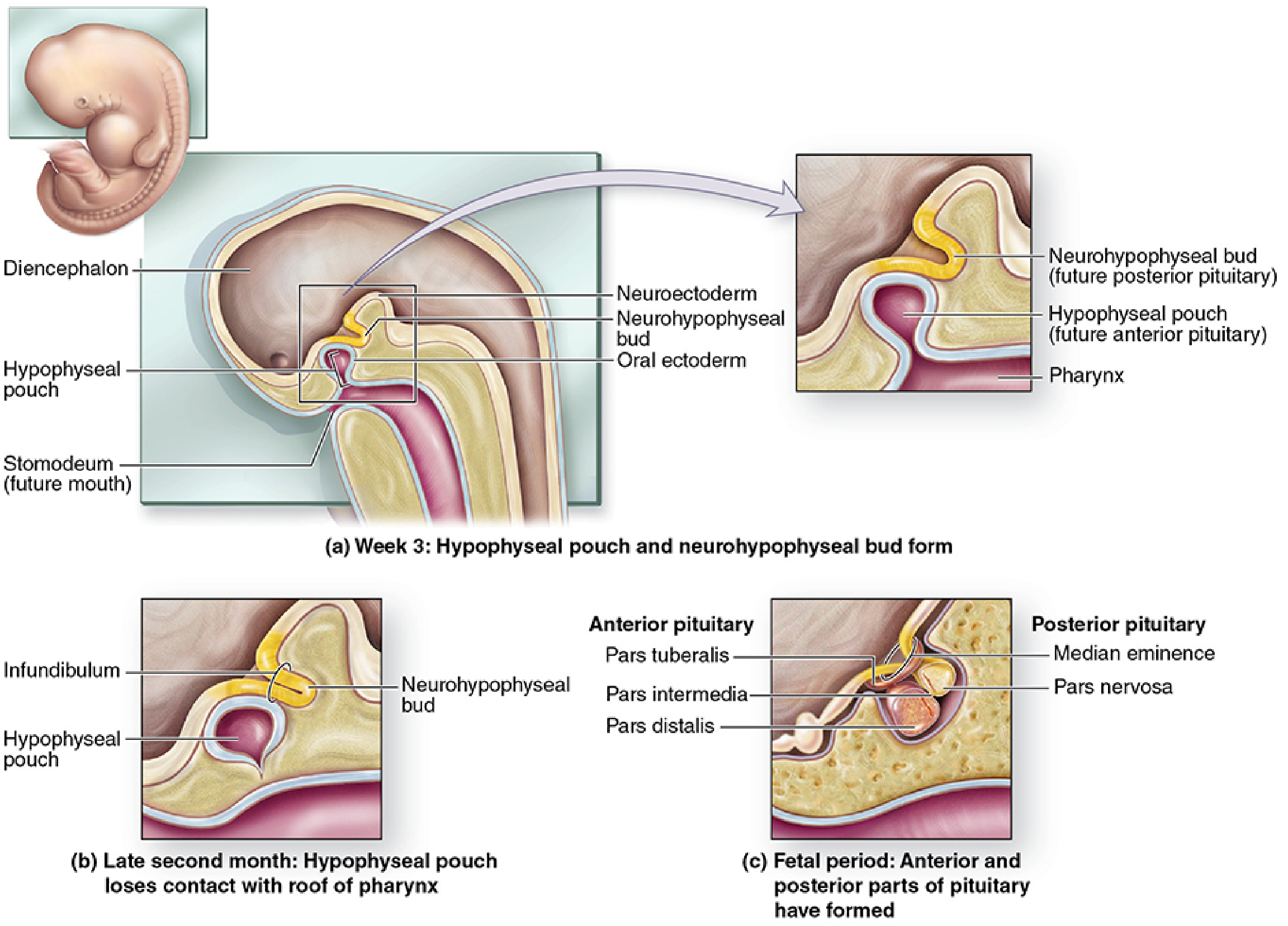
Week 3: Initiation
- Rathke's pouch appears as an upward evagination from the roof of the stomodeum (primitive oral cavity), immediately in front of the oropharyngeal membrane.
- Simultaneously, the neurohypophyseal bud (infundibulum) begins as a downward extension of the floor of the diencephalon.
- Both structures grow toward each other.
(Langman's Medical Embryology)
End of Month 2: Separation
- Rathke's pouch loses its connection with the oral cavity (the stalk connecting it to the pharynx degenerates).
- The pouch is now in close contact with the infundibulum.
Fetal Period: Differentiation into Parts
- Anterior wall of Rathke's pouch proliferates extensively → forms the pars distalis (main bulk of the anterior lobe).
- A small extension from the anterior wall grows upward along the infundibular stalk → forms the pars tuberalis (wraps around the stalk).
- Posterior wall of Rathke's pouch → forms the thin pars intermedia (intermediate lobe), adjacent to the posterior lobe.
- The residual lumen of Rathke's pouch persists as the residual cleft (Rathke's cleft), separating the anterior and intermediate lobes.
- The infundibulum gives rise to:
- The pars nervosa (posterior lobe proper)
- The median eminence
- The infundibular stalk
(Langman's; Junqueira's Basic Histology)
Parts of the Adult Pituitary
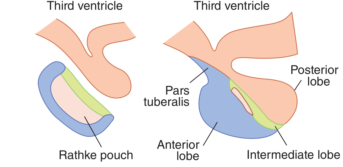
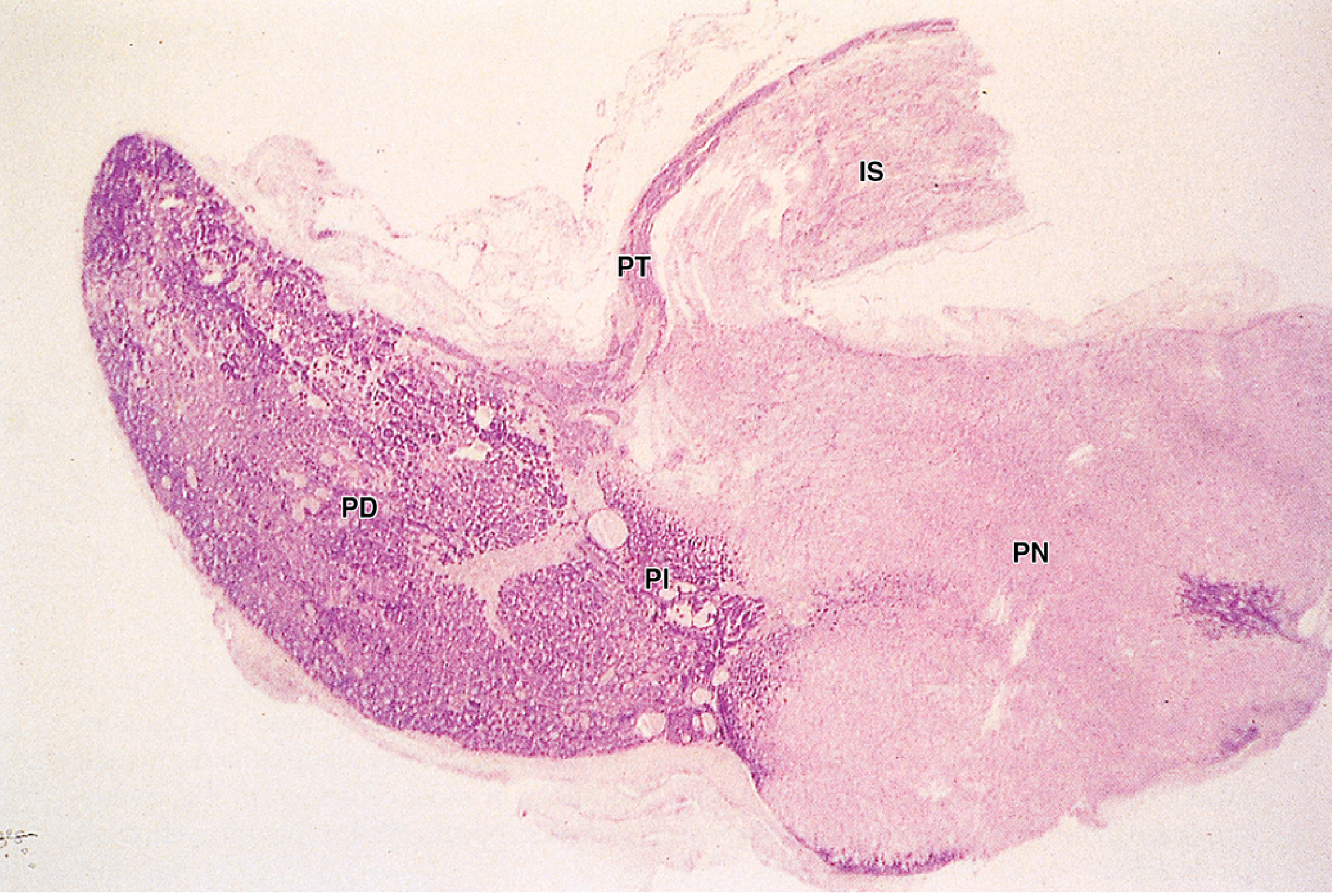
Adenohypophysis (from Rathke's pouch - oral ectoderm)
- Pars distalis - the large anterior lobe; ~80% of gland mass
- Pars tuberalis - wraps around the pituitary stalk
- Pars intermedia - rudimentary in adult humans; most cells are incorporated into the anterior lobe
Neurohypophysis (from infundibulum - neuroectoderm)
- Pars nervosa (posterior lobe)
- Infundibular stalk
- Median eminence
Transcription Factors Governing Anterior Pituitary Cell Differentiation
The anterior pituitary arises from pluripotent Sox2-expressing precursor cells within Rathke's pouch. Their differentiation into six terminal cell types is orchestrated by a cascade of lineage-specific transcription factors:
| Transcription Factor | Role |
|---|---|
| HESX1 | Early pituitary specification; mutations → septo-optic dysplasia |
| LHX3, LHX4 | Early organogenesis and cell survival |
| SOX3 | Infundibulum development |
| FGFR1 | Pituitary morphogenesis |
| PROP-1 | Induces development of Pit-1 lineages AND gonadotropes |
| PIT-1 (POU1F1) | Determines expression of GH, PRL, TSH → somatotropes, lactotropes, thyrotropes |
| TEF (Thyrotrope Embryonic Factor) | Induces TSH expression in thyrotropes |
| SF-1 (Steroidogenic Factor-1) | Gonadotrope cell specification |
| DAX-1 | Further defines gonadotropes |
| T-Pit | Required for corticotrope differentiation (POMC expression) |
(Harrison's 22e; Tietz Textbook of Laboratory Medicine; Robbins Pathology)
Key Lineage Relationships
- Somatotropes, mammosomatotropes, lactotropes, and thyrotropes all share a common PIT-1-expressing precursor
- Corticotropes are derived separately via T-Pit
- Gonadotropes require PROP-1 + SF-1 + DAX-1
Cell Types of the Anterior Pituitary
Six terminally differentiated secretory cell types are present in the adult anterior pituitary:
| Cell Type | Hormone Secreted | Staining (H&E) |
|---|---|---|
| Somatotropes | Growth Hormone (GH) | Acidophil |
| Mammosomatotropes | GH + Prolactin | Acidophil |
| Lactotropes | Prolactin (PRL) | Acidophil |
| Corticotropes | ACTH (from POMC) | Basophil |
| Thyrotropes | TSH | Basophil |
| Gonadotropes | FSH + LH | Basophil |
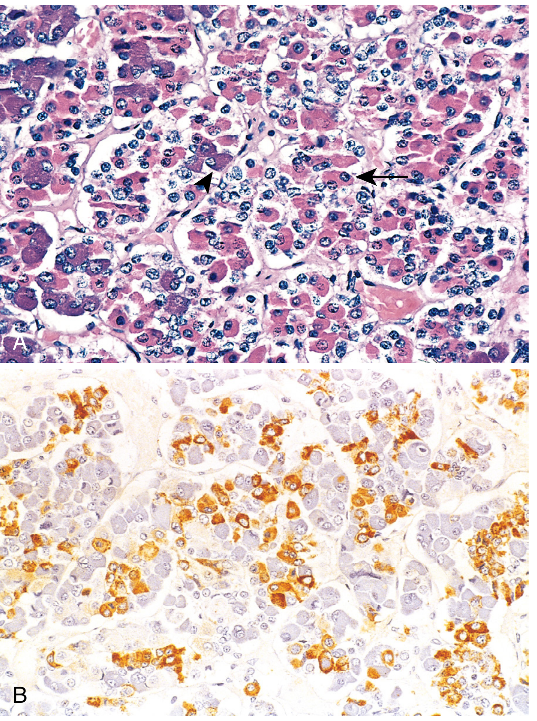
Classically, anterior pituitary cells were divided into acidophils, basophils, and chromophobes based on H&E staining. Modern immunocytochemistry assigns them more precisely by hormone content.
(Robbins; Ganong's)
Posterior Pituitary Histology
The neurohypophysis is essentially brain tissue and consists of:
- Pituicytes - modified astrocytes (stellate glial cells), the dominant cell type
- Axon terminals from magnocellular neurons in the supraoptic and paraventricular nuclei of the hypothalamus - these axons travel down the stalk and release oxytocin and ADH (vasopressin) directly into the systemic circulation from the posterior lobe
The posterior pituitary does not synthesize these hormones - they are made in the hypothalamus and merely stored/released from the posterior lobe.
(Ganong's; Robbins)
Pars Intermedia
In humans, the pars intermedia is rudimentary. It is active primarily in utero and late in pregnancy, secreting:
- α-, β-, γ-MSH (melanocyte-stimulating hormones)
- Corticotropin-like intermediate lobe peptide (CLIP)
- γ-Lipotropin
- β-Endorphin
(Tietz Textbook of Laboratory Medicine)
Clinical Correlations
Craniopharyngioma
Remnants of Rathke's pouch that fail to fully regress can give rise to craniopharyngiomas - benign but locally destructive tumors derived from Rathke's pouch epithelium. They are a key cause of hypopituitarism in children.
Rathke's Cleft Cysts
Persistence of the lumen of Rathke's pouch as a symptomatic cyst between the anterior and posterior lobes.
Transcription Factor Mutations → Combined Pituitary Hormone Deficiency (CPHD)
- PIT-1 mutations → combined GH + PRL + TSH deficiency
- PROP-1 mutations → most common genetic cause of CPHD (GH, PRL, TSH, and variable gonadotropin/ACTH deficiency)
- HESX1 mutations → septo-optic dysplasia (pituitary hypoplasia + optic nerve hypoplasia + absent septum pellucidum)
- T-Pit mutations → isolated ACTH deficiency
(Harrison's 22e; Tietz)
Pituitary Dysplasia
Because pituitary development follows midline cell migration from the nasopharyngeal Rathke's pouch, midline defects (e.g., cleft lip/palate, holoprosencephaly) are often associated with pituitary aplasia or hypoplasia. (Harrison's 22e)
Sources: Langman's Medical Embryology | Junqueira's Basic Histology (17e) | Ganong's Review of Medical Physiology (26e) | Robbins & Cotran Pathologic Basis of Disease | Harrison's Principles of Internal Medicine (22e, 2025) | Tietz Textbook of Laboratory Medicine (7e)
This is a shared conversation. Sign in to Orris to start your own chat.