
Actinomycosis
I'll start with a quick overview while I pull from the medical library.
Actinomycosis is a chronic, slowly progressive bacterial infection caused by Actinomyces species (most commonly A. israelii), gram-positive anaerobic/microaerophilic filamentous bacteria that are normal oral/GI flora. It produces characteristic "sulfur granules" in purulent discharge and forms indurated, woody masses that cross tissue planes. The three classic forms are cervicofacial, thoracic, and abdominopelvic.
Searching sources...
Reading File
Now I'll search the library and images in parallel.
Finding Sources
Searching Images
actinomycosis sulfur granules histology cervicofacial
Searching PubMed
actinomycosis
Good results. Now let me read from the most content-rich sources in parallel.
Reading File
Reading File
Reading File
Reading File
Excellent data. Now let me get the pulmonary section and the GI section, plus search for images.
I now have comprehensive material from multiple authoritative textbooks. Let me compile the full answer.
Actinomycosis
Organism and Microbiology
Actinomyces species are gram-positive, non-acid-fast, anaerobic to microaerophilic, filamentous branching bacteria - not true fungi, despite the name "ray fungus." The most common pathogenic species is A. israelii, followed by A. bovis and A. naeslundii. They are normal commensals found in high concentrations in the tonsillar crypts, carious teeth, colon, and urogenital tract.
- Cummings Otolaryngology, p. 1496
Pathogenesis
The organism is non-invasive under normal conditions. Disruption of mucosal barriers (dental trauma, surgery, GI perforation, aspiration) allows tissue invasion, producing a chronic suppurative granulomatous reaction. Key predisposing factors:
- Poor oral hygiene / dental disease
- Diabetes mellitus
- Immunosuppression, long-term steroid use
- Malnutrition and alcoholism
- Prior GI/abdominal surgery or perforation
Once established, the infection crosses natural tissue planes (fascial, lobar fissures) - a hallmark that distinguishes actinomycosis from other infections.
Classic Forms and Clinical Features
| Form | Frequency | Key Features |
|---|---|---|
| Cervicofacial | ~55% | "Lumpy jaw"; indurated woody mass around mandible/submandibular region; draining sinuses; mandible > maxilla (4:1) |
| Abdominopelvic | ~20% | Often post-appendiceal rupture or surgery; appendix most common site; fistula formation (enteroenteric, enterocutaneous, enterovesical) |
| Pulmonary/Thoracic | ~15% | Infiltrate crossing lobar fissures; pleural involvement >50%; chest wall sinuses; periosteal rib destruction |
| Primary gastric | Rare | Mimics gastric carcinoma; epigastric mass with fistula/UGI bleed |
- Cummings Otolaryngology, p. 1496; Sleisenger & Fordtran's GI Disease, p. block12
Cervicofacial - the Prototype
The classic presentation is a painless, indurated ("woody") swelling of the jaw/neck/submandibular region that softens centrally to form multiple draining cutaneous fistulae. Constitutional symptoms (fever, lymphadenopathy) are typically absent. CT shows obliteration of tissue planes and extensive soft tissue destruction.
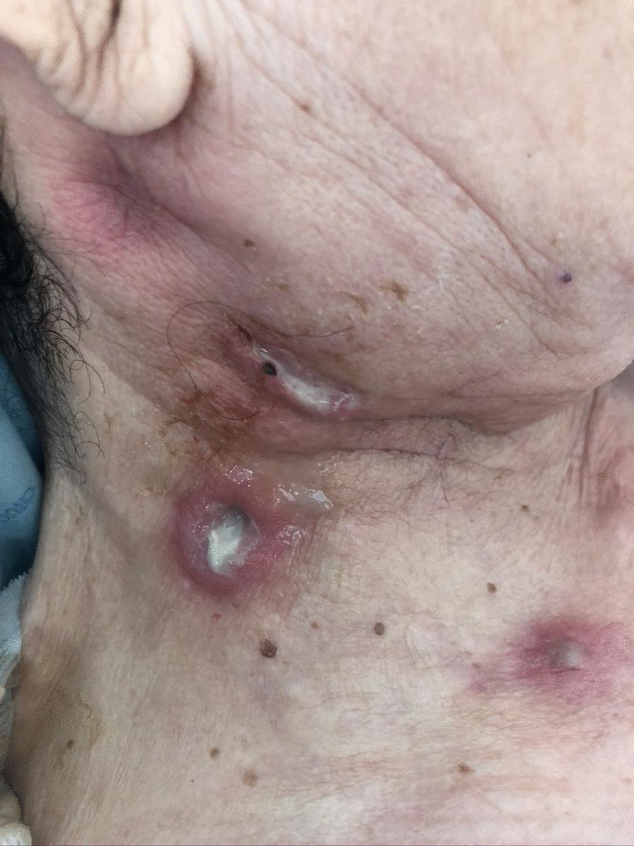
Cervicofacial actinomycosis: multiple suppurative nodules and draining sinus tracts in the submandibular/cervical region. Note the whitish-yellow purulent discharge - characteristic sulfur granules may be visible within.
Pathological Hallmark: Sulfur Granules
"Sulfur granules" are yellow, gritty, 1-2 mm grains visible in pus or discharge. Microscopically they consist of:
- Central mass of fine, delicate branching gram-positive filaments
- Peripheral eosinophilic clubs (immunoglobulin deposits = Splendore-Hoeppli phenomenon), giving the appearance of "rays" - hence "ray fungus"
They also occur in nocardiosis, but filamentous gram-positive rods in the granule confirm actinomycosis (Nocardia is weakly acid-fast).
- Andrews' Diseases of the Skin, p. 316
Diagnosis
| Method | Detail |
|---|---|
| Microscopy | Crushed granule → gram stain shows long gram-positive filaments; most important test |
| Anaerobic culture | Brain-heart infusion blood agar at 37°C; culture recovery < 50%; takes weeks |
| Histopathology | Fibrous encasement of multiloculated abscesses with sulfur granules |
| Fine needle aspiration / fistula swab | Rapid identification of granules |
| Imaging (CT/MRI) | Mass crossing tissue planes; periosteal proliferation or bone destruction; no respect for anatomical boundaries |
The condition is frequently misdiagnosed as malignancy due to the indurated mass appearance - histology is essential.
Treatment
Antibiotics (cornerstone)
- First-line: Penicillin G - high doses (10-20 MU/day IV) for 4-6 weeks, then oral penicillin 4-6 g/day for 2-3 more months (total ~3-6 months minimum; some sources say up to 12 months for deep infection)
- Actinomyces species have no known resistance to penicillin
- Alternatives: Amoxicillin, ampicillin, clindamycin, doxycycline, erythromycin, ceftriaxone, tetracyclines
- Gastric actinomycosis: amoxicillin/clavulanic acid for 6-12 months
Surgery
-
Incision and drainage of abscesses
-
Excision of devitalized tissue, sinus tracts, and dense fibrotic masses (especially when antibiotic response is poor)
-
Cure rates approach ~90% despite frequent late diagnosis
-
Cummings Otolaryngology, p. 1496; Andrews' Diseases of the Skin, p. 316; Sleisenger & Fordtran's, block12
Thoracic Actinomycosis (Pulmonary)
-
50% have pleural involvement (effusion or marked thickening)
-
Chest radiograph: localized infiltrate extending across fissures to adjacent lobes + pleural thickening
-
Chest wall abscess or draining sinus tract is pathognomonic
-
Bone changes: periosteal proliferation or rib destruction
-
Pleural fluid: may be frank empyema (PMN-predominant) or serous (lymphocyte-predominant)
-
Treatment: high-dose penicillin for prolonged periods; pleural effusion managed as any parapneumonic effusion
-
Murray & Nadel's Respiratory Medicine, block25
Abdominal/Pelvic Actinomycosis
- Most common site: appendix (often masquerades as appendiceal abscess)
- CT/MRI may show a mass compressing the ileocaecum; sinus and fistula tracts (enteroenteric, enterocolic, enterocutaneous, enterovesical)
- Associated with IUD use in women (pelvic actinomycosis)
- Grainger & Allison's Diagnostic Radiology, p. block4
Key Differentials
| Mimicker | How to distinguish |
|---|---|
| Malignancy | Actinomycosis crosses tissue planes, has sinus tracts; biopsy/histology decisive |
| Nocardiosis | Sulfur granules present but organism is weakly acid-fast (Actinomyces is not) |
| Tuberculosis | AFB positive; caseating granulomas without sulfur granules |
| Fungal mycetoma | Eumycetoma has larger granules; different organism |
Summary Points
- Gram-positive anaerobic commensal - NOT a fungus
- Disrupted mucosal barriers are the trigger
- Crosses tissue planes - hallmark
- Sulfur granules + gram-positive filaments = diagnostic
- Culture sensitivity < 50%; direct microscopy more reliable
- Penicillin is the drug of choice; prolonged therapy (months) is required
- Often mistaken for malignancy - biopsy is key
- Three main forms: cervicofacial (55%), abdominopelvic (20%), pulmonary (15%)
This is a shared conversation. Sign in to Orris to start your own chat.