
Hypoglossal nerve stimulation. What is it ? How do we do it ? Why do we do it ?
Reading File
Finding Sources
Finding Sources
Searching PubMed
"hypoglossal nerve stimulation" AND "sleep apnea"
Searching the Web
hypoglossal nerve stimulation obstructive sleep apnea patient selection procedure FDA professional guidance
Reading File
Reading File
Reading Web Page
https://aasm.org/overview-of-fda-approved-hypoglossal-nerve-…
Searching Images
diagram hypoglossal nerve stimulation implant system tongue airway obstructive sleep apnea
Hypoglossal nerve stimulation (HNS), also called upper-airway stimulation, is an implanted treatment for selected people with obstructive sleep apnea (OSA) who cannot use or do not benefit enough from CPAP.
What is it?
OSA occurs when throat muscles relax during sleep and the upper airway repeatedly narrows or closes. The tongue can fall backward and contribute to this blockage.
HNS uses a small implanted electrical stimulator to activate selected branches of the hypoglossal nerve (cranial nerve XII), the nerve that controls tongue muscles. Stimulation makes the tongue move forward and stiffen during inspiration, helping keep the airway open. It is synchronized with breathing and is normally used only during sleep.
Murray & Nadel's Textbook of Respiratory Medicine, pp. 2691-2693.
Murray & Nadel's Textbook of Respiratory Medicine, pp. 2691-2693.
How do we do it?
1. Select suitable patients
This is not first-line treatment. Assessment usually includes:
- A sleep study to confirm moderate-to-severe, predominantly obstructive OSA
- Documentation that CPAP has failed or is not tolerated
- Review of weight/BMI, comorbidities, anatomy, and central-apnea burden
- Drug-induced sleep endoscopy (DISE) to see where and how the airway collapses
A key exclusion is complete concentric collapse of the soft palate, because this pattern responds poorly to HNS.
Murray & Nadel's Textbook of Respiratory Medicine, p. 2707. Coverage and device eligibility requirements vary by country, insurer, and device. The CMS coverage guidance illustrates commonly used selection factors.
Murray & Nadel's Textbook of Respiratory Medicine, p. 2707. Coverage and device eligibility requirements vary by country, insurer, and device. The CMS coverage guidance illustrates commonly used selection factors.
2. Implant the system
Under general anesthesia, typically as an outpatient operation, the surgeon implants:
- A stimulation lead around selected hypoglossal-nerve branches in the upper neck
- A small pulse generator beneath the skin of the upper chest
- Depending on the system, a breathing-sensing lead near the chest wall to coordinate stimulation with inhalation
The patient later uses a remote or wearable controller to turn therapy on before sleep.
3. Activate and adjust it
The device is not generally activated immediately. After surgical healing, the sleep team activates it and gradually programs a comfortable level of tongue stimulation. A follow-up sleep study or home sleep test is used to optimize settings and check effectiveness.
Why do we do it?
The purpose is to reduce obstruction, improve oxygenation and sleep continuity, and lessen daytime sleepiness and other OSA-related symptoms in a person who cannot adequately use CPAP.
The physiological rationale is direct: stimulating tongue-protruding muscles, particularly the genioglossus, moves and stiffens the tongue forward, reduces pharyngeal collapse, and improves airflow without surgically removing or rearranging airway tissue.
Cummings Otolaryngology Head and Neck Surgery, pp. 1491-1500.
Cummings Otolaryngology Head and Neck Surgery, pp. 1491-1500.
Important limitations and risks
- It treats obstructive apnea, not primarily central sleep apnea.
- It is surgery, so risks include pain, infection, bleeding, wound problems, temporary tongue weakness or altered tongue sensation, discomfort from stimulation, and need for device revision or replacement.
- Some people do not respond sufficiently, even after careful selection and programming.
- MRI compatibility depends on the exact implanted device and model.
- It does not replace weight management, addressing nasal obstruction, positional therapy, or other aspects of OSA care when those are relevant.
Recent systematic reviews and meta-analyses report sustained improvements in OSA metrics and quality of life in selected patients, but the evidence should not be interpreted as meaning it is appropriate for everyone with OSA: 2024 systematic review and 2024 meta-analysis.
In one line: HNS is a surgically implanted, breathing-timed tongue-nerve stimulator used as a second-line option to prevent airway collapse during sleep in carefully selected patients with OSA.
What are the surgical steps, complications, and follow-up?
Reading File
Finding Sources
Finding Sources
Searching PubMed
"hypoglossal nerve stimulation" AND complications
Searching the Web
hypoglossal nerve stimulation implantation surgical procedure postoperative activation follow-up complications clinical practice
Reading File
Reading File
Reading File
Reading Web Page
https://pmc.ncbi.nlm.nih.gov/articles/PMC8116425
Surgical steps
The exact technique varies by device, but respiration-synchronized HNS is generally implanted under general anesthesia as a 2 to 3 hour outpatient procedure.
-
Preoperative confirmation
- Confirm CPAP-intolerant, predominantly obstructive OSA.
- Review drug-induced sleep endoscopy (DISE) to exclude complete concentric palatal collapse.
- Plan device side and obtain informed consent regarding surgery, device programming, imaging compatibility, and future battery replacement.
-
Neck incision and hypoglossal-nerve lead
- A small upper-neck incision is made, usually below the mandibular border.
- The surgeon identifies the distal hypoglossal nerve and uses intraoperative stimulation and/or nerve monitoring to identify the branches that activate tongue protrusion, especially the genioglossus.
- A cuff electrode is placed around the appropriate protrusor-muscle branches. Branches that retract the tongue are avoided, because stimulating them could worsen obstruction.
-
Chest pulse-generator pocket
- A subcutaneous pocket is created in the upper chest and the implantable pulse generator (IPG) is positioned there.
-
Respiratory sensing lead
- In systems using a sensing lead, a chest-wall incision allows placement of a sensor between intercostal muscle layers.
- The sensor detects the breathing cycle, allowing stimulation to be timed with inspiration.
-
Connection and testing
- Leads are tunneled beneath the skin and connected to the IPG.
- The system is tested to confirm an appropriate respiratory waveform and forward tongue movement without tongue retraction.
- Incisions are closed and dressed. A postoperative chest radiograph may be obtained, particularly when a chest-wall sensing lead has been placed.
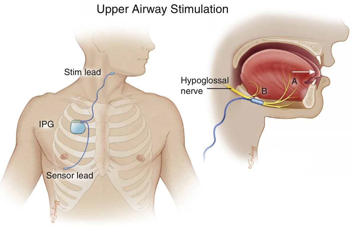
The stimulation cuff targets medial hypoglossal branches for tongue protrusion, while the sensing lead coordinates stimulation with breathing. Murray & Nadel's Textbook of Respiratory Medicine, p. 2725.
Complications
Common, usually temporary
- Incisional pain, neck or chest discomfort, swelling, bruising
- Sore throat after intubation
- Temporary tongue weakness, altered tongue movement, or tongue discomfort
- Stimulation discomfort during early use
- Tongue abrasion or soreness from repetitive tongue movement
- Dry mouth, sleep disruption, headache
Many treatment-related effects occur during the first months while settings are being adjusted. Stimulation discomfort often improves with reprogramming.
Less common but important
- Hematoma or bleeding
- Wound infection, device infection, skin erosion or device exposure
- Pneumothorax or pleural effusion related to placement of the chest sensing lead
- Hypoglossal-nerve injury, persistent tongue weakness, dysarthria, dysphagia, or neuropraxia
- Lead displacement, lead fracture, device malfunction, inadequate respiratory sensing, or ineffective tongue motion
- Need for reoperation, revision, replacement, or rarely explantation
- Problems around MRI or external procedures, depending on the specific model and its conditions for use
Serious events are uncommon in selected patients. The initial STAR trial reported serious device-related events in fewer than 2% of participants, primarily requiring device repositioning. A 2024 systematic review of adverse events found that most reported treatment effects were transient and clustered during early acclimatization.
Follow-up pathway
| Time after implant | Main purpose |
|---|---|
| Same day to 1 week | Recovery assessment, analgesia, wound care, review for bleeding, infection, respiratory symptoms, or pneumothorax. Limit vigorous shoulder/arm movement as directed by the surgical team. |
| 1 to 2 weeks | Incision and device-pocket examination. Remove sutures if needed; assess tongue movement, swallowing, speech, and infection. |
| About 3 to 6 weeks | Device activation after wounds have healed. The patient is taught to use the remote or controller, including start delay, nightly use, and warning signs. Initial stimulation is deliberately conservative. |
| Following weeks to 2 to 3 months | Gradual home adjustment within prescribed limits plus clinic programming. The aim is effective tongue protrusion that remains comfortable and does not fragment sleep. |
| About 2 to 4 months after activation | Objective efficacy evaluation with in-lab titration polysomnography or a home sleep apnea test, depending on local practice. Voltage and other parameters are adjusted to reduce residual obstruction. |
| Long term | At least annual sleep and device review: adherence, snoring, Epworth Sleepiness Scale, symptoms, weight change, residual AHI/oxygenation, side effects, battery status, and need for reprogramming. |
The textbook pathway describes titration at approximately two months after implantation, adjusting voltage to control residual obstructive apneas and hypopneas. Murray & Nadel's Textbook of Respiratory Medicine, p. 2727. Actual timing differs across centers and device platforms.
When to contact the surgical or sleep team urgently
- Increasing neck or chest swelling, bleeding, fever, wound redness or drainage
- Chest pain, shortness of breath, or sudden cough after surgery
- New persistent tongue weakness, trouble swallowing, aspiration, or speech difficulty
- Painful stimulation, failure of the device to function, or inability to sleep with therapy
HNS is not “set and forget.” Good outcomes depend on correct surgical placement, careful activation, repeated programming, and repeat sleep testing to verify that obstructive events are actually controlled.
This is a shared conversation. Sign in to Orris to start your own chat.