
Amelogenesis
Reading File
Amelogenesis is the process of enamel formation by ameloblasts during tooth development. Let me pull comprehensive textbook content on this topic.
Searching sources...
Finding Sources
Finding Sources
The Histology textbook has a dedicated section on amelogenesis. Let me read it fully, along with the embryology content.
Reading File
Reading File
Let me read the full amelogenesis section from the histology textbook, and also grab any images.
Reading File
Reading File
Reading File
Reading File
Loading Image
I now have comprehensive information to write a thorough answer. Let me also fetch the Tomes process / enamel rod diagram.
Loading Image
I now have all the material needed for a comprehensive, well-illustrated answer.
Amelogenesis
Amelogenesis is the process of dental enamel formation by specialized epithelial cells called ameloblasts. It is a matrix-mediated biomineralization process that produces the hardest substance in the human body. Enamel formation is irreversible - ameloblasts degenerate after enamel is fully formed at the time of tooth eruption, meaning enamel cannot regenerate once lost.
1. Origin of Ameloblasts
Ameloblasts derive from the inner enamel epithelium of the enamel organ, which itself develops from oral ectoderm during tooth development:
- Bud stage: A tooth bud grows into the underlying mesenchyme from the dental lamina
- Cap stage: The cap's concave surface differentiates into tall columnar cells (ameloblasts) forming the inner enamel epithelium. Neural crest-derived mesenchyme invaginates to form the dental papilla
- Bell stage: The enamel organ now has four distinct layers:
- Outer enamel epithelium - convex surface
- Inner enamel epithelium - concave surface (future ameloblasts)
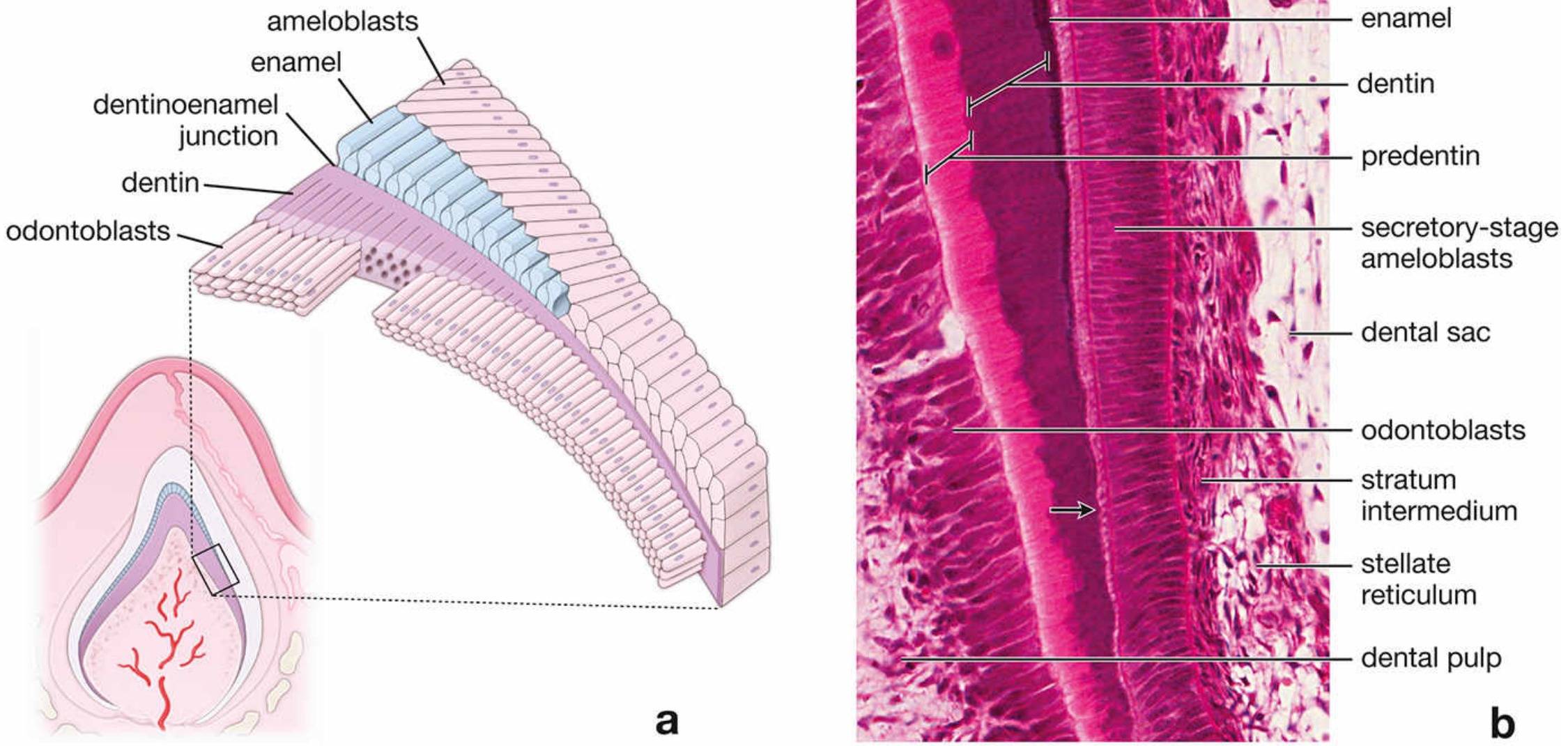
- Stratum intermedium - internal to the inner enamel epithelium; contains alkaline phosphatase and supports ameloblast function
- Stellate reticulum - star-shaped cells occupying the inner portion
At the early bell stage, the dental lamina degenerates, detaching the tooth primordium from the oral epithelium. Preodontoblasts (neural crest-derived) adjacent to the inner enamel epithelium differentiate into odontoblasts and begin dentinogenesis first - dentin must be laid down before enamel formation can start.
2. Stages of Amelogenesis
Stage 1: Matrix Production (Secretory Stage)
- Dentin forms first; then partially mineralized enamel matrix is deposited directly onto the dentin surface
- Secretory-stage ameloblasts are polarized, tall columnar cells that use rER, Golgi apparatus, and secretory granules to produce and secrete an organic proteinaceous matrix
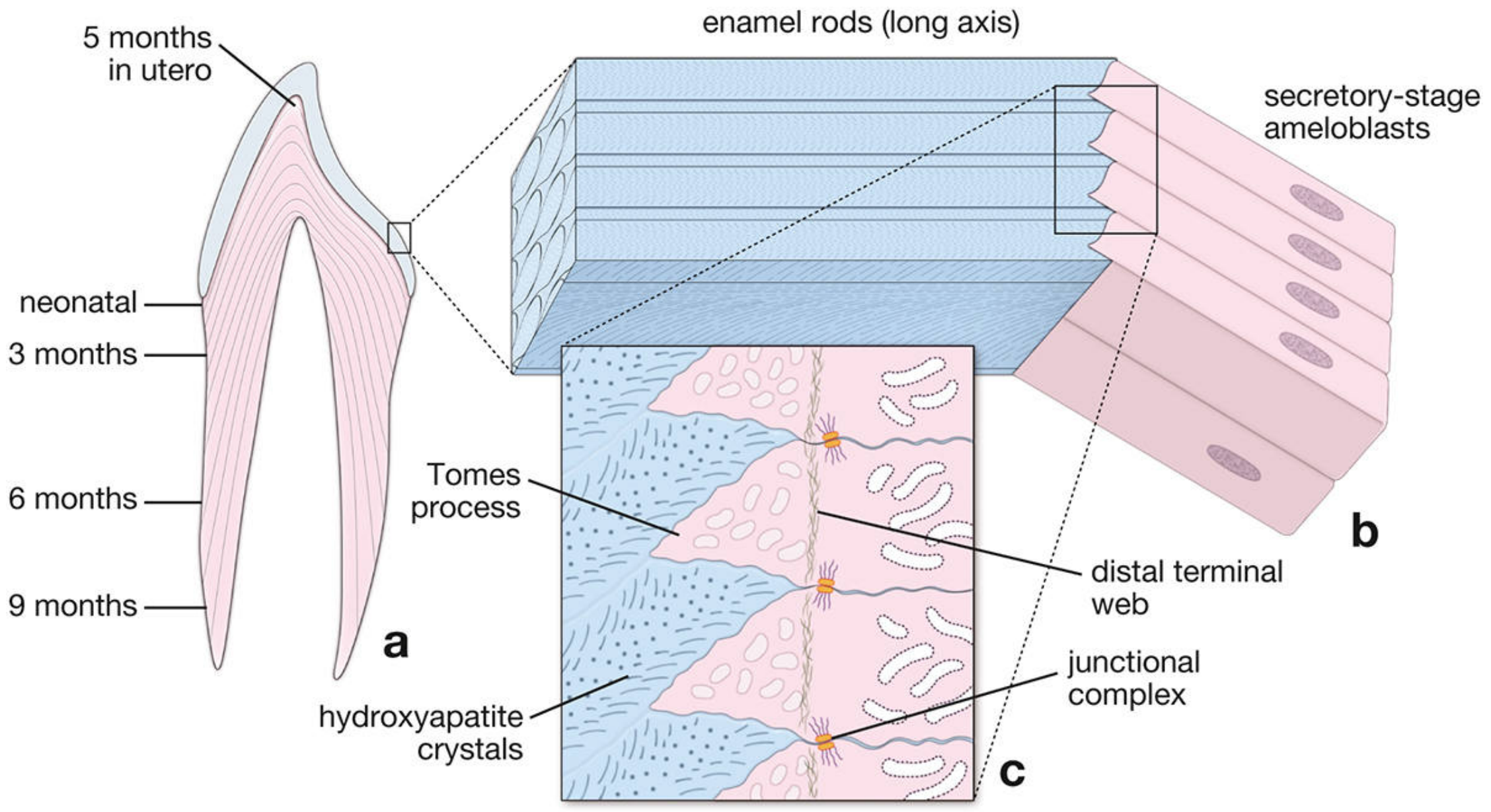
- Each ameloblast bears an apical cytoplasmic extension called the Tomes process, which is surrounded by developing enamel
- The rod produced by each ameloblast forms in the wake of the cell as it moves outward - so the direction of the enamel rod records the path the ameloblast once took
- Secretory-stage ameloblasts continue producing matrix until the full thickness of future enamel is achieved
- Stratum intermedium cells adjacent to the base of ameloblasts express alkaline phosphatase, supporting calcification
Stage 2: Matrix Maturation
- Maturation-stage ameloblasts differentiate from secretory-stage ameloblasts
- They function primarily as transporting epithelium, moving substances into and out of maturing enamel
- Key feature: a striated (ruffled) border on the apical surface
- They undergo cyclical modulation - alternating between striated and smooth-bordered morphology (~70% striated, ~30% smooth at any time)
- Striated-bordered cells: secrete bicarbonate ions (HCO3-) and contain plasma membrane Ca²⁺-ATPases (PMCA) that pump Ca²⁺ into maturing enamel
- Smooth-bordered cells: produce and secrete enzymes to degrade and reabsorb the now-unnecessary organic matrix
- During maturation, the stratum intermedium, stellate reticulum, and outer enamel epithelium collapse together, reorganizing into the papillary layer (containing blood vessels and stellate papillary cells adjacent to maturation-stage ameloblasts)
- Both maturation-stage ameloblasts and papillary cells are rich in mitochondria, reflecting their high energy demands as a transport epithelium
3. Enamel Matrix Proteins
The enamel matrix is highly heterogeneous and contains several key proteins:
| Protein | Function |
|---|---|
| Amelogenins | Establish and maintain spacing between enamel rods in early development. Removed during maturation |
| Ameloblastins | Signaling proteins produced from early secretory to late maturation stages. Control elongation of enamel crystals and form junctional complexes between crystals |
| Enamelins | Distributed throughout the enamel layer; undergo proteolytic cleavage as enamel matures. Low-MW cleavage products remain in mature enamel, localized on crystal surfaces |
| Tuftelins | Earliest detected proteins, located near the dentinoenamel junction. Acidic and insoluble - aid in nucleation of enamel crystals. Account for enamel tufts (regions of hypomineralization) |
After maturation, amelogenins and ameloblastins are fully removed. Mature enamel retains only enamelins and tuftelins.
4. Fate of Ameloblasts
Ameloblasts degenerate at approximately the time of tooth eruption through the gum, after enamel is fully formed. This is why enamel cannot self-repair - there are no remaining cells to produce new enamel matrix.
5. Clinical Correlation: Amelogenesis Imperfecta
Amelogenesis imperfecta is a group of at least 14 inherited clinical entities involving defective enamel formation in the absence of systemic disease. It is an ectodermal birth defect affecting enamel exclusively.
- Enamel phenotypes: may be hypoplastic (thin), hypocalcified (soft), or hypomature (incompletely developed)
- Appearance: enamel may be hard or soft, pitted or smooth, thin or normal thickness
- Incidence: 1 in 700 (Sweden) to 1 in 1200 (USA)
- Inheritance: multiple modes (autosomal dominant, autosomal recessive, X-linked)
- Genes involved: AMELX (amelogenin), ENAM (enamelin), MMP20 (matrix metalloproteinase 20 / enamelysin)
- Classification: based on clinical and radiographic findings plus mode of inheritance
Summary Table
| Feature | Detail |
|---|---|
| Cell of origin | Ameloblast (from inner enamel epithelium / oral ectoderm) |
| Sequence | Dentinogenesis first, then amelogenesis |
| Stage 1 | Secretory - matrix deposition via Tomes process |
| Stage 2 | Maturation - organic removal, Ca²⁺/HCO3⁻ transport |
| Key organelles | rER, Golgi, secretory granules, mitochondria |
| Structural unit | Enamel rod (formed in wake of each ameloblast) |
| Mature enamel proteins | Enamelins + Tuftelins only |
| Post-eruption | Ameloblasts degenerate - no enamel regeneration possible |
- Histology: A Text and Atlas with Correlated Cell and Molecular Biology, pp. 1443-1451
- The Developing Human: Clinically Oriented Embryology, pp. 1204-1207
This is a shared conversation. Sign in to Orris to start your own chat.