
Nervous system and its significance in the organism. Classification of nervous system. Inter relationship of its parts
nervous system classification diagram CNS PNS autonomic somatic
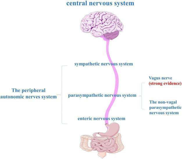
An anatomical diagram illustrating the components of the gut-brain axis and the peripheral autonomic nervous system (ANS). At the top, a human brain represents the central nervous system (CNS), which is connected via a long neural pathway to the gastrointestinal tract (stomach and intestines) at the bottom. The peripheral autonomic nervous system is categorized into three major divisions: the sympathetic nervous system, the parasympathetic nervous system, and the enteric nervous system (ENS). The diagram highlights the role of the vagus nerve within the parasympathetic division, noted with 'strong evidence' as a primary bidirectional communication route between the brainstem and the gut. It also distinguishes between vagal and non-vagal parasympathetic pathways. This illustration is used to explain the neuroanatomical basis of the gut-brain axis, particularly in the context of neurodegenerative research like Parkinson's disease, where alpha-synuclein may propagate from the ENS to the CNS via these autonomic pathways.
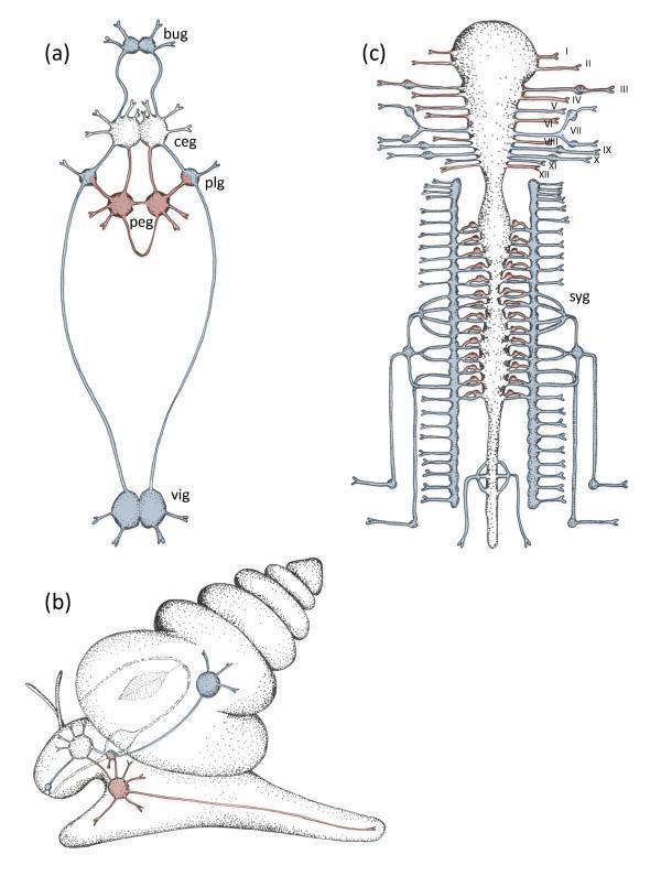
This comparative anatomical diagram illustrates the organizational duality of the somatic and visceral nervous systems across different taxa. Panel (a) and (b) depict the molluscan nervous system, using a snail as a model. The somatic system (red) includes the cerebral (ceg) and pedal ganglia (peg) forming a pedal cord for locomotion. The visceral system (blue) consists of buccal (bug), pleural (plg), and visceral ganglia (vig), which form a ring around the gut to regulate autonomic functions. Panel (c) shows the vertebrate nervous system (human/mammalian model), highlighting the brainstem, spinal cord, and peripheral nerves. The cranial nerves are labeled I-XII, representing the somatosensory and motor pathways. Flanking the spinal cord is the sympathetic chain (syg), color-coded blue to represent the autonomic/visceral components, demonstrating the evolutionary conservation of the viscero-somatic functional split. This resource serves as an educational tool for understanding comparative neurobiology and the structural organization of the autonomic versus somatic nervous systems.
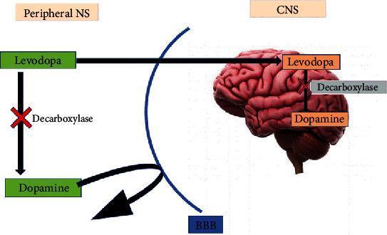
This pathophysiology diagram illustrates the pharmacological pathway of levodopa (L-DOPA) from the peripheral nervous system (PNS) to the central nervous system (CNS), focusing on its interaction with the blood-brain barrier (BBB). The diagram is divided into two sections: the 'Peripheral NS' on the left and the 'CNS' on the right, separated by a blue arc representing the BBB. In the PNS, a red 'X' indicates the inhibition of the enzyme decarboxylase, which prevents the peripheral conversion of levodopa into dopamine; a curved black arrow shows that dopamine itself cannot cross the BBB. Levodopa, however, is shown crossing the BBB into the brain (depicted as a 3D anatomical model). Inside the CNS, levodopa is successfully converted into dopamine. The diagram highlights the clinical strategy of preventing premature peripheral metabolism of levodopa to ensure it reaches the brain, where it can be converted to the active neurotransmitter dopamine for the treatment of conditions like Parkinson's disease. The context suggests that certain substances, like coffee, may influence this decarboxylase inhibition process.
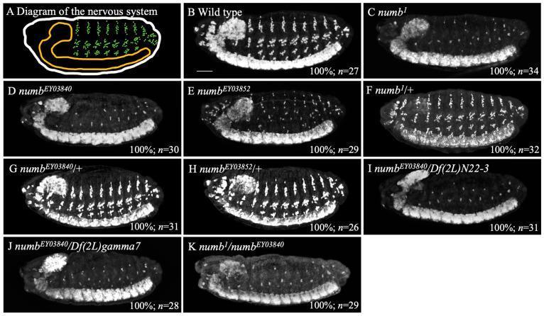
This image is a composite of a schematic diagram and several diagnostic immunofluorescence images (labeled A-K) illustrating the nervous system development in Drosophila embryos at stage 14. Panel A provides an anatomical diagram highlighting the central nervous system (CNS, orange) and the peripheral nervous system (PNS, green dots). Panels B through K are lateral view fluorescence micrographs where neurons are stained white using an anti-Elav antibody. Panel B shows the wild-type phenotype with a dense, organized distribution of neurons in both the CNS and PNS. Panels C, D, E, I, J, and K demonstrate the 'antineurogenic phenotype' associated with numb loss-of-function alleles (numb1, numbEY03840, and various transheterozygotes), characterized by a marked reduction in white-stained PNS neurons while the CNS remains relatively intact. Panels F, G, and H show heterozygous genotypes (numb/+) which maintain a neuron distribution comparable to the wild type, indicating the recessive nature of the mutation. The scale bar represents 25 µm.
central peripheral nervous system brain spinal cord anatomy overview
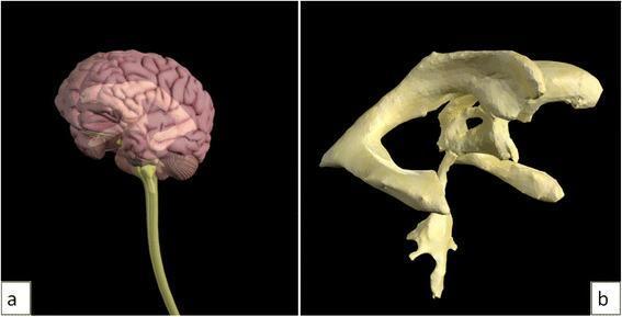
This dual-panel image features 3D anatomical models of the central nervous system and ventricular system, used for neuroanatomical education. Image (a) depicts a lateral view of the human brain and spinal cord. The cerebral cortex shows distinct gyri and sulci, with the cerebellum positioned posteriorly and the brainstem tapering into a long, cylindrical spinal cord. The model utilizes varying opacities to suggest internal depth. Image (b) provides a detailed, isolated 3D reconstruction of the cerebral ventricular system. Visible structures include the bilateral lateral ventricles with their characteristic C-shape (comprising the anterior, posterior, and inferior horns), the midline third ventricle, the narrow cerebral aqueduct of Sylvius, and the fourth ventricle located inferiorly. The model demonstrates the spatial relationships and interconnected pathways essential for understanding cerebrospinal fluid (CSF) flow. This visual material is designed for medical students and clinicians to study intracranial anatomy and the pathophysiology of conditions like hydrocephalus.
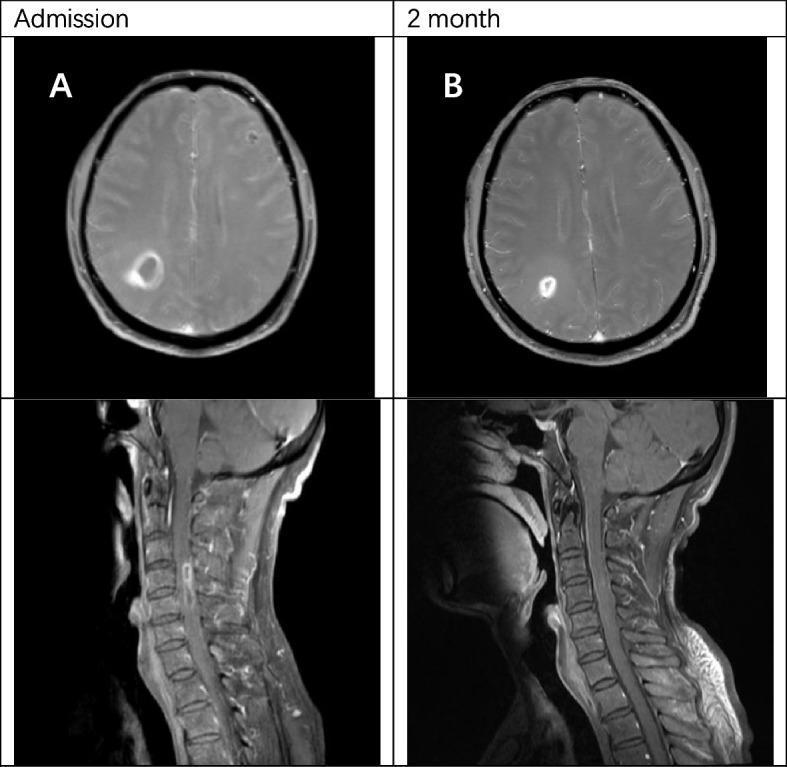
This diagnostic image comparison displays axial brain MRIs and sagittal cervical spinal cord MRIs from a patient with central nervous system infection, likely secondary to infective endocarditis. Panel A (Admission) illustrates post-contrast T1-weighted images showing a prominent ring-enhancing lesion in the right parietal lobe of the brain and a similar ring-enhancing lesion within the spinal cord at the C4-C5 level, accompanied by surrounding edema. Panel B (2 months) demonstrates the follow-up imaging after a course of antibiotic therapy, including ceftriaxone and linezolid. Visually, there is a significant reduction in the size and enhancement intensity of both the cerebral and spinal cord abscesses, indicating a positive response to treatment. The imaging highlights the classic radiological appearance of pyogenic abscesses, characterized by peripheral rim enhancement and central necrosis, and emphasizes the importance of longitudinal neuroimaging in monitoring therapeutic efficacy in infectious CNS diseases.
Diagnostic Imaging: This composite clinical image displays T1-weighted Magnetic Resonance Imaging (MRI) sequences of the central nervous system. Panel A features a sagittal view of the brain, demonstrating normal intracranial anatomy. Key visible structures include the cerebral cortex with preserved sulcation and gyration, the corpus callosum, the brainstem (pons and medulla), and the cerebellum. The brain parenchyma shows no evidence of masses, midline shift, or signal abnormalities. Panel B presents a sagittal MRI of the spinal column. The alignment of the vertebral bodies is preserved, and the intervertebral disc spaces appear intact. The spinal cord is clearly visualized within the spinal canal, showing no signs of extrinsic compression or significant nerve root thickening. While the clinical context mentions possible mild pial enhancement, the primary educational value of this image set lies in demonstrating baseline normal neurological imaging in a patient undergoing workup for demyelinating polyneuropathy (CIDP). These images serve as a reference for negative diagnostic findings in cases of progressive sensory-motor neuropathy.
The Nervous System: Significance, Classification, and Interrelationship of Parts
1. Significance of the Nervous System in the Organism
"The human brain is the most complex tissue in the body. It mediates behavior ranging from simple movements and sensory perception to learning, memory, and consciousness. It is the organ of the mind and accounts for the human capacity for invention, discovery, and language."
| Function | Role |
|---|---|
| Homeostasis | Regulates heart rate, blood pressure, respiration, temperature, digestion, and fluid balance |
| Voluntary movement | Commands skeletal muscle for locomotion and fine motor skills |
| Higher cognition | Enables thought, language, problem-solving, creativity |
| Reflexes | Rapid, involuntary protective responses (e.g., withdrawal from pain) |
| Integration of systems | Coordinates cardiovascular, respiratory, endocrine, immune, and digestive systems |
| Reproduction | Regulates hormonal cycles and reproductive behavior |
2. Classification of the Nervous System
A. Anatomical Classification
- Comprises the brain and spinal cord
- Located within the cranial cavity and spinal canal respectively
- Protected by three meningeal layers: dura mater (outer), arachnoid (middle), pia mater (inner)
- Internally organized into gray matter (neuron cell bodies, nuclei) and white matter (myelinated axons)
- Myelin is produced by oligodendrocytes (unique to CNS)
- CNS axons cannot regenerate after injury
- Includes CN II (optic nerve) and the retina as CNS extensions
- All neural tissue lying outside the dura mater
- Includes: sensory receptors, spinal nerves, cranial nerves (except CN II), peripheral ganglia, and peripheral autonomic nerves
- Myelin is produced by Schwann cells (unique to PNS)
- PNS axons can regenerate after injury
- Afferent (sensory) nerves carry impulses toward the CNS
- Efferent (motor) nerves carry impulses away from the CNS
- Ganglia = clusters of neuron cell bodies outside the CNS
B. Functional Classification
- Controls functions under conscious, voluntary control (with the exception of reflexes)
- Provides sensory and motor innervation to the body wall, limbs, and skin - everything except viscera, smooth muscle, cardiac muscle, and glands
- Motor pathway: one neuron from CNS directly to skeletal muscle (effector)
- Controls involuntary, visceral functions: heart rate, blood pressure, digestion, temperature regulation, reproductive function, glandular secretion
- Anatomically spans both CNS and PNS
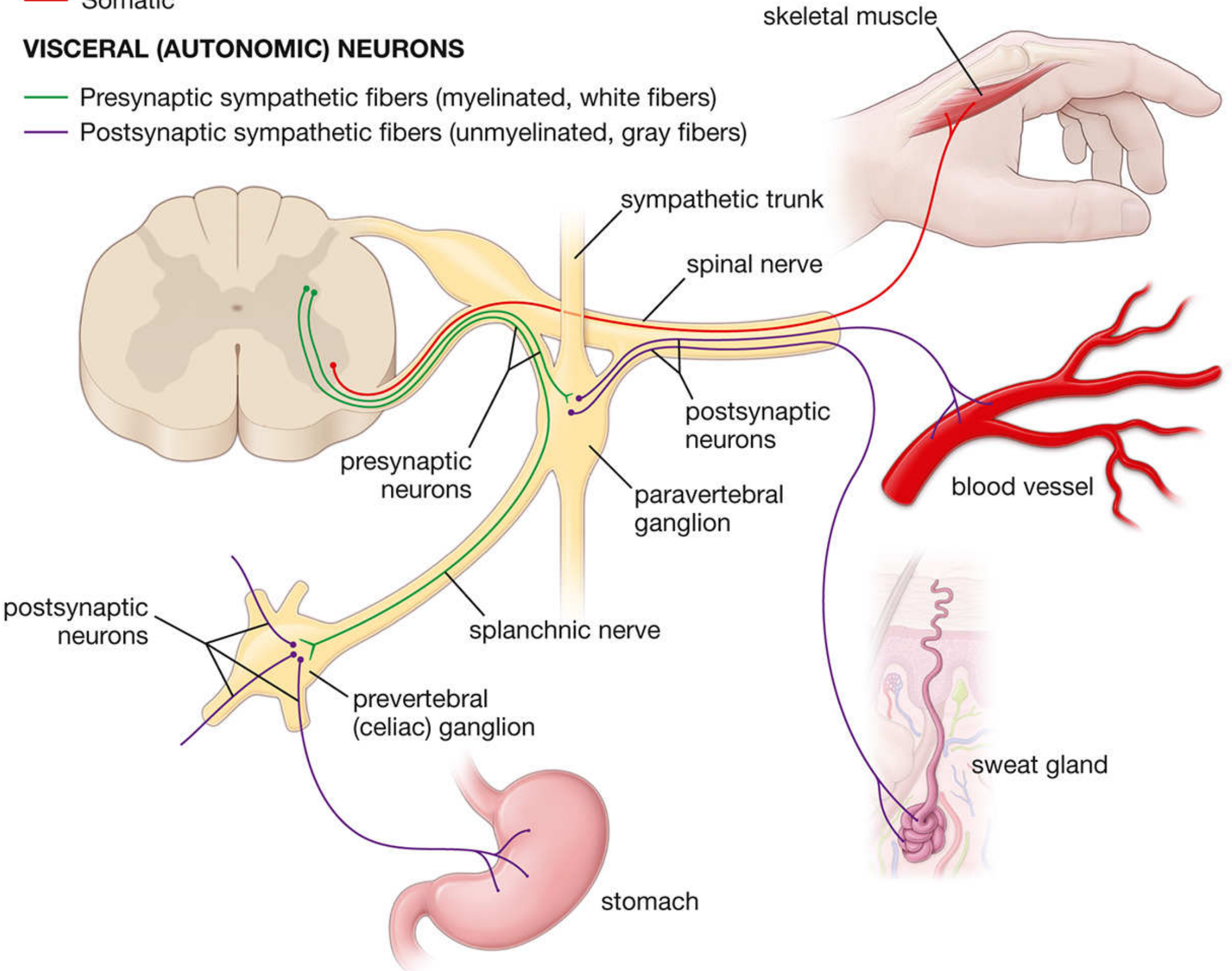
- Motor pathway: two-neuron chain - a presynaptic neuron (within CNS) synapses onto a postsynaptic neuron (in a peripheral ganglion), which then innervates the effector (smooth muscle, cardiac muscle, or gland)
- Each presynaptic neuron synapses with several postsynaptic neurons (divergence)
| Division | Origin | Ganglia Location | Key Functions |
|---|---|---|---|
| Sympathetic | Thoracolumbar spinal cord (T1-L2) | Paravertebral (sympathetic trunk) or prevertebral ganglia | "Fight or flight" - increases HR, BP, diverts blood to muscles |
| Parasympathetic | Craniosacral (brainstem + S2-S4) | Terminal ganglia near/in target organs | "Rest and digest" - slows HR, promotes digestion, salivation |
| Enteric | Embedded in GI tract wall | Myenteric & submucosal plexuses | Autonomous regulation of GI motility and secretion |
3. Summary Classification Scheme
NERVOUS SYSTEM
│
├── ANATOMICAL
│ ├── Central Nervous System (CNS)
│ │ ├── Brain (cerebrum, cerebellum, brainstem)
│ │ └── Spinal cord
│ └── Peripheral Nervous System (PNS)
│ ├── Cranial nerves (I–XII, except CN II)
│ ├── Spinal nerves (31 pairs)
│ ├── Peripheral ganglia
│ └── Sensory receptors
│
└── FUNCTIONAL
├── Somatic Nervous System
│ ├── Somatic sensory (afferent)
│ └── Somatic motor (efferent) → Skeletal muscle
└── Autonomic Nervous System (ANS)
├── Sympathetic division
├── Parasympathetic division
└── Enteric division
4. Interrelationship of Parts
CNS ↔ PNS
- The CNS is the processing center; the PNS is the communication highway
- Sensory information flows via PNS afferents into the CNS; motor commands flow via PNS efferents out to effectors
- Reflex arcs integrate both: sensory receptor (PNS) → afferent nerve (PNS) → integration center (CNS) → efferent nerve (PNS) → effector
Somatic ↔ Autonomic
- Both are subdivisions of the overall nervous system, sharing CNS integration centers
- The hypothalamus (CNS) is the primary autonomic control center, receiving somatic and visceral sensory inputs and modulating ANS output
- Many responses involve both systems simultaneously (e.g., pain triggers both somatic withdrawal reflex AND sympathetic activation)
ANS Divisions (Sympathetic ↔ Parasympathetic ↔ Enteric)
- Sympathetic and parasympathetic divisions often antagonize each other on the same target organ (e.g., heart: sympathetic increases rate, parasympathetic decreases rate) - this dual innervation allows fine-tuned control
- The enteric nervous system can function semi-autonomously but is modulated by both sympathetic and parasympathetic inputs
- Visceral afferents travel alongside ANS efferent fibers, closing the feedback loop for visceral reflex control
Key Unifying Principle
- Medical Physiology, Boron & Boulpaep (9780323319737) - Organization of the Nervous System, Ch. 10
- Histology: A Text and Atlas with Correlated Cell and Molecular Biology, Pawlina (9781975181512) - Overview and Organization of the Nervous System, Ch. 12
- Gray's Anatomy for Students (9780323934237) - Nervous System Functional Organization
Concept of neuron. Nervous fibres, roots and fascicles, neural nodes their classification and structure. Answer this question based on anatomy
The Neuron, Nerve Fibres, Roots, Fascicles, and Neural Nodes - An Anatomical Account
1. The Concept of the Neuron
Definition
Structural Components of a Neuron
- Contains the nucleus (usually large, pale-staining, with prominent nucleolus) and the metabolic machinery of the cell
- Houses Nissl bodies (rough endoplasmic reticulum + ribosomes) - sites of protein synthesis; they extend into dendrites but are absent from the axon hillock
- Contains Golgi apparatus, mitochondria, neurofilaments, and microtubules
- Cannot be replaced if lost (post-mitotic in adults)
- Multiple, shorter, branching processes that carry impulses toward the cell body (afferent to soma)
- Increase receptive surface area enormously
- Microtubules in dendrites show mixed polarity (some plus-end distal, some minus-end distal) - enabling bidirectional transport
- Many dendrites bear dendritic spines - tiny protrusions that are postsynaptic sites
- Usually a single, long process that carries impulses away from the cell body (efferent from soma) to a terminal
- Arises from the axon hillock - a cone-shaped eminence on the cell body that lacks Nissl bodies
- The axon initial segment (AIS) - the bare region between the hillock and the start of myelin - is where the action potential is generated
- AIS acts as a molecular checkpoint ("border crossing") that prevents axonal plasma membrane proteins from mixing with somatic/dendritic proteins
- Microtubules in axons have uniform polarity (all plus-ends pointing distally) - enabling unidirectional kinesin-driven anterograde transport
- The axon terminates in synaptic endings (boutons) containing neurotransmitter vesicles
- The specialized junction where the axon terminal contacts another neuron or an effector cell
- Chemical synapses: impulse transmission via neurotransmitter release across a synaptic cleft
- Electrical synapses: gap junctions allow direct ionic current flow between cells (common in invertebrates, some CNS locations in vertebrates)
Classification of Neurons
A. By Number of Processes (Anatomical)
| Type | Processes | Location / Examples |
|---|---|---|
| Multipolar | Many dendrites + 1 axon | Most common; motor neurons, interneurons, Purkinje cells, pyramidal cells |
| Bipolar | 1 dendrite + 1 axon | Rare; retina, cochlear/vestibular ganglia (CN VIII), olfactory epithelium |
| Pseudounipolar (Unipolar) | Single process that bifurcates close to cell body into peripheral and central branches | Primary sensory neurons; cell bodies in dorsal root ganglia and cranial nerve ganglia |
B. By Function
| Type | Direction | Function |
|---|---|---|
| Sensory (afferent) | Periphery → CNS | Convey pain, temperature, touch, proprioception, special senses |
| Motor (efferent) | CNS → effectors | Somatic: voluntary skeletal muscle; Visceral: smooth muscle, cardiac, glands |
| Interneurons (intercalated) | Within CNS | Integration network; >99.9% of all neurons belong to this group |
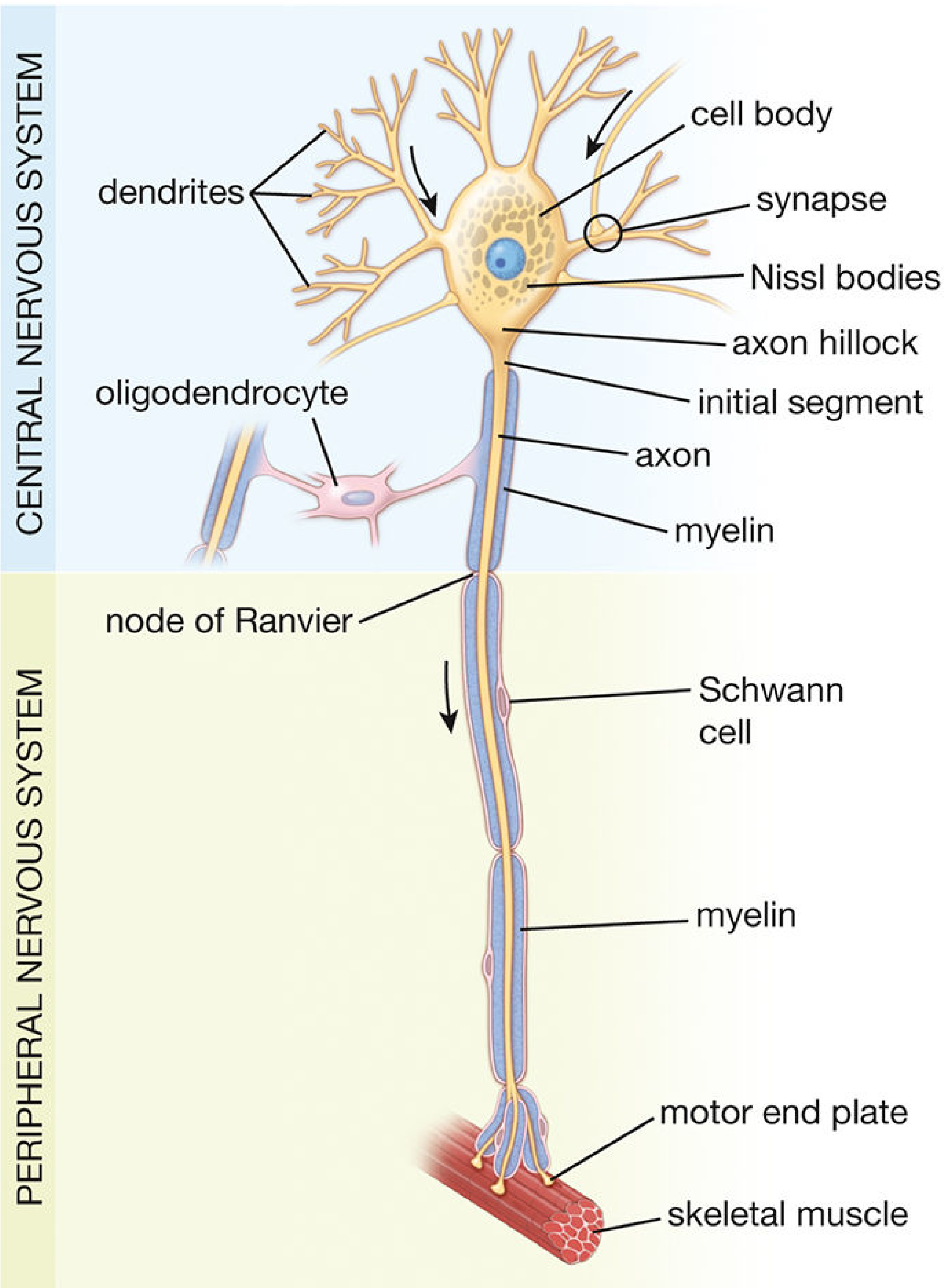
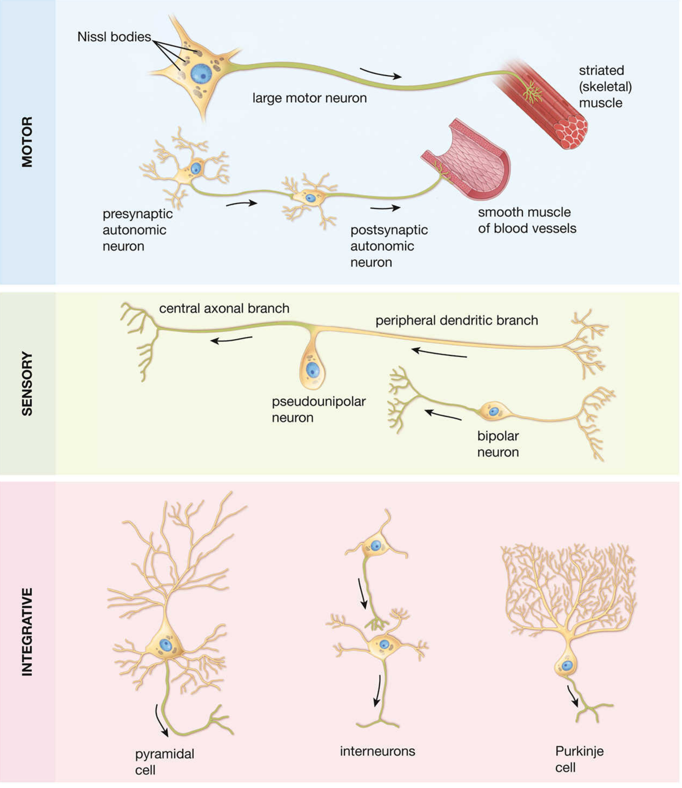
2. Nerve Fibres
Definition
Myelinated vs. Unmyelinated Fibres
- Each axon is wrapped by a single Schwann cell that spirally winds its membrane around the axon, extruding cytoplasm to form compacted myelin
- Myelin is ~80% lipid
- The junction between two adjacent Schwann cells is the Node of Ranvier (see below)
- Conduction is saltatory (jumps node to node) - fast
- Thickness of myelin is determined by axon diameter (not the Schwann cell)
- Multiple axons (up to 20) are simply enfolded in grooves in a single Schwann cell without wrapping
- Conduction is continuous along the entire axon membrane - slow
Classification of Nerve Fibres
| Fibre Type | Function | Diameter (μm) | Conduction Velocity (m/s) |
|---|---|---|---|
| Aα | Proprioception; somatic motor | 12-20 | 70-120 |
| Aβ | Touch, pressure | 5-12 | 30-70 |
| Aγ | Motor to muscle spindles (fusimotor) | 3-6 | 15-30 |
| Aδ | Pain (fast/sharp), temperature | 2-5 | 12-30 |
| B | Preganglionic autonomic (myelinated) | <3 | 3-15 |
| C (dorsal root) | Pain (slow/burning), temperature | 0.4-1.2 | 0.5-2 |
| C (sympathetic) | Postganglionic sympathetic (unmyelinated) | 0.3-1.3 | 0.7-2.3 |
| Group | Equivalent | Fibre source |
|---|---|---|
| Ia | Aα | Primary afferents from muscle spindles (annulospiral endings) |
| Ib | Aα | Golgi tendon organs |
| II | Aβ | Secondary muscle spindle afferents; cutaneous touch/pressure |
| III | Aδ | Free nerve endings; sharp pain, temperature |
| IV | C | Free nerve endings; slow pain, temperature, itch |
3. Structure of a Peripheral Nerve - Roots, Fascicles, and Connective Tissue Sheaths
Spinal Nerve Roots
- Dorsal (posterior) root: carries sensory (afferent) fibres. Contains the dorsal root ganglion (housing pseudounipolar sensory neuron cell bodies). The peripheral branch of the pseudounipolar neuron extends to receptors in skin/muscle; the central branch enters the dorsal horn of the spinal cord.
- Ventral (anterior) root: carries motor (efferent) fibres. Motor neuron cell bodies are in the ventral horn of the spinal cord gray matter; their axons exit via the ventral root to innervate skeletal muscle.
Fascicles (Funiculi)
- A fascicle is a discrete bundle of nerve fibres wrapped by perineurium
- Fascicles vary in size and number along the nerve course
- In the proximal portion of a nerve, fascicles form an intertwining plexiform pattern (Sunderland plexus) - providing mechanical resilience
- In the distal portion, fascicles run more parallel - enabling more somatotopic organization
Connective Tissue Sheaths (from innermost to outermost)
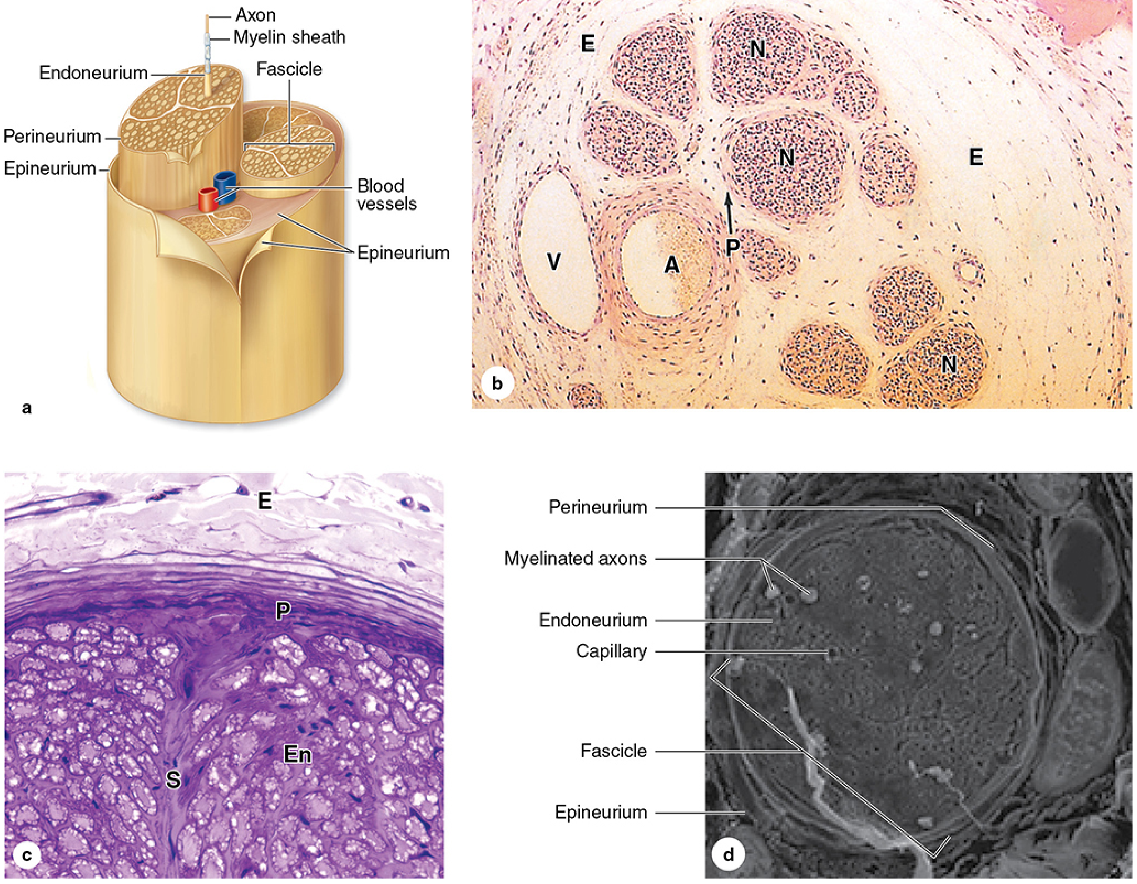
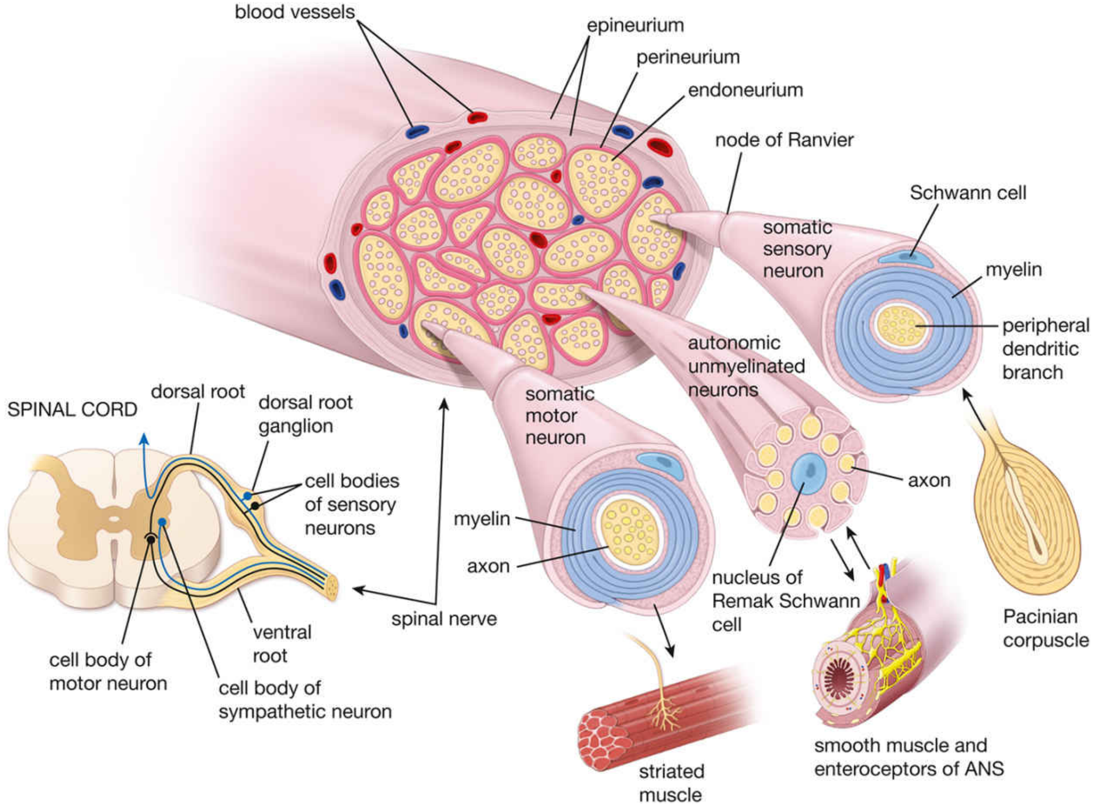
Endoneurium (innermost)
- Composition: Loose connective tissue (mainly collagen fibrils running parallel to and circumferentially around individual nerve fibres) + occasional fibroblasts, macrophages, mast cells, and capillaries
- Location: Surrounds each individual nerve fibre (axon + Schwann cell)
- Cells: 90% of nuclei visible in cross-sections belong to Schwann cells; 10% = fibroblasts + endothelial cells + macrophages
- Collagen fibrils are largely secreted by Schwann cells
- Contributes to the blood-nerve barrier (along with perineurium) via tight junctions of capillary endothelial cells within it
Perineurium
- Composition: Specialized squamous connective tissue cells (perineurial cells) arranged in concentric layers (1-6 layers depending on nerve size), with collagen fibrils between layers
- Location: Surrounds each nerve fascicle as a whole
- Key feature: Perineurial cells are joined by tight junctions - this forms the blood-nerve barrier that maintains the ionic and immunological milieu within the fascicle (analogous to blood-brain barrier)
- Cells are also contractile (contain actin filaments, similar to smooth muscle)
- Has an external (basal) lamina on both surfaces of each cell layer
Epineurium (outermost)
- Composition: Dense irregular connective tissue
- Location: Surrounds the entire peripheral nerve trunk and fills the spaces between fascicles (interfascicular epineurium)
- Contains the major blood vessels supplying the nerve (arteriae nervorum), adipose tissue, and fibroblasts
- The interfascicular portion is an extension of the dura mater
- An outermost loose areolar layer, the mesoneurium, allows passive movement of the nerve in both transverse and longitudinal planes
| Layer | Surrounds | Main Component | Special Role |
|---|---|---|---|
| Endoneurium | Individual nerve fibre | Loose collagen fibrils | Supports axon-Schwann unit |
| Perineurium | Fascicle | Squamous perineurial cells + tight junctions | Blood-nerve barrier |
| Epineurium | Whole nerve + interfascicular spaces | Dense irregular CT | Mechanical protection, vascular supply |
4. Neural Nodes - The Node of Ranvier
Structure
- The nodal gap itself: a short (~1 µm) segment of bare axolemma, exposed to the extracellular space
- Highest density of voltage-gated Na⁺ channels in the entire nervous system - this is critical for impulse propagation
- Flanked on each side by paranodal loops - cytoplasmic tongue-like extensions of the Schwann cell that spiral down and attach to the axolemma via specialized junctions
- The juxtaparanodal region (just outside the paranodes) is rich in voltage-gated K⁺ channels - these help repolarize the membrane
Functional Significance: Saltatory Conduction
- Much higher conduction velocity without requiring a proportionally larger axon diameter
- Greater energy efficiency (Na⁺/K⁺ ATPase need only pump ions at nodes)
5. Neural Ganglia (Neural Nodes in the Broader Anatomical Sense)
A. Sensory (Afferent) Ganglia
- Dorsal root ganglia (spinal ganglia): Located on each dorsal root just before it enters the spinal cord; contain pseudounipolar neuron cell bodies for somatic and visceral sensation
- Cranial nerve ganglia: Similar sensory ganglia for cranial nerves (e.g., trigeminal ganglion for CN V; geniculate ganglion for CN VII; spiral ganglion for CN VIII)
- Neurons are pseudounipolar (DRG) or bipolar (special sense ganglia)
- Each neuron cell body is enveloped by satellite cells (a type of glial cell), which regulate the neuron's microenvironment
B. Autonomic (Motor/Efferent) Ganglia
- Sympathetic ganglia:
- Paravertebral ganglia: Form the sympathetic trunk (chain) running bilaterally alongside the vertebral column (T1-L2)
- Prevertebral (collateral) ganglia: Lie anterior to the vertebral column (celiac ganglion, superior/inferior mesenteric ganglia)
- Parasympathetic ganglia: Located close to or within the wall of target organs (terminal ganglia) - e.g., ciliary ganglion, pterygopalatine ganglion, submandibular ganglion, otic ganglion in the head; pelvic ganglia for pelvic viscera
- Enteric ganglia: Embedded in the wall of the GI tract (Auerbach's/myenteric plexus, Meissner's/submucosal plexus)
Structure of a Ganglion
- Neuronal cell bodies surrounded by satellite cells (flattened glial cells forming a continuous envelope - analogous to astrocytes in the CNS)
- Connective tissue capsule (continuous with perineurium of the nerve)
- Each cell body + satellite cell complex is surrounded by a thin basal lamina
- Histology: A Text and Atlas with Correlated Cell and Molecular Biology, Pawlina (9781975181512) - Chapter 12: Nervous Tissue
- Junqueira's Basic Histology - Text and Atlas, 17e (9781264930395) - Nerve Organization, Chapter 9
- Bradley and Daroff's Neurology in Clinical Practice (9780323642613) - Peripheral Nerve Trunks, Chapter 64
- Ganong's Review of Medical Physiology, 26e (9781260122404) - Nerve Fiber Types & Function, Chapter 4
- Neuroscience: Exploring the Brain, 5e (9781284286878) - The Prototypical Neuron, Chapter 2