
Nephritis edema short notes easy language
Reading File
Now let me search the medical library for nephritis edema content.
Finding Sources
Finding Sources
Finding Sources
Finding Sources
Reading File
Reading File
Finding Sources
Reading File
I now have enough information from authoritative textbooks. Let me put together a clear, easy-to-understand short note.
Nephritis Edema - Short Notes (Easy Language)
What is Nephritis?
Nephritis means inflammation of the kidneys (mainly the glomeruli - the tiny filters inside the kidney). It leads to a condition called Nephritic Syndrome.
What is Nephritis Edema?
Edema in nephritis = swelling due to fluid accumulation in the body, caused by kidney inflammation.
Key point: Nephritis edema is less severe than nephrotic edema. You won't see as much puffiness.
How Does Edema Happen in Nephritis? (Mechanism)
The main reason is sodium (salt) and water retention - NOT protein loss (unlike nephrotic syndrome).
Step-by-step:
- Kidney gets inflamed (e.g., after a strep throat infection)
- Inflammation reduces blood flow through the glomeruli (GFR falls)
- Kidneys hold onto sodium and water - they don't excrete it normally
- This extra fluid builds up in the blood vessels, raising blood pressure
- Fluid then leaks out into body tissues = edema
- The circulating blood volume increases (unlike nephrotic syndrome where it decreases)
This is why nephritis edema comes with high BP and raised neck veins (JVP)
Where Does the Swelling Appear?
- Puffiness around eyes (periorbital edema) - especially in the morning
- Swelling of legs/ankles
- In severe cases: pulmonary edema (fluid in lungs) - can happen without any heart disease
Classic Presentation of Nephritic Syndrome (Remember as "PHAROH")
| Feature | Details |
|---|---|
| Proteinuria | Mild to moderate (not heavy like nephrotic) |
| Hematuria | Blood in urine - cola/tea/brown colored urine |
| Azotemia | Raised blood urea, creatinine (reduced GFR) |
| RBC casts | Red blood cell casts in urine (pathognomonic) |
| Oliguria | Reduced urine output |
| Hypertension | High BP (due to salt/water retention) |
| Edema | Mild to moderate swelling |
Comparison: Nephritis Edema vs. Nephrotic Edema
| Feature | Nephritis Edema | Nephrotic Edema |
|---|---|---|
| Severity | Mild (++) | Severe (++++) |
| Onset | Sudden/Abrupt | Gradual/Insidious |
| Mechanism | Na/water retention | Low albumin (protein loss) |
| Blood pressure | HIGH | Normal |
| JVP (neck veins) | RAISED | Normal/Low |
| Serum albumin | Normal/slightly low | Very LOW |
| Hematuria | Present (+++) | May or may not be present |
| RBC casts in urine | PRESENT | Absent |
(Source: Comprehensive Clinical Nephrology, 7th Edition)
Common Causes of Nephritic Syndrome (with Edema)
- Post-streptococcal GN - most classic cause in children (after throat/skin infection by Group A Strep)
- IgA nephropathy - most common worldwide
- Lupus nephritis (SLE)
- Goodpasture syndrome (anti-GBM disease)
- ANCA-associated vasculitis
- Membranoproliferative GN
Classic Example - Post-Strep GN (The Textbook Case)
- Child gets strep throat infection
- After a latent period of ~2 weeks, develops:
- Sudden facial puffiness (especially morning)
- Brown/cola-colored urine
- High blood pressure
- Reduced urine output
- Weight gain (due to fluid retention)
- Blood tests: Low complement (C3), raised ASO titer
- Urine: protein + RBCs + RBC casts
(Source: Tietz Textbook of Laboratory Medicine, 7th Edition)
Key Points to Remember
- Nephritis edema = sodium/water retention (not protein loss)
- Blood volume is increased (so BP goes up)
- Edema is mild, not anasarca (whole body swelling)
- Always look for hematuria + RBC casts - this confirms nephritis
- Can progress to pulmonary edema if severe (fluid overload)
- If kidney function deteriorates rapidly over days-weeks = called RPGN (Rapidly Progressive GN) - a medical emergency
Spinal cord lesion short note easy language
Finding Sources
Finding Sources
Reading File
Finding Sources
Reading File
Finding Sources
Reading File
Reading File
Reading File
Finding Sources
I now have rich content from multiple authoritative sources. Let me fetch the diagram image too.
Loading Image
Excellent! I have all I need. Here is the complete short note in easy language.
Spinal Cord Lesion - Short Notes (Easy Language)
What is the Spinal Cord?
The spinal cord is like a telephone cable running from the brain down through the backbone (vertebral column). It carries:
- Motor signals (brain → muscles) = so you can move
- Sensory signals (body → brain) = so you can feel
When this cable is damaged at any level, you get a spinal cord lesion.
Key Concept: Effects are BELOW the Level of Lesion
Whatever level the damage is at, everything below that level gets affected.
Example: Damage at chest level (T10) = legs are affected, arms are fine.
Two Types of Spinal Cord Injury
1. Complete Lesion
- ALL pathways are cut
- Total loss of movement AND sensation below the lesion
- Bladder/bowel also affected
2. Incomplete Lesion
- SOME pathways are spared
- Partial function preserved below the lesion
- Better prognosis for recovery
Important Syndromes (Types of Spinal Cord Lesions)
A. Complete Transection (Full cord cut)
- Everything below = paralyzed + no feeling
- Paraplegia (if thoracic level) or Quadriplegia (if cervical level)
- Bladder & bowel incontinence
B. Brown-Sequard Syndrome (Half cord cut - Hemisection)
Think of it as: "Same side motor, Opposite side sensory"
| Side | What is Lost |
|---|---|
| SAME side (ipsilateral) | Weakness/paralysis + loss of vibration & position sense |
| OPPOSITE side (contralateral) | Loss of pain & temperature sensation |
- Cause: stab wound, bullet injury
- (Source: Bradley and Daroff's Neurology in Clinical Practice)
C. Central Cord Syndrome
- Most common incomplete spinal cord injury (usually in elderly with neck injury)
- Arms are weaker than legs (upper limbs more affected)
- Bladder dysfunction (urinary retention)
- Sensory loss below lesion
- Cause: hyperextension neck injury in elderly + cervical spondylosis
D. Anterior Cord Syndrome (Front half of cord damaged)
- Loss of motor function below lesion (paralysis)
- Loss of pain and temperature below lesion
- Preserved: vibration and position sense (posterior columns safe)
- Cause: anterior spinal artery occlusion, disc compression, aortic surgery
E. Posterior Cord Syndrome (Rear half damaged - Rare)
- Loss of vibration, position sense (proprioception) below lesion
- Motor and pain/temperature sensation PRESERVED
- Cause: Vitamin B12 deficiency (subacute combined degeneration), tabes dorsalis
F. Cauda Equina Syndrome (Below L1 level - Not true cord lesion)
- Flaccid (floppy) paralysis of both legs
- Saddle anesthesia (loss of sensation in inner thighs, buttocks, genitals)
- Bladder and bowel incontinence/retention
- Surgical emergency!
Visual Guide - Sensory Loss Patterns in Different Lesions
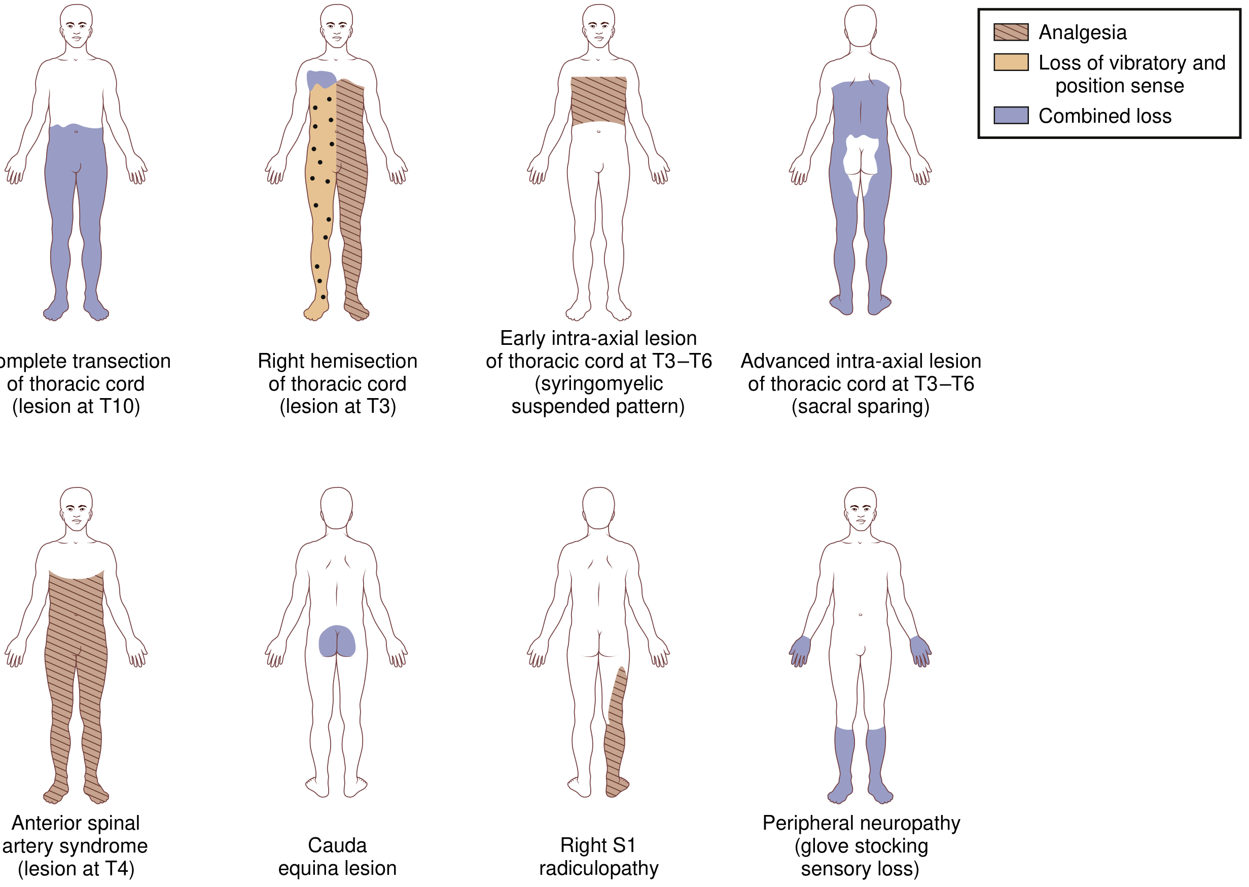
Spinal Shock (After Acute Injury)
When the spinal cord is suddenly injured, there is an initial phase called spinal shock:
| Phase | Features |
|---|---|
| Immediately after injury | Flaccid (floppy) paralysis, NO reflexes, low BP, no bladder reflex |
| After weeks-months | Spasticity develops, reflexes return (HYPERREFLEXIA = exaggerated reflexes) |
This is important - early flaccid paralysis does NOT mean permanent lower motor neuron damage.
UMN vs. LMN Signs in Spinal Cord Lesions
| Feature | At the level of lesion | Below the level |
|---|---|---|
| Motor neuron type | LMN (Lower Motor Neuron) | UMN (Upper Motor Neuron) |
| Muscle tone | Decreased/flaccid | Increased/spastic |
| Reflexes | Absent/decreased | Exaggerated (hyperreflexia) |
| Plantar reflex | - | Upgoing (Babinski +ve) |
Common Causes
| Category | Examples |
|---|---|
| Trauma | Road accidents, falls, stab wounds |
| Compression | Tumors (metastatic cancer), disc herniation, degenerative spine |
| Vascular | Anterior spinal artery occlusion (cord stroke) |
| Infection | Epidural abscess, TB spine, viral myelitis |
| Inflammatory | Multiple sclerosis, transverse myelitis |
| Nutritional | Vitamin B12 deficiency (subacute combined degeneration) |
| Developmental | Syringomyelia, Chiari malformation |
Expected Function at Different Levels (Cervical Cord)
| Level | What the patient can do |
|---|---|
| C3-C4 | Needs ventilator support, power wheelchair with mouth/chin control |
| C5 | Power wheelchair, dress upper body with help |
| C6 | Manual wheelchair with effort, some independence |
| C7 | Manual wheelchair, independent transfers, can drive |
| C8-T4 | Nearly independent with daily activities |
| T5-T12 | Fully independent, walks with braces |
| L1-L5 | Walks with short/long leg braces |
(Source: Bailey and Love's Short Practice of Surgery, 28th Edition)
Key Points to Remember
- Damage above C4 = may stop breathing (needs ventilator)
- Sensory level = find the lowest point where feeling is normal - lesion is at or above that level
- Spinal cord lesion is a medical emergency - act fast, get MRI urgently
- 80% rule for tumors: Patients treated before losing the ability to walk = 80% remain walking; treated after = 80% remain permanently unable to walk
- Brown-Sequard = same side motor loss, opposite side pain/temp loss (crossed pattern)
- Central cord = arms weaker than legs (opposite of what you expect)
- Anterior cord = motor + pain/temp lost, but position/vibration SPARED
Pho to transduction short notes easy language
Finding Sources
Finding Sources
Reading File
Reading File
Reading File
Reading File
Loading Image
Reading File
Loading Image
Reading File
I now have everything needed for a thorough, easy-language short note with diagrams. Here it is:
Phototransduction - Short Notes (Easy Language)
What is Phototransduction?
Phototransduction = Converting light energy into an electrical signal (nerve impulse) that the brain can understand.
Simple analogy: It's like a solar panel converting sunlight into electricity - your eye cells convert light into electrical signals.
It happens in the photoreceptor cells (rods and cones) of the retina.
The Players (Key Molecules)
| Molecule | Role |
|---|---|
| Rhodopsin | The light-sensitive pigment in rods (= Opsin protein + Retinal) |
| Retinal | A Vitamin A derivative - the actual "light catcher" |
| Opsin | The protein part of rhodopsin (7 transmembrane helices - G protein coupled receptor) |
| Transducin | A G-protein - the messenger |
| Phosphodiesterase (PDE) | An enzyme that breaks down cGMP |
| cGMP | Second messenger that keeps Na+ channels open |
| cGMP-gated Na+ channels | Ion channels that are open in the dark, close in light |
The Core Cascade - Step by Step
IN THE DARK (No light hitting the eye):
- cGMP levels in the rod are HIGH
- cGMP keeps the Na+ channels OPEN
- Na+ flows into the cell = "Dark current"
- Cell membrane potential = -30 mV (relatively depolarized)
- The depolarized cell continuously releases neurotransmitter (glutamate) onto bipolar neurons
Think: Dark = cGMP high = Na+ channels open = cell always releasing signals
WHEN LIGHT HITS THE EYE:
Step 1: Light hits Rhodopsin
- Light photon hits retinal (the chromophore part of rhodopsin)
- Retinal shape changes from 11-cis-retinal → all-trans-retinal (isomerization)
- This activates the opsin protein = Rhodopsin is now "bleached" (colour changes from purple to yellow)
Step 2: Rhodopsin activates Transducin (G-protein)
- Activated rhodopsin stimulates Transducin
- Transducin releases its alpha subunit
Step 3: Transducin activates Phosphodiesterase (PDE)
- Transducin alpha subunit activates PDE enzyme
- PDE breaks down cGMP → 5'-GMP
- Result: cGMP levels FALL
Step 4: Na+ channels CLOSE
- With less cGMP, the cGMP-gated Na+ channels close
- Na+ can no longer enter the cell
- K+ continues to leave normally
Step 5: Hyperpolarization
- Cell becomes more negative (membrane potential goes from -30 mV → -70 mV)
- This is HYPERPOLARIZATION (unusual! - most sensory receptors depolarize when stimulated)
Step 6: Less neurotransmitter released
- Hyperpolarized rod releases less glutamate to bipolar neurons
- This change in signal is detected and transmitted to the brain → you see light!
Visual Summary of the Cascade
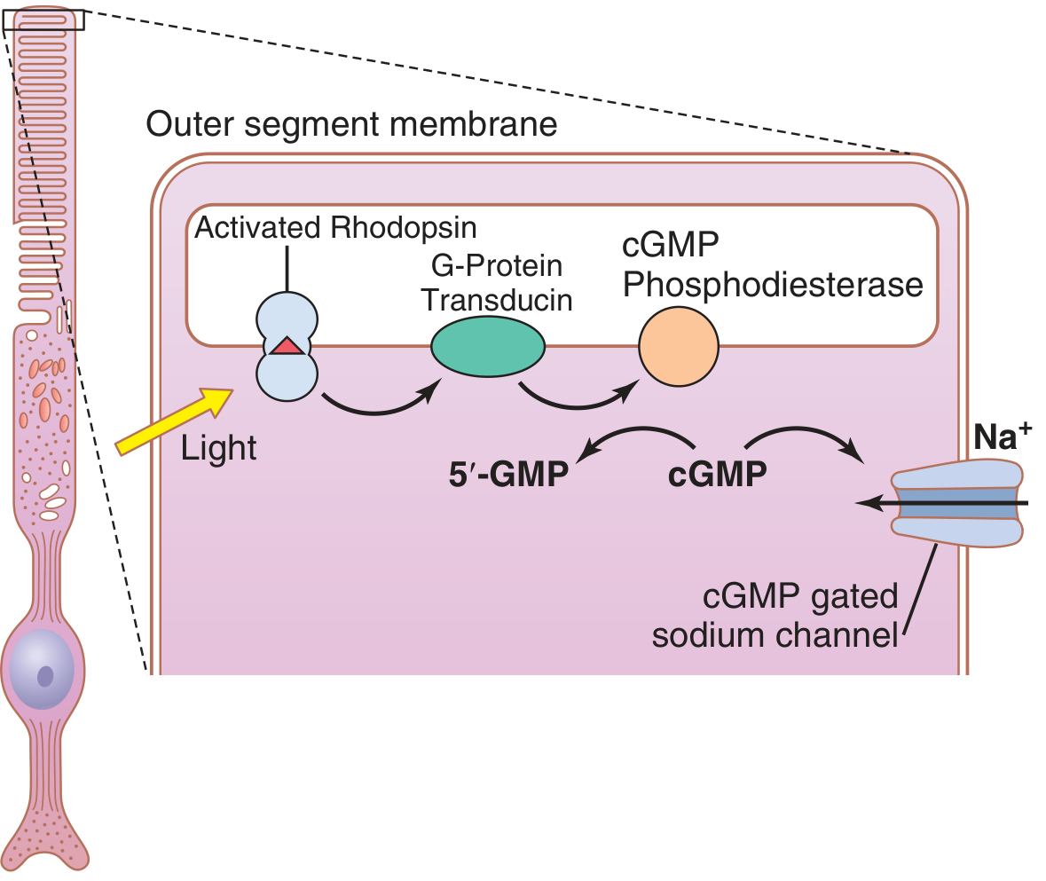
Dark vs Light - Side by Side
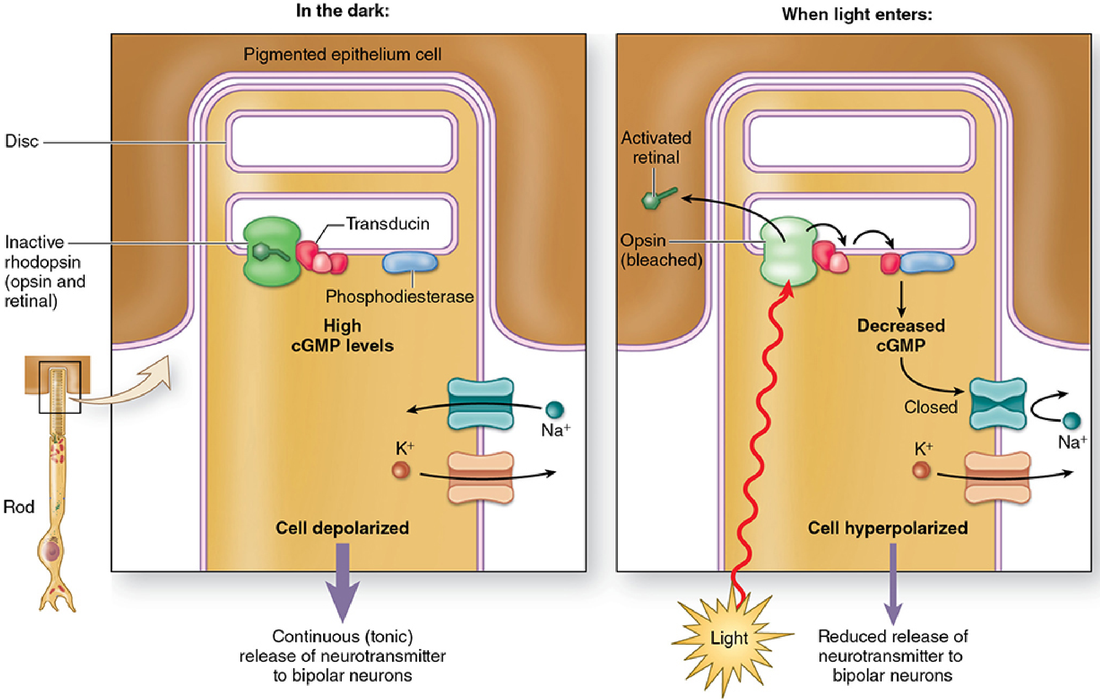
Easy Memory Flow Chart
LIGHT
  ↓
Rhodopsin activated (11-cis → all-trans retinal = BLEACHING)
  ↓
Transducin (G-protein) activated
  ↓
Phosphodiesterase (PDE) activated
  ↓
cGMP broken down → cGMP levels FALL
  ↓
Na+ channels CLOSE
  ↓
Cell HYPERPOLARIZES (becomes more negative)
  ↓
Less glutamate released to bipolar cells
  ↓
Signal to ganglion cells → optic nerve → BRAIN → YOU SEE!
Rods vs Cones in Phototransduction
| Feature | Rods | Cones |
|---|---|---|
| Pigment | Rhodopsin | Iodopsin (3 types) |
| Function | Dim light / night vision | Bright light / color vision |
| Sensitivity | Very high (1 photon enough) | Lower (needs more light) |
| Colors detected | Only black/white | Red, Green, Blue |
| Mechanism | Same cascade (cGMP/transducin) | Same cascade - different opsin |
| Location | Periphery of retina | Mainly fovea (center) |
Color Vision - The 3 Types of Cones
The 3 cone opsins respond to different wavelengths:
- Blue cones - peak at ~430 nm
- Green cones - peak at ~530 nm
- Red cones - peak at ~560 nm
The brain compares the activity of all three cone types to perceive different colors. This is the Young-Helmholtz Trichromacy Theory.
Recovery After Light (Regeneration of Rhodopsin)
After light bleaches rhodopsin:
- all-trans-retinal leaves the rod and enters pigment epithelial cells
- In the pigment epithelium, it is converted back to 11-cis-retinal
- It returns to the rod and recombines with opsin → Rhodopsin regenerated
- Ready for the next photon!
This regeneration takes time - explains why you are temporarily blind entering a dark room from bright light (dark adaptation)
Key Points to Remember
- Phototransduction = Light → Electrical signal
- The photoreceptor is unusual - it's depolarized in the dark and hyperpolarizes in light
- Key messenger: cGMP - high in dark (channels open), low in light (channels closed)
- Key G-protein: Transducin
- Key enzyme: Phosphodiesterase (PDE) - breaks down cGMP
- Rhodopsin = Opsin + Retinal (Vitamin A derivative)
- Bleaching = rhodopsin loses its color when light hits retinal (11-cis → all-trans)
- Defects in phototransduction cascade → Retinitis Pigmentosa and other visual diseases
(Sources: Guyton & Hall Textbook of Medical Physiology; Neuroscience: Exploring the Brain, 5th Ed; Junqueira's Basic Histology, 17e)
Bitemporal short notes
I want to make sure I give you the most useful answer. "Bitemporal" can refer to a few different medical topics.
Asking for Preferences
What topic are you asking about?
Finding Sources
Finding Sources
Reading File
Reading File
Reading File
Finding Sources
Finding Sources
Reading File
Finding Sources
Finding Sources
Loading Image
Finding Sources
I now have all the content needed. Here is the complete short note:
Bitemporal Hemianopia - Short Notes (Easy Language)
What Does it Mean?
Break it down:
- Bi = Both sides
- Temporal = Outer/side (temporal) visual fields
- Hemianopia = Loss of half the visual field
So Bitemporal Hemianopia = Loss of the outer (side) vision in BOTH eyes - you cannot see things coming from the left or right sides.
Imagine wearing a costume horse mask - you can only see what is straight ahead, not to the sides. That is what this feels like.
The Key Anatomy You Must Know
The visual pathway has a critical crossover point called the Optic Chiasm.
How the optic chiasm works normally:
LEFT EYE                    RIGHT EYE
  |                              |
  Optic nerve               Optic nerve
        \                    /
         \                  /
          OPTIC CHIASM (X)
         /                  \
        /                    \
   Left optic tract      Right optic tract
        ↓                         ↓
  Left brain (sees        Right brain (sees
  right visual field)     left visual field)
The critical rule at the chiasm:
- Nasal (inner) fibers CROSS over to the other side
- Temporal (outer) fibers stay on the same side
The nasal fibers of each eye carry information from the temporal (outer) visual field.
Why Does Chiasm Damage Cause Bitemporal Hemianopia?
When something compresses the middle of the optic chiasm, it squeezes and damages the crossing nasal fibers from BOTH eyes.
- Nasal fibers from LEFT eye (seeing left temporal field) are damaged
- Nasal fibers from RIGHT eye (seeing right temporal field) are damaged
- Result: Both temporal fields are lost = Bitemporal hemianopia
Easy memory: "Chiasm is hit in the middle → Crossing fibers go → Both outer fields lost"
Causes (Most to Least Common)
| Cause | Notes |
|---|---|
| Pituitary adenoma | #1 most common cause - grows upward from the sella turcica and compresses the chiasm from below |
| Craniopharyngioma | Especially in children |
| Meningioma | Tuberculum sellae or diaphragma sellae |
| Optic/hypothalamic glioma | Especially children |
| Aneurysm | Anterior communicating artery, internal carotid artery |
| Rathke cleft cyst | Benign sellar cyst |
| Sarcoidosis / lymphoma | Rare infiltrative causes |
| Hydrocephalus | Dilated 3rd ventricle pushing down on chiasm |
The golden rule: Bitemporal hemianopia = Think Pituitary first!
Clinical Features
Visual symptoms:
- Gradual, insidious onset - patients often don't notice it for a long time
- Cannot see objects approaching from both outer sides
- May bump into things on the sides
- Often goes undetected unless each eye is tested separately
Associated symptoms (with pituitary adenoma):
- Headache (compressive)
- Hormonal disturbances (pituitary compressed):
- Women: menstrual irregularity, infertility, galactorrhea (prolactinoma)
- Men: reduced libido, impotence
- Both: growth hormone excess (acromegaly) or Cushing's disease
- Hypopituitarism if large tumor destroys pituitary tissue
Variations by Level of Compression
The exact pattern depends on WHERE the chiasm is hit:
| Pattern | What it means |
|---|---|
| Classic bitemporal hemianopia | Symmetric compression of middle chiasm (pituitary adenoma) |
| Superior bitemporal quadrantanopia | Compression from below (pituitary) hits inferior chiasm first = upper outer fields lost first |
| Inferior bitemporal quadrantanopia | Compression from above hits superior chiasm = lower outer fields lost |
| Junctional scotoma | Lesion at junction of optic nerve and chiasm = optic neuropathy in one eye + superior temporal cut in the other |
Pituitary adenoma typically starts with superior temporal quadrantanopia (upper outer fields go first) before becoming full bitemporal hemianopia.
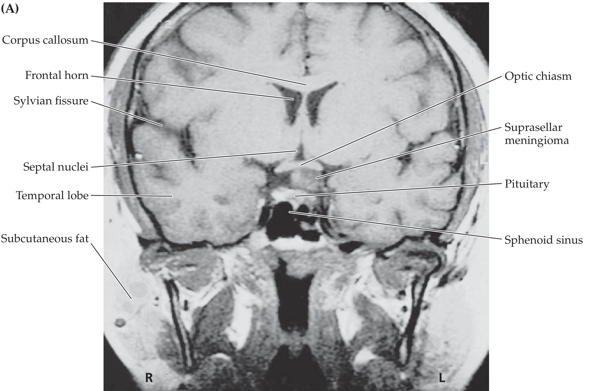
MRI Finding - Classic Image
This is what a suprasellar meningioma compressing the optic chiasm looks like on coronal MRI:
Diagnosis
- Clinical examination - Visual field testing (confrontation, perimetry)
- Test each eye separately - binocular testing misses it!
- Formal visual field testing - Humphrey/Goldmann perimetry
- MRI brain - with contrast (sellar/suprasellar region)
- Hormonal blood tests - Prolactin, GH, IGF-1, cortisol, thyroid function, FSH, LH
- Ophthalmology review - Optic disc may show pallor (atrophy) in longstanding cases
Treatment
Depends on the cause:
| Cause | Treatment |
|---|---|
| Prolactinoma (most common pituitary adenoma) | Medical: Dopamine agonist (Cabergoline/Bromocriptine) - often shrinks tumor |
| Non-functioning adenoma / other adenomas | Transsphenoidal surgery (through the nose) |
| Meningioma | Surgery |
| Craniopharyngioma | Surgery ± radiation |
Important: Vision can partially or fully recover after decompression of the chiasm if treated early, before permanent optic atrophy sets in.
Summary - Key Points
| Point | Fact |
|---|---|
| Site of lesion | Optic Chiasm |
| Fibers affected | Crossing nasal fibers from both eyes |
| Field lost | Both temporal (outer) fields |
| #1 cause | Pituitary adenoma |
| Associated features | Headache + hormonal symptoms |
| Onset | Insidious - often missed |
| Diagnosis | MRI brain + visual field test |
| Treatment | Surgery (transsphenoidal) or dopamine agonist |
(Sources: Harrison's Principles of Internal Medicine, 22nd Ed; Adams and Victor's Principles of Neurology, 12th Ed; Neuroanatomy through Clinical Cases, 3rd Ed)
Brain blood barrier short notes
Finding Sources
Finding Sources
Reading File
Reading File
Reading File
Reading File
Loading Image
Reading File
I have everything I need. Here is the complete short note:
Blood-Brain Barrier (BBB) - Short Notes (Easy Language)
What is the BBB?
The Blood-Brain Barrier (BBB) is a highly selective protective wall between the blood vessels and the brain tissue. It controls exactly what enters and exits the brain.
Easy analogy: Think of the BBB as a strict security checkpoint at the brain's entrance - only approved substances with the right "pass" are allowed in.
How Was it Discovered?
In the 1800s, scientists noticed that when a coloured dye was injected into the bloodstream, it stained every organ in the body except the brain. This was the first proof that the brain has a special protective barrier.
Where is the BBB Located?
The BBB is located in the capillary walls (tiny blood vessels) inside the brain.
Structure of Brain Capillaries vs. Body Capillaries:
| Feature | Normal body capillaries | Brain capillaries (BBB) |
|---|---|---|
| Gaps between cells | Have fenestrations (gaps/holes) | No gaps - sealed by tight junctions |
| Passage of substances | Relatively free | Strictly controlled |
| Pinocytosis (vesicle transport) | Active | Very little |
Cellular Components of the BBB
The BBB is made of 4 key components working together:
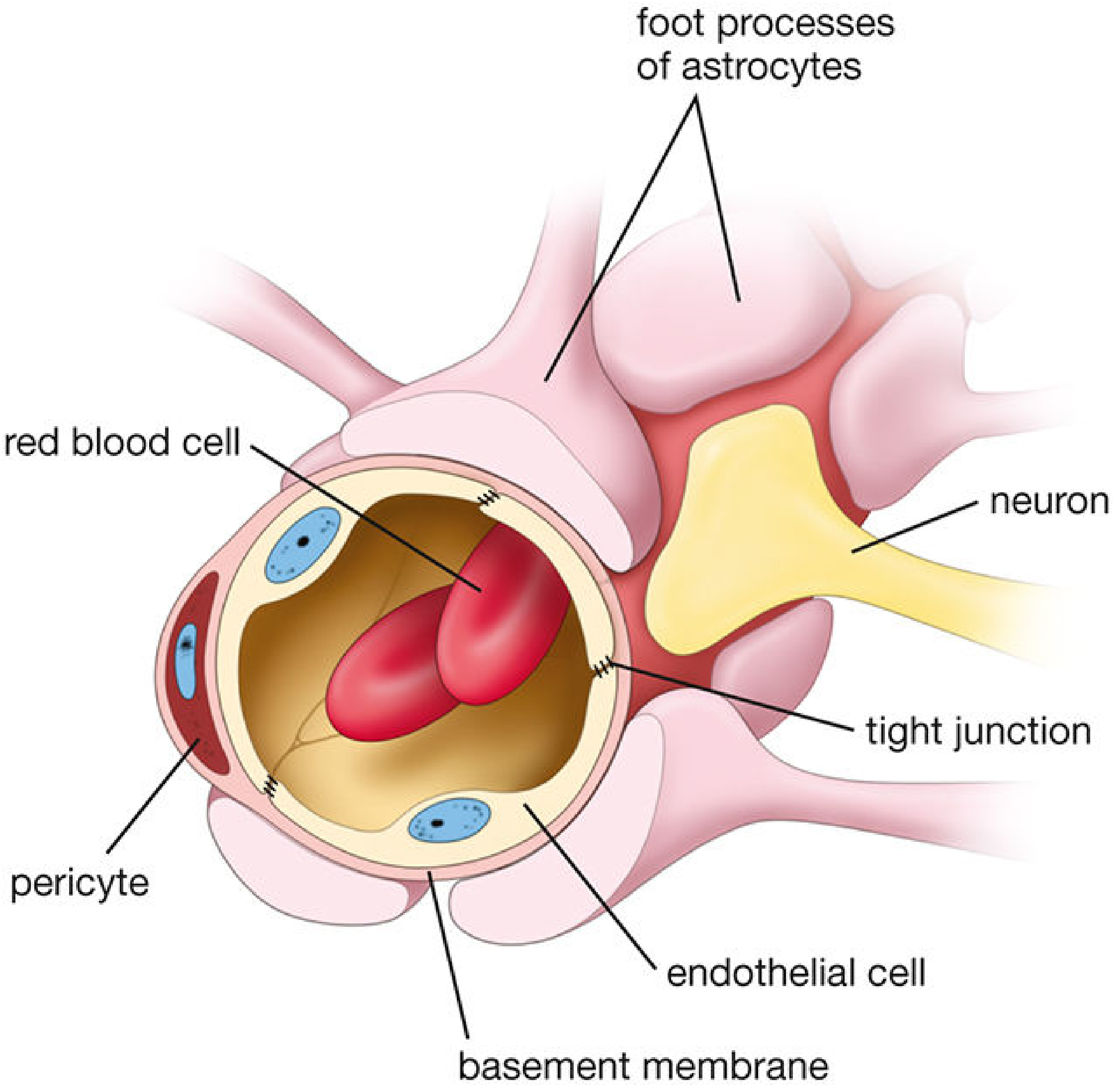
| Component | Role |
|---|---|
| Endothelial cells | Form the inner wall of the capillary; sealed by tight junctions - the main barrier |
| Tight junctions | Glue the endothelial cells tightly together - NO gaps allowed |
| Basement membrane | Structural support layer under the endothelium |
| Astrocyte foot processes | Wrap around the outside of the capillary; maintain and regulate tight junctions, help with water transport (via Aquaporin-4 channels) |
| Pericytes | Sit embedded in the basement membrane; help regulate blood flow and barrier integrity |
The tight junctions are the real gatekeepers. They are more elaborate than tight junctions found anywhere else in the body.
Functions of the BBB
- Protect the brain from blood-borne toxins, pathogens, and harmful chemicals
- Meet metabolic demands - let in what the brain needs (glucose, oxygen, amino acids)
- Maintain homeostasis - regulate the perfect chemical environment for neurons to function
- Buffer ion levels - especially K+ (neurons are very sensitive to potassium changes)
What CAN and CANNOT Cross the BBB?
Freely CROSS (no barrier needed):
- O2 and CO2 (small gases - pass right through)
- Lipid-soluble molecules (e.g., ethanol, steroid hormones, anaesthetics)
- Water (via aquaporin-4 channels in astrocyte feet)
CANNOT cross freely (need transport proteins):
- Water-soluble substances
- Large molecules (>500 Daltons generally cannot cross)
- Most drugs
- Proteins and antibodies
- Most pathogens (bacteria, etc.)
Cross via ACTIVE TRANSPORT (need a carrier):
- Glucose (via GLUT1 transporter) - brain's main fuel
- Amino acids (via SLC transporters)
- Vitamins and ions
- L-DOPA (used in Parkinson's treatment)
Important clinical example: Dopamine CANNOT cross the BBB, so it cannot be given as a drug for Parkinson's disease. Instead, L-DOPA is given because it crosses the BBB and is then converted to dopamine inside the brain.
Efflux Transporters (Pumping Things OUT)
The BBB also has ABC transporters that actively pump toxins and drugs OUT of the brain back into the blood. This is a major reason why many drugs fail to reach the brain - they get pumped back out.
Circumventricular Organs - Areas WITHOUT BBB
Not all parts of the brain are protected by the BBB. Certain regions are intentionally left "open" to monitor blood chemistry. These are called Circumventricular Organs (CVOs):
| Organ | Function |
|---|---|
| Area postrema | Detects toxins in blood → triggers vomiting (the "chemoreceptor trigger zone") |
| Median eminence | Releases hypothalamic hormones into blood |
| Subfornical organ | Regulates fluid balance |
| Pineal gland | Melatonin secretion (circadian rhythms) |
| Posterior pituitary | Releases ADH and oxytocin |
| Organum vasculosum of lamina terminalis | Neuroendocrine functions |
When the BBB Breaks Down
In disease, the BBB can be disrupted, allowing fluid to leak into the brain:
| Condition | What happens |
|---|---|
| Brain tumors | Disrupt tight junctions → leaky capillaries |
| Meningitis/Encephalitis | Inflammation breaks down the barrier |
| Stroke / Cerebral infarction | BBB breaks → Vasogenic edema (fluid leaks into interstitial space) |
| Traumatic brain injury | Mechanical disruption of tight junctions |
| Multiple sclerosis | Inflammatory breakdown → immune cells cross into CNS |
Two types of brain edema from BBB failure:
| Type | Mechanism | Cause |
|---|---|---|
| Vasogenic edema | BBB breaks → fluid leaks into extracellular space | Tumors, abscess, trauma, inflammation |
| Cytotoxic edema | Cell damage → fluid builds up INSIDE cells | Stroke/ischemia |
Blood-CSF Barrier - Related Concept
There is also a Blood-CSF barrier at the choroid plexus:
- Choroid plexus capillaries are leaky (fenestrated)
- But the choroid epithelial cells are joined by tight junctions → forming the Blood-CSF barrier
- Substances must be transported through the epithelial cells to reach the CSF
Key Points to Remember
| Point | Fact |
|---|---|
| Location | Capillary endothelial cells of brain |
| Physical basis | Tight junctions between endothelial cells |
| Supporting cells | Astrocyte foot processes (regulate), Pericytes |
| Freely crosses | O2, CO2, lipid-soluble substances, ethanol |
| Needs transport | Glucose (GLUT1), amino acids, L-DOPA |
| Cannot cross | Dopamine, most drugs, proteins, bacteria |
| No BBB areas | Circumventricular organs (area postrema, pineal, etc.) |
| BBB breakdown | → Vasogenic edema |
| Clinical importance | Why most CNS drugs are hard to design (must be lipid-soluble or use active transport) |
(Sources: Neuroanatomy through Clinical Cases, 3rd Ed; Histology: A Text and Atlas, Wolters Kluwer; Junqueira's Basic Histology, 17th Ed)
This is a shared conversation. Sign in to Orris to start your own chat.