
Levels of neck lymph node , their embryology and drainage content. As per cummings and scott brown
Reading File
Finding Sources
Finding Sources
Searching PubMed
"neck dissection" AND "lymph nodes"
Searching the Web
Cummings Otolaryngology neck lymph node levels drainage embryology Scott-Brown
Finding Sources
Finding Sources
Finding Sources
Finding Sources
Reading File
Reading File
Reading File
Finding Sources
Reading File
Reading File
Finding Sources
Reading File
Finding Sources
Finding Sources
Loading Image
Cervical lymph-node levels: contents, boundaries, drainage and embryological basis
The standard surgical classification is Levels I to VI. Level VII is the superior mediastinal continuation of the central compartment, below the suprasternal notch. Levels I, II and V have sublevels. The scheme identifies predictable first-echelon nodal basins for head and neck malignancy.
Scott-Brown's Otorhinolaryngology Head & Neck Surgery, p. 350; Cummings Otolaryngology Head and Neck Surgery, p. 2289.
Scott-Brown's Otorhinolaryngology Head & Neck Surgery, p. 350; Cummings Otolaryngology Head and Neck Surgery, p. 2289.
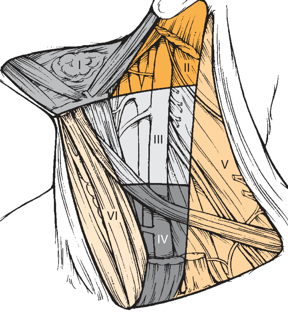
1. Level I: Submental and submandibular groups
| Sublevel | Boundaries | Contents | Main afferent drainage / primary sites |
|---|---|---|---|
| IA: Submental | Between both anterior bellies of digastric; from mandibular symphysis to hyoid | Submental nodes | Midline lower lip, chin, anterior floor of mouth, mandibular incisors, tip of tongue and anterior midline oral tongue |
| IB: Submandibular | Body of mandible superiorly; anterior and posterior bellies of digastric; stylohyoid posteriorly | Submandibular nodes, periglandular nodes, facial/perifacial nodes may be relevant; submandibular gland is removed in surgical clearance | Lateral lower lip, cheek, lateral anterior tongue, floor of mouth, anterior nasal cavity, gingiva, mandibular/maxillary alveolus, submandibular gland |
Clinical point: Level IA involvement is particularly associated with cancers of the lip, anterior floor of mouth and midface.
Scott-Brown's Otorhinolaryngology Head & Neck Surgery, p. 350; Cummings Otolaryngology Head and Neck Surgery, p. 2289.
Scott-Brown's Otorhinolaryngology Head & Neck Surgery, p. 350; Cummings Otolaryngology Head and Neck Surgery, p. 2289.
2. Level II: Upper jugular group
| Sublevel | Boundaries | Contents | Main afferent drainage / primary sites |
|---|---|---|---|
| IIA | Skull base or jugular fossa superiorly to hyoid inferiorly; anterior to the spinal accessory nerve plane | Upper internal jugular nodes, especially jugulodigastric node | Nasal cavity, nasopharynx, parotid, oral cavity, oropharynx, tonsil, base of tongue, supraglottic larynx |
| IIB | Same superior-inferior extent; posterior to spinal accessory nerve, anterior to posterior SCM border | Upper jugular nodes posterior to the spinal accessory nerve | Similar upper aerodigestive sources, but less commonly involved in some oral cavity cancers |
- The spinal accessory nerve (SAN) separates IIA from IIB.
- The hallmark node is the jugulodigastric node, often enlarged in tonsillitis and tonsillar/oropharyngeal cancer.
- Medial nodes behind the carotid vessels are retropharyngeal nodes, not level II.
Scott-Brown's Otorhinolaryngology Head & Neck Surgery, p. 350; Cummings Otolaryngology Head and Neck Surgery, p. 2289.
3. Level III: Middle jugular group
| Boundaries | Contents | Main afferent drainage / primary sites |
|---|---|---|
| Hyoid bone or carotid bifurcation superiorly to lower border of cricoid or omohyoid-IJV junction inferiorly; sternohyoid medially and posterior SCM laterally | Nodes around the middle third of the internal jugular vein | Oral tongue and floor of mouth, oropharynx, nasopharynx, hypopharynx and larynx |
Important pattern: Oral cavity malignancy commonly spreads through levels I to III, particularly from tongue and floor of mouth.
Cummings Otolaryngology Head and Neck Surgery, p. 2289.
4. Level IV: Lower jugular group
| Boundaries | Contents | Main afferent drainage / primary sites |
|---|---|---|
| Lower border of cricoid or omohyoid-IJV junction superiorly to clavicle inferiorly; sternohyoid medially and posterior SCM laterally | Nodes around lower third of internal jugular vein; lower deep cervical nodes | Hypopharynx, larynx, cervical oesophagus, thyroid, lower pharynx |
- Left supraclavicular node, at the termination of the thoracic duct, may enlarge from abdominal or thoracic malignancy. It is the classic Virchow node.
- Laryngeal and hypopharyngeal cancers commonly involve levels II to IV.
Cummings Otolaryngology Head and Neck Surgery, p. 2289.
5. Level V: Posterior-triangle group
| Sublevel | Boundaries | Contents | Main afferent drainage / primary sites |
|---|---|---|---|
| VA | Posterior triangle above the lower border of cricoid | Nodes along the spinal accessory nerve | Posterior scalp and neck, nasopharynx, and some oropharyngeal primaries |
| VB | Posterior triangle below lower border of cricoid to clavicle | Nodes along transverse cervical vessels and supraclavicular nodes | Posterior scalp/neck, thyroid, thoracic and infraclavicular sources; may receive lower-neck drainage |
Overall boundaries: posterior border of SCM anteriorly/medially, anterior border of trapezius posteriorly/laterally, and clavicle inferiorly.
Clinical point: Level VB disease often implies more advanced regional disease. A left supraclavicular node may also represent systemic or infradiaphragmatic malignancy.
Scott-Brown's Otorhinolaryngology Head & Neck Surgery, p. 350; Cummings Otolaryngology Head and Neck Surgery, p. 2289.
Scott-Brown's Otorhinolaryngology Head & Neck Surgery, p. 350; Cummings Otolaryngology Head and Neck Surgery, p. 2289.
6. Level VI: Central/anterior compartment
| Boundaries | Contents | Main afferent drainage / primary sites |
|---|---|---|
| Hyoid superiorly to suprasternal notch inferiorly; medial borders of carotid sheaths laterally | Prelaryngeal/precricoid (Delphian) node, pretracheal nodes, paratracheal nodes, perithyroidal nodes, nodes along recurrent laryngeal nerves | Thyroid, subglottic larynx, apex of piriform sinus, cervical trachea and cervical oesophagus |
- Level VI is a midline compartment. Unlike II to IV, it is generally not designated right versus left.
- The Delphian node is clinically important in thyroid and laryngeal malignancy.
Cummings Otolaryngology Head and Neck Surgery, p. 2289.
7. Level VII: Superior mediastinal nodes
| Boundaries | Contents | Main afferent drainage / primary sites |
|---|---|---|
| From suprasternal notch/manubrium to the upper border of the aortic arch or innominate artery | Superior mediastinal paratracheal nodes | Inferior thyroid drainage, subglottis, cervical trachea and cervical oesophagus |
Scott-Brown notes that this is anatomically below the neck, extending from the suprasternal notch to the innominate artery.
Scott-Brown's Otorhinolaryngology Head & Neck Surgery, p. 350.
Scott-Brown's Otorhinolaryngology Head & Neck Surgery, p. 350.
Embryology relevant to cervical lymphatics
Development of the lymphatic system
- Around the 5th to 6th embryonic week, lymphatic endothelial precursors arise mainly from venous endothelium.
- Paired jugular lymph sacs develop beside the internal jugular veins. They are the key embryological origin for cervical lymphatic channels.
- Lymphatic vessels extend from these sacs through the head and neck. Mesenchymal tissue invades and partitions the sacs, forming lymph-node architecture.
- Efferent lymph ultimately enters the venous system:
- Right lymphatic duct at the right venous angle
- Thoracic duct at the left venous angle, explaining the significance of the left supraclavicular/Virchow node.
The primordial lymphatic system is identifiable by about the sixth week, initially as lymph sacs adjacent to the jugular veins.
Sabiston Textbook of Surgery, “Embryology and Anatomy,” pp. 536-538.
Sabiston Textbook of Surgery, “Embryology and Anatomy,” pp. 536-538.
Relation to pharyngeal-arch embryology
The neck levels themselves are not embryological derivatives of individual pharyngeal arches. They are surgical-anatomical compartments. However, drainage patterns relate to the embryological origin and final anatomy of the head and neck organs:
- First arch derivatives and oral cavity structures tend to drain first to Levels I to III.
- Pharyngeal structures, especially oropharynx and nasopharynx, commonly drain to Level II and retropharyngeal nodes.
- The larynx has a dual developmental and lymphatic pattern:
- Supraglottis: rich lymphatics, mainly levels II to III
- Glottis: relatively sparse lymphatics
- Subglottis: pretracheal/paratracheal drainage, mainly level VI and VII
- Thyroid and lower visceral neck structures commonly drain to the central compartment, levels VI and VII.
Cummings emphasizes that head and neck structures develop in close relation to the pharyngeal apparatus, while lymph-node groups are classified clinically by their predictable metastatic drainage pathways.
Cummings Otolaryngology Head and Neck Surgery, pp. 2289 and 3434-3435.
Cummings Otolaryngology Head and Neck Surgery, pp. 2289 and 3434-3435.
High-yield drainage summary
- Lip, anterior oral tongue, floor of mouth → I, then II and III
- Tonsil, base of tongue, oropharynx → II, often bilateral for midline lesions
- Nasopharynx → retropharyngeal nodes and II, often bilateral
- Larynx/hypopharynx → II, III, IV
- Thyroid, subglottis, trachea, cervical oesophagus → VI, then VII
- Posterior scalp and posterior neck skin → VA/VB
- Midline lesions can drain bilaterally, especially base of tongue, soft palate, pharyngeal wall, nasopharynx and larynx.
This is a shared conversation. Sign in to Orris to start your own chat.