
ulnar nerve course branches hand claw hand deformity
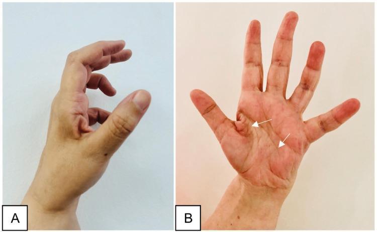
Two-panel clinical photograph demonstrating the late sequelae of a peripheral nerve injury in the hand, specifically illustrating features of ulnar nerve palsy. Image A (lateral view) shows a classic 'claw hand' deformity, characterized by hyperextension at the metacarpophalangeal (MCP) joints and flexion at the interphalangeal (IP) joints, most prominent in the ring and little fingers. The thumb appears adducted and flexed. Image B (anterior/palmar view) displays generalized muscle atrophy of the hand, with significant hollowing of the hypothenar eminence and interosseous spaces (indicated by white arrows). Notably, the thenar eminence remains relatively spared, suggesting the median nerve's innervation to the thenar muscles is intact. This combination of clawing and selective intrinsic muscle wasting is an important educational marker for localizing nerve lesions to the distal ulnar nerve (e.g., Guyon's canal or post-surgical complication). The images illustrate the clinical presentation of chronic motor deficit and the characteristic postural changes resulting from unbalanced muscle pull in the hand.
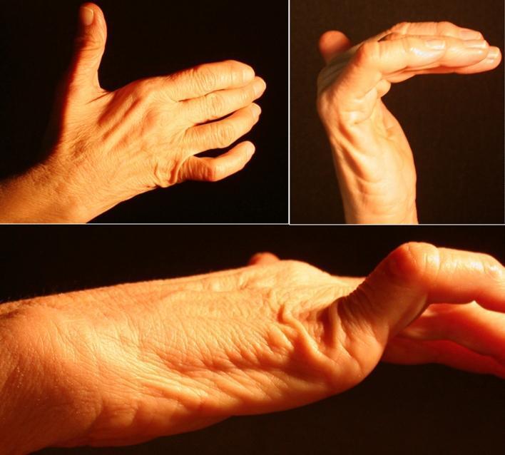
This composite of clinical photographs demonstrates a 'claw hand' (main en griffe) deformity, a characteristic sign of ulnar nerve neuropathy. The images show a right hand from dorsal, palmar, and lateral perspectives. Key visual features include hyperextension at the metacarpophalangeal (MCP) joints and flexion at the proximal and distal interphalangeal (IP) joints, most pronounced in the fourth and fifth digits. There is noticeable atrophy of the hypothenar eminence and hollowing of the interosseous spaces on the dorsal surface, indicating wasting of the intrinsic hand muscles (interossei and ulnar-innervated lumbricals). The thenar eminence appears relatively preserved, though subtle flattening of the adductor pollicis region may be present. This presentation is consistent with distal ulnar nerve compression, such as within Guyon’s canal, leading to motor deficit and muscle denervation. These clinical signs are essential for diagnosing peripheral nerve entrapment syndromes and distinguishing them from proximal lesions or central nervous system pathologies.
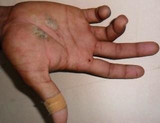
Clinical photograph of a right hand demonstrating ulnar claw hand (main en griffe) deformity, secondary to traumatic ulnar nerve injury. The image shows characteristic hyperextension at the fourth and fifth metacarpophalangeal (MCP) joints, accompanied by fixed flexion at the proximal and distal interphalangeal (PIP and DIP) joints of the ring and little fingers. In addition to the neurological deficit, the palmar surface exhibits multiple signs of acute trauma, including two irregular areas of pale/abrasive discoloration on the palm and a small punctate wound near the base of the index finger. An adhesive bandage is visible on the thumb. This visual presentation is classic for a distal or high ulnar nerve palsy where the loss of intrinsic muscle function (lumbricals and interossei) leads to an imbalance in the hand's extrinsic musculature. The image serves as an educational tool for identifying physical signs of peripheral nerve compression or transection following orthopedic trauma, such as a humeral shaft fracture.
medium sized artery microstructure layers tunica intima media adventitia histology
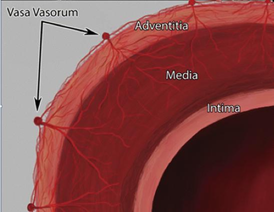
This anatomical diagram provides a cross-sectional view of a large systemic artery wall, illustrating the structural layers and the microvascular supply. The diagram identifies three distinct histological layers: the tunica intima (innermost lining), the tunica media (thick muscular middle layer), and the tunica adventitia (outermost connective tissue layer). A primary focus of the illustration is the 'vasa vasorum'—the 'vessels of the vessels.' The vasa vasorum are depicted as a branching network of small capillaries originating from larger nutrient vessels on the external surface of the adventitia. These microvessels penetrate through the adventitia and extend into the outer portions of the tunica media, highlighting the physiological mechanism for delivering oxygen and nutrients to thick-walled vessels where simple diffusion from the lumen is insufficient. The image serves as a pedagogical tool for understanding vascular histology and the pathophysiology of arterial wall maintenance in conditions such as atherosclerosis or aortic disease.
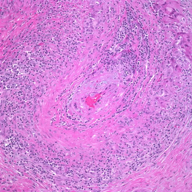
High-power hematoxylin and eosin stained transverse section of a medium-sized artery demonstrating granulomatous arteritis. The lumen is narrowed by edema and fibrosis of the intima, while the tunica media is uniformly expanded by a mixed inflammatory infiltrate. Lymphocytes, plasma cells, and histiocytes predominate, with several multinucleated giant cells visible in the upper right quadrant. The internal elastic lamina is variably destroyed, with fragmentation evident at the media- adventitia interface. Transmural inflammation produces concentric thickening of the vessel wall and marked luminal compromise. Admixed eosinophils are sparse; occasional foamy histiocytes emphasize granulomatous architecture. The adventitia shows mild edema and vascular remodeling. Overall features are classic for granulomatous vasculitis, most consistent with giant cell arteritis (temporal arteritis) or other large-vessel vasculitides such as Takayasu arteritis. Diagnostic significance: histology provides definitive confirmation when clinical suspicion is high and ESR/CRP elevations are present. Differential considerations include polyarteritis nodosa, infectious vasculitis, and eosinophilic granulomatosis with polyangiitis depending on clinical context. Clinically relevant correlations include age >50 years, new headaches, jaw claudication, visual symptoms, and constitutional signs. This image is valuable for pathology education, residency training, and radiologic-pathologic correlation studies in vasculitis. Useful for teaching differential diagnosis and correlating clinical features with histologic patterns in practice.
Anatomy Exam - Complete Answers
SECTION I (visible partial) - Ulnar Nerve Palsy
SECTION II - Reasoning Out Short Notes
1. Anatomical Basis of Ulnar Claw Hand (2 marks)
- The ulnar nerve (C8, T1) innervates the interossei, the two medial lumbricals (ring and little fingers), and the hypothenar muscles.
- The lumbricals and interossei normally flex the metacarpophalangeal (MCP) joints and extend the interphalangeal (IP) joints.
- When these muscles are paralyzed, the long flexors (flexor digitorum profundus and superficialis - supplied by the median nerve) and long extensors remain unopposed.
- Result: hyperextension at the MCPs (extensor digitorum dominates) and flexion at the PIP and DIP joints (FDP dominates), producing the claw posture.
- The lateral two lumbricals (index and middle) are innervated by the median nerve, so they are preserved.
- The medial two lumbricals (ring and little) are supplied by the ulnar nerve, so clawing is most pronounced in these two digits.
2. Inflammation of the Gallbladder - Pain at the Tip of the 9th Costal Cartilage (Reason Out) (2 marks)
- The gallbladder receives its visceral sensory innervation from T7-T9 spinal cord segments via the greater splanchnic nerve.
- The tip of the 9th costal cartilage is the surface landmark of McBurney's equivalent for the gallbladder - the point of maximum tenderness in cholecystitis, also known as Murphy's point.
- Afferent pain impulses from the inflamed gallbladder travel via splanchnic nerves to the dorsal horn at T7-T9.
- At those same segments, somatic afferents from the skin and fascia overlying the right upper quadrant (including the 9th costal cartilage area) also converge.
- The brain cannot distinguish the source and interprets the pain as coming from the somatic area - i.e., the tip of the 9th costal cartilage.
- Phrenic nerve irritation (C3-C5) from an inflamed gallbladder may also produce referred pain to the right shoulder tip (C4 dermatome).
SECTION I (Full Question) - Ulnar Nerve Palsy (1+3+4 = 8 marks)
a) Root Value
b) Course and Relations
- Arises from the medial cord, enters the arm alongside the median nerve and axillary artery.
- Passes through proximal regions medial to the axillary artery.
- In the middle of the arm, it pierces the medial intermuscular septum and enters the posterior compartment.
- Lies anterior to the medial head of triceps brachii.
- Passes posterior to the medial epicondyle of the humerus (the "funny bone" groove).
- Has no major branches in the arm.
- Enters the anterior compartment by passing between the humeral and ulnar heads of flexor carpi ulnaris (FCU).
- Runs down the medial forearm in the plane between FCU and flexor digitorum profundus (FDP).
- In the distal two-thirds, the ulnar artery lies lateral to the ulnar nerve.
- Both enter the hand by passing superficial to the flexor retinaculum, lateral to the pisiform bone (through Guyon's canal).
- Enters lateral to the pisiform, posteromedially to the ulnar artery.
- Divides into a deep branch (mainly motor) and a superficial branch (mainly sensory).
c) Branches and Distribution (C7, C8, T1)
| Region | Branch | Supply |
|---|---|---|
| Forearm | Muscular branches | FCU, medial half of FDP (ring and little fingers) |
| Forearm | Palmar cutaneous branch | Skin over medial palm |
| Forearm | Dorsal cutaneous branch | Skin over posteromedial dorsum of hand and medial 1.5 digits |
| Hand - Deep branch | Motor | Hypothenar muscles (abductor, flexor, opponens digiti minimi), all interossei (4 dorsal, 3 palmar), medial 2 lumbricals, adductor pollicis, medial head of flexor pollicis brevis |
| Hand - Superficial branch | Sensory | Anterior little finger, ulnar side of ring finger, hypothenar skin |
d) Anatomical Basis of Ulnar Claw Hand
SECTION III - Write Short Notes
1. Supination and Pronation (10 marks)
- Supination = rotation of the forearm to bring the palm anteriorly (palm up, "carrying a bowl of soup")
- Pronation = rotation to bring the palm posteriorly (palm down)
- Total range ~180 degrees
- Superior (proximal) radioulnar joint
- Inferior (distal) radioulnar joint
- The radius pivots around the ulna; because the hand articulates predominantly with the radius, the hand moves with the radius.
- Biceps brachii - most powerful supinator; most effective when elbow is flexed. Wraps around the proximal radius in pronation; contraction "unwraps" and produces supination.
- Supinator - located in the posterior compartment; originates from the supinator crest of the ulna and lateral epicondyle of the humerus; wraps around the posterior and lateral surface of the upper third of the radius to attach to the radial shaft above the oblique line; innervated by the radial nerve (posterior interosseous nerve).
- Pronator teres - runs from medial epicondyle of humerus to lateral surface of the radius (midshaft); innervated by median nerve.
- Pronator quadratus - spans between the anterior surfaces of the distal ends of radius and ulna; deepest muscle; innervated by anterior interosseous nerve (branch of median).
2. Microstructure of a Medium-Sized Artery (10 marks)
1. Tunica Intima (Innermost)
- Endothelium: single layer of flat endothelial cells lining the lumen; acts as a selective barrier; produces NO, prostacyclin.
- Subendothelial connective tissue: thin layer of loose connective tissue.
- Internal elastic lamina (IEL): a prominent, fenestrated elastic membrane separating intima from media; allows diffusion of nutrients; appears wavy on histology due to vessel constriction during fixation.
2. Tunica Media (Middle - thickest layer in muscular arteries)
- Predominantly smooth muscle cells arranged in circular/spiral layers - typically 10-40 layers.
- Smooth muscle regulates vasomotor tone (vasoconstriction and vasodilation).
- Also contains elastic fibres, reticular fibres (collagen type III), and proteoglycans.
- External elastic lamina (EEL): another fenestrated elastic sheet separating media from adventitia (thinner than IEL, sometimes absent in smaller arteries).
3. Tunica Adventitia (Outermost)
- Collagen fibres (type I) - predominantly longitudinally oriented.
- Elastic fibres.
- Fibroblasts.
- Vasa vasorum - small nutrient vessels that supply the outer wall (adventitia and outer media cannot be supplied by diffusion from the lumen alone).
- Lymphatics and nerve fibres (vasomotor innervation to smooth muscle in media).
3. Cadaver Ethics (10 marks)
- Most countries regulate cadaver use under Anatomy Acts (e.g., the Indian Anatomy Act 1984 governs cadaver procurement in India).
- Bodies may be sourced through voluntary donation (willed body programs) or unclaimed bodies from government institutions.
-
Respect and Dignity: The cadaver must be treated with the same respect accorded to a living person. Dissection rooms should have a solemn atmosphere; students are expected to maintain professional conduct.
-
Informed Consent: Voluntary body donation requires full prior written informed consent from the donor during their lifetime, or from the next of kin. No coercion or financial inducement is permissible.
-
Confidentiality: The identity of the donor must be protected. Cadavers are typically anonymized (numbered) before use.
-
Beneficence and Non-maleficence: Bodies should be used only for legitimate educational, research, or surgical training purposes. Commercial use or misuse is prohibited.
-
Minimal use and proportionality: No more tissue should be used than is necessary for the educational objective.
-
Proper Disposal: After dissection, the remains must be disposed of respectfully - either returned to the family or cremated/buried with dignity, according to the donor's wishes.
-
Cultural and Religious Sensitivity: Religious and cultural beliefs of donors regarding bodily integrity after death must be acknowledged and respected.
-
Non-commodification: Selling human bodies or body parts is illegal and unethical worldwide.
-
Student responsibilities: Medical students must:
- Maintain confidentiality
- Not photograph or record without permission
- Not discuss cases outside appropriate professional settings
- Treat the cadaver as a "first patient" and "silent teacher"
-
Institutional oversight: Medical institutions should have ethics committees reviewing cadaver use protocols, storage conditions (formalin preservation, cold storage), and donation programs.
Summary Table (Quick Reference)
| Question | Key Points |
|---|---|
| Ulnar nerve root | C8, T1 (medial cord) |
| Ulnar claw | MCP hyperextension + IP flexion; worse in ring/little; due to loss of lumbricals + interossei |
| Gallbladder pain at 9th costal cartilage | Referred pain - visceral T7-T9 converges with somatic T7-T9 at spinal cord |
| Varicocele on left | Left testicular vein drains at 90° into left renal vein (right drains at acute angle into IVC); higher hydrostatic pressure on left |
| Supination muscles | Biceps brachii + Supinator |
| Pronation muscles | Pronator teres + Pronator quadratus |
| Medium artery layers | Intima (endothelium + IEL), Media (smooth muscle + EEL), Adventitia (collagen + vasa vasorum) |
| Cadaver ethics | Consent, dignity, confidentiality, legal framework, proper disposal |