
Interal capsule
I'll look up the internal capsule from the medical library for you right away.
Searching sources...
Reading File
Finding Sources
Finding Sources
Good results. Now let me read the key sections from the neuroanatomy and neurology textbooks in parallel.
Reading File
Reading File
Reading File
Excellent content. Now let me get the clinical sections (lesion effects, blood supply, hemorrhage) and fetch the key diagrams.
Reading File
Reading File
Loading Image
I now have comprehensive information. Let me compile the full answer.
Internal Capsule
Overview
The internal capsule is a compact lamina of white matter representing the largest projection fiber tract in the brain. It contains both afferent and efferent nerve fibers passing to and from the brainstem and cerebral hemispheres. Superiorly it continues as the corona radiata; inferiorly it continues as the cerebral peduncles (basis pedunculi) of the midbrain.
Shape and Location
In a horizontal section, the internal capsule appears V-shaped (like two letter Vs with their points facing inward), with its convexity directed inward.
Medial neighbors: Caudate nucleus + Thalamus
Lateral neighbor: Lenticular nucleus (Putamen + Globus pallidus)
A useful memory rule: The caudate and thalamus are ALWAYS medial to the internal capsule; the lentiform nucleus is ALWAYS lateral.
Parts (5 Segments)
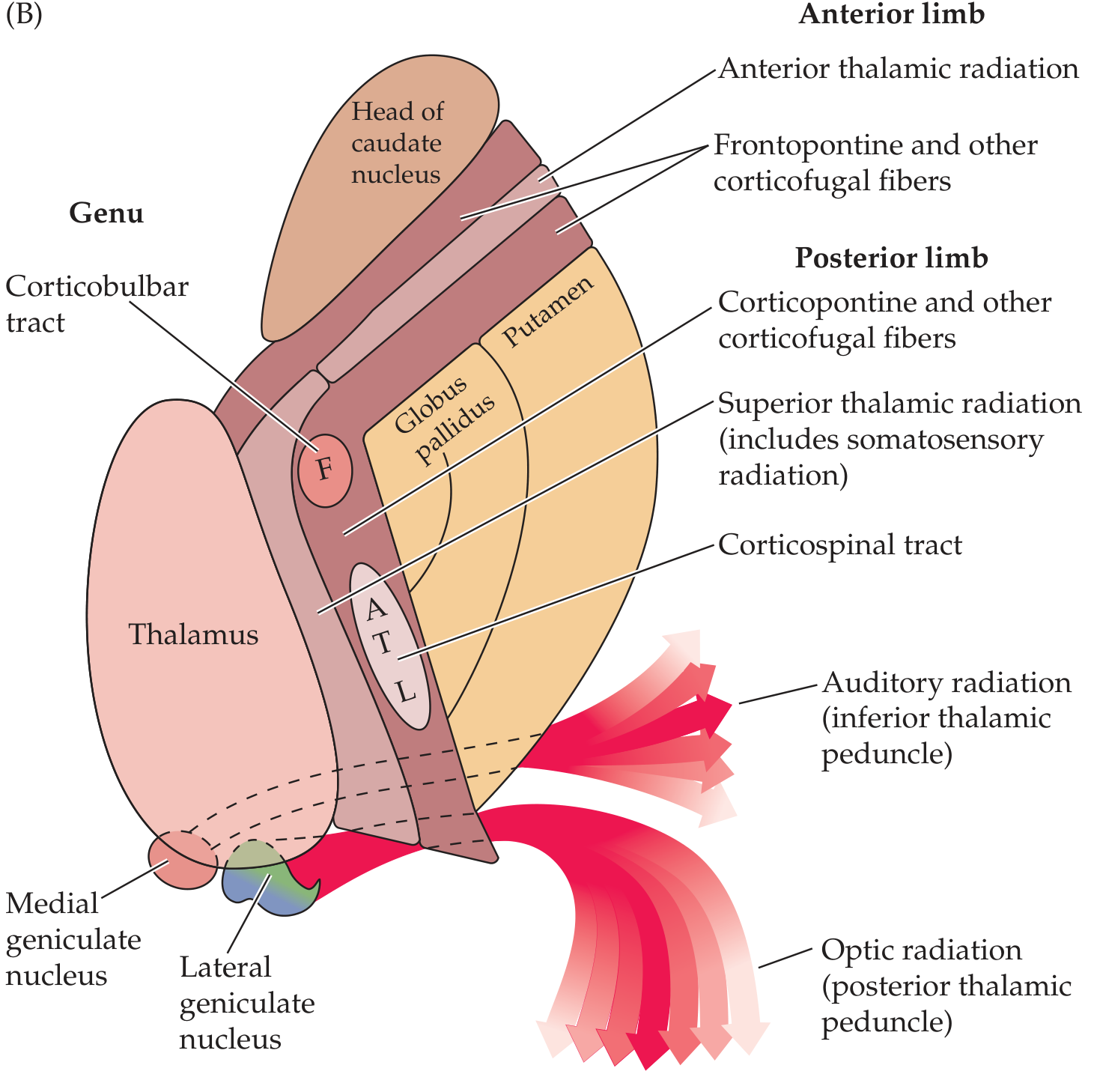
| Segment | Location | Fibers Contained |
|---|---|---|
| Anterior limb | Between caudate head (medial) and lentiform nucleus (lateral) | Frontopontine fibers; anterior thalamic radiation (thalamocortical/corticothalamic fibers connecting frontal lobe to thalamus); caudate-putaminal fibers |
| Genu | At the "knee" (level of foramen of Monro), between caudate and thalamus | Corticobulbar tract (motor fibers to cranial nerve nuclei in brainstem); motor corticopontine fibers; corticoreticular pathway |
| Posterior limb | Between lentiform nucleus (lateral) and thalamus (medial) | Corticospinal tract (somatotopically organized: face most anterior, then arm, trunk, leg most posterior); superior thalamic radiation (somatosensory); corticorubral, corticothalamic fibers |
| Retrolenticular segment | Behind the lentiform nucleus | Optic radiations (geniculo-calcarine tract / radiation of Gratiolet); corticotectal, corticonigral fibers |
| Sublenticular segment | Below the lentiform nucleus | Auditory radiation; temporopontine fibers |
Somatotopic Organization of Corticospinal Fibers in the Posterior Limb
Moving from anterior to posterior:
Face (F) → Arm (A) → Trunk (T) → Leg (L)
Because these fibers are so compact, even a small lesion in the posterior limb can cause complete contralateral hemiplegia (face + arm + leg).
Blood Supply
| Segment | Arterial Supply |
|---|---|
| Anterior limb | Recurrent artery of Heubner (branch of anterior cerebral artery) |
| Genu + middle/inferior posterior limb | Anterior choroidal artery (branch of ICA) |
| Superior anterior + posterior limb | Lenticulostriate arteries (branches of middle cerebral artery) |
These are all small perforating arteries - particularly prone to hypertensive damage and lacunar infarction.
Clinical Syndromes
Posterior Limb Lesion (most common capsular stroke)
- Contralateral pure motor hemiplegia (face, arm, leg equally affected) - classic "capsular" pattern
- Because fibers are so compact, a tiny lacunar infarct causes complete hemiplegia
Genu Lesion - "Capsular Genu Syndrome"
- Facial and lingual hemiparesis (corticobulbar involvement)
- Dysarthria
- Mild limb involvement
Lateral and Posterior Capsule (e.g., anterior choroidal artery territory)
- Contralateral hemiparesis
- Contralateral hemisensory loss (diminished pain and temperature)
- Dysarthria
- Homonymous hemianopia (usually with a tongue of sparing along the horizontal meridian, due to optic radiation involvement)
- Memory impairment
- With right-sided lesions: visuoperceptual abnormalities
Internal Capsular Hemorrhage
- Small hemorrhages in the genu or posterior limb cause pure motor hemiplegia (PMH) or a mixed sensorimotor syndrome
- Bilateral posterior limb hemorrhages can cause paraparesis (rare)
- Hypertensive hemorrhage classically ruptures the lenticulostriate arteries in this region
Decoricate Rigidity
- Seen on the hemiplegic side after hemorrhages or thromboses in the internal capsule; the small arteries here are especially prone to rupture or thrombotic occlusion in hypertension
Key Relationships Summary
Lateral ventricle
      |
Caudate nucleus  ←  ANTERIOR LIMB  →  Lentiform nucleus
      |                                      |
    GENU (corticobulbar)
      |
  Thalamus  ←  POSTERIOR LIMB  →  Lentiform nucleus
      |
Cerebral peduncle (below)
Sources:
- Neuroanatomy through Clinical Cases, 3rd Edition, pp. 257-258, 590+
- Localization in Clinical Neurology, 8e, pp. 31-32
- Bradley and Daroff's Neurology in Clinical Practice, p. 1395
cortical and sup cortical area
Searching sources...
Finding Sources
Finding Sources
Let me search more specifically for cortical areas (Brodmann), motor cortex, sensory cortex, and subcortical structures.
Finding Sources
Finding Sources
Finding Sources
Your credits are exhausted. You can purchase a top-up or subscribe to a plan to continue using Orris. If you're already subscribed, your daily credits will reset tomorrow.
Create a cortical and subcortical areas reference guide
Your credits are exhausted. You can purchase a top-up or subscribe to a plan to continue using Orris. If you're already subscribed, your daily credits will reset tomorrow.
This is a shared conversation. Sign in to Orris to start your own chat.