
Rebuilding the Base: Pelvic Floor Rehabilitation in Cervical Cancer Survivorship Presentation with evidence base to medical staffs
Rebuilding the Base: Pelvic Floor Rehabilitation in Cervical Cancer Survivorship Presentation with evidence base to medical staffs
Rebuilding the Base: Pelvic Floor Rehabilitation in Cervical Cancer Survivorship Presentation to medical staffs- please prepare professional ppt with images around 1 1/2 hrs presentation
"pelvic floor" AND "cervical cancer" AND rehabilitation
"pelvic floor muscle training" AND "cancer survivors"
"vaginal dilator" AND "pelvic radiotherapy"
"lower limb lymphedema" AND "cervical cancer"
"sexual dysfunction" AND "gynecologic cancer" AND rehabilitation
pelvic floor muscle anatomy diagram female
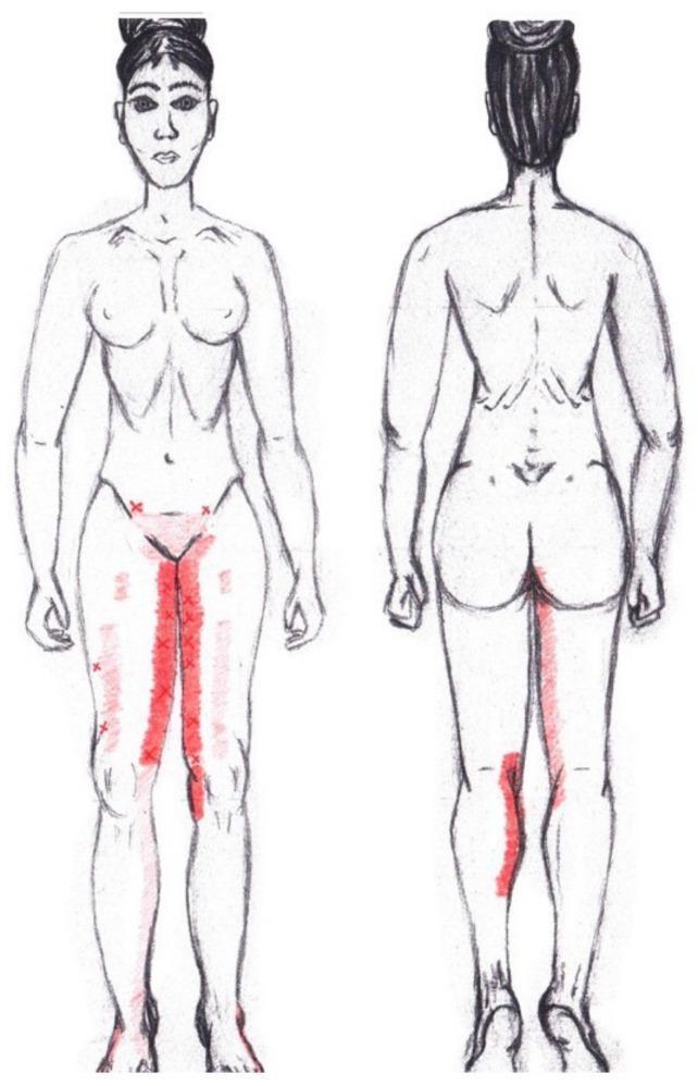
This medical illustration is a graphical simulation of myofascial referred pain patterns associated with the adductor magnus muscle, presented on a female body schematic in anterior and posterior views. The diagram uses red color mapping to indicate pain distribution and frequency. In the anterior view, primary referred pain is depicted as intense, vertical red bands along the bilateral medial thighs, extending from the groin to the superior aspect of the knees. Faint red patches and 'X' markers on the lateral and superior thighs indicate areas of less frequent or secondary referred pain. In the posterior view, the simulation identifies a significant referred pain area on the superior-medial aspect of the right calf. The color intensity serves as a semi-quantitative scale, where more vibrant red highlights common clinical trigger point referral zones. This educational visual is designed for physical therapy, anatomy education, and clinical diagnostic training related to musculoskeletal pain syndromes and pelvic floor dysfunction.
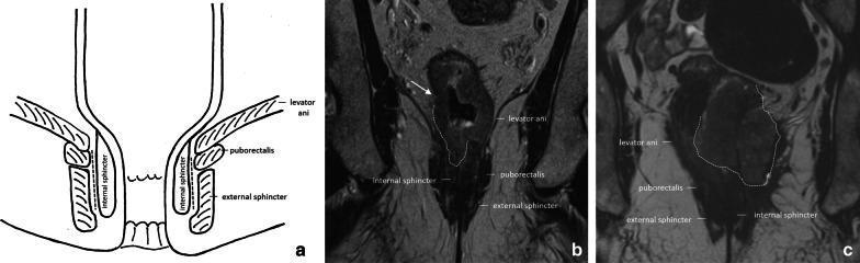
This composite educational image illustrates the pelvic floor and anal canal anatomy and its clinical relevance in rectal cancer staging. Panel (a) is an anatomical diagram depicting the coronal view of the anal canal, highlighting the levator ani, puborectalis muscle, external sphincter, and internal sphincter, with a dashed line marking the intersphincteric plane. Panel (b) is a coronal T2-weighted MRI showing a cT3a rectal tumor in a male patient. An arrow indicates irregular wall thickening with minimal (±1 mm) perirectal extension invading the right internal anal sphincter. Panel (c) is a coronal T2-weighted MRI of a female patient with a cT4b rectal tumor. The tumor (outlined by a dashed line) shows extensive infiltration, invading both internal sphincters and extending into the external sphincter, levator ani, and puborectalis muscles on the left side. Muscles and sphincters appear characteristically hypointense on T2 imaging, while the tumor presents as an intermediate-signal mass disrupting normal anatomical boundaries, essential for determining surgical management such as abdominoperineal resection.
biofeedback pelvic floor physical therapy
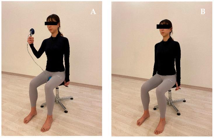
Clinical photograph consisting of two panels (A and B) demonstrating different feedback methods for pelvic floor muscle contraction (PFMC) in a rehabilitative or physical therapy setting. In both panels, a female subject is shown seated in a lumbo-pelvic upright position on a height-adjustable stool, characterized by anterior pelvic rotation, maintained lumbar lordosis, and a relaxed thorax. Panel (A) illustrates pressure biofeedback training: the subject holds a digital pressure gauge connected by tubing to a sensor placed at the perineal region, allowing for visual feedback of muscle contraction intensity. Panel (B) illustrates verbal feedback training, where the subject maintains the same standardized posture without external instrumentation, relying solely on clinician instructions. This comparison highlights the visual and equipment-based differences between instrumental biofeedback and traditional verbal cues in pelvic floor rehabilitation. The content is relevant for musculoskeletal and urogynecological physical therapy training, focusing on body mechanics and neuromuscular retraining techniques.
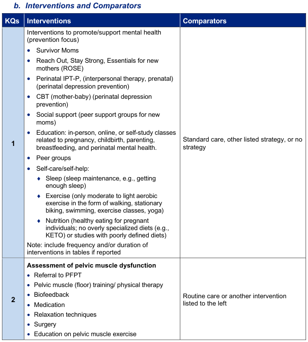
b. Interventions and Comparators
![<table><thead><tr><th>Question</th><th>Set #</th><th>Concept</th><th>Strategy</th></tr></thead><tbody><tr><td rowspan="5">KQ 1 (cont.)</td><td>10.</td><td>Limit to Meta Analyses and Systematic Reviews</td><td>#9 AND ('meta analysis'/exp OR 'systematic review'/de OR cochrane:jt OR [cochrane review]/lim OR systematic*:ti,ab OR cochrane*:ti,ab OR metaanaly*:ti,ab OR 'meta analy*':ti,ab OR (search*:ti,ab AND (cinahl*:ti,ab OR databases:ti,ab OR ebsco*:ti,ab OR embase*:ti,ab OR psychinfo*:ti,ab OR psycinfo*:ti,ab OR 'science direct*':ti,ab OR sciencedirect*:ti,ab OR scopus*:ti,ab OR systematic*:ti,ab OR 'web of knowledge*':ti,ab OR 'web of science':ti,ab)) OR ((systematic* NEAR/3 review*):ti,ab)) NOT (((protocol NEXT/3 review):ti) OR 'review protocol':ti OR 'scoping review':ti)</td></tr><tr><td>11.</td><td>Limit to Randomized Controlled Trials</td><td>#9 AND ('random sample'/de OR 'randomization'/de OR 'randomized controlled trial'/exp OR 'phase 3':ti,ab OR 'phase iii':ti,ab OR random*:ti,ab OR rct:ti,ab)</td></tr><tr><td>12.</td><td>Combine Concepts</td><td>#10 OR #11</td></tr><tr><td>13.</td><td>Apply Date Limits</td><td>#12 AND [2017-2022]/py AND [04-02-2017]/sd NOT [01-06-2022]/sd</td></tr><tr><td>14.</td><td>Limit to English</td><td>#13 AND [english]/lim</td></tr><tr><td rowspan="5">KQ 2</td><td>15.</td><td>Pregnancy</td><td>'expectant mother'/exp OR 'parameters concerning the fetus, newborn and pregnancy'/exp OR 'perinatal care'/exp OR 'perinatal period'/exp OR 'pregnancy'/exp OR 'pregnancy disorder'/exp OR 'pregnant woman'/exp OR 'prenatal care'/exp OR 'prenatal period'/exp OR antenatal:ti,kw OR antepartum:ti,kw OR gestation*:ti,kw OR matern*:ti,kw OR obstet*:ti,kw OR 'peri natal':ti,kw OR perinatal:ti,kw OR 'pre natal':ti,kw OR prenatal:ti,kw OR pregnan*:ti,kw OR trimester*:ti,kw</td></tr><tr><td>16.</td><td>Labor/Delivery/ Childbirth</td><td>'birth setting'/exp OR 'cesarean section'/exp OR 'childbirth'/exp OR 'intrapartum care'/exp OR 'labor'/exp OR 'labor complication'/exp OR 'labor induction'/exp OR 'natural childbirth'/exp OR 'obstetric delivery'/exp OR 'vaginal delivery'/exp OR 'vaginal birth after cesarean'/exp OR 'cesarean'/exp OR birth*:ti,kw OR caesarean:ti,kw OR caesarian:ti,kw OR cesarian:ti,kw OR childbirth*:ti,kw OR hypnobirth*:ti,kw OR intrapartum:ti,kw OR labor:ti,kw OR 'vaginal delivery':ti,kw OR vbac:ti,kw</td></tr><tr><td>17.</td><td>Postpartum</td><td>'postnatal care'/exp OR 'pregnancy outcome'/exp OR 'puerperium'/exp OR 'after delivery':ti,kw OR 'following delivery':ti,kw OR 'post natal':ti,kw OR postnatal:ti,kw OR 'post neonatal':ti,kw OR postneonatal:ti,kw OR 'post partum':ti,kw OR postpartum:ti,kw OR puerperal:ti,kw OR puerperium:ti,kw</td></tr><tr><td>18.</td><td>Pelvic Floor Muscle Dysfunction</td><td>'pelvic floor disorder'/exp OR 'pelvic floor muscle training'/de OR 'pelvic floor prolapse'/exp OR 'pelvis floor'/exp OR 'pelvis muscle'/exp OR ((pelvic:ti OR pelvis:ti) AND floor:ti) OR ((biofeedback:ti,ab OR electromanipulation:ti,ab OR kegel:ti,ab OR exercise*:ti,ab OR neurofeedback:ti,ab OR 'physical therapy':ti,ab OR physiotherapy:ti,ab OR rehabilitation:ti,ab OR stimulation:ti,ab OR training:ti,ab) AND 'pelvic floor':ti,ab)</td></tr><tr><td>19.</td><td>Combine Concepts</td><td>(#15 OR #16 OR #17) AND #18</td></tr></tbody></table>](https://cdn.orris.care/cdss_images/GLGCA_21618_1763314235741_3b1c3680-6b7b-42e0-aafa-145e0b1c0b69.png)
<table><thead><tr><th>Question</th><th>Set #</th><th>Concept</th><th>Strategy</th></tr></thead><tbody><tr><td rowspan="5">KQ 1 (cont.)</td><td>10.</td><td>Limit to Meta Analyses and Systematic Reviews</td><td>#9 AND ('meta analysis'/exp OR 'systematic review'/de OR cochrane:jt OR [cochrane review]/lim OR systematic*:ti,ab OR cochrane*:ti,ab OR metaanaly*:ti,ab OR 'meta analy*':ti,ab OR (search*:ti,ab AND (cinahl*:ti,ab OR databases:ti,ab OR ebsco*:ti,ab OR embase*:ti,ab OR psychinfo*:ti,ab OR psycinfo*:ti,ab OR 'science direct*':ti,ab OR sciencedirect*:ti,ab OR scopus*:ti,ab OR systematic*:ti,ab OR 'web of knowledge*':ti,ab OR 'web of science':ti,ab)) OR ((systematic* NEAR/3 review*):ti,ab)) NOT (((protocol NEXT/3 review):ti) OR 'review protocol':ti OR 'scoping review':ti)</td></tr><tr><td>11.</td><td>Limit to Randomized Controlled Trials</td><td>#9 AND ('random sample'/de OR 'randomization'/de OR 'randomized controlled trial'/exp OR 'phase 3':ti,ab OR 'phase iii':ti,ab OR random*:ti,ab OR rct:ti,ab)</td></tr><tr><td>12.</td><td>Combine Concepts</td><td>#10 OR #11</td></tr><tr><td>13.</td><td>Apply Date Limits</td><td>#12 AND [2017-2022]/py AND [04-02-2017]/sd NOT [01-06-2022]/sd</td></tr><tr><td>14.</td><td>Limit to English</td><td>#13 AND [english]/lim</td></tr><tr><td rowspan="5">KQ 2</td><td>15.</td><td>Pregnancy</td><td>'expectant mother'/exp OR 'parameters concerning the fetus, newborn and pregnancy'/exp OR 'perinatal care'/exp OR 'perinatal period'/exp OR 'pregnancy'/exp OR 'pregnancy disorder'/exp OR 'pregnant woman'/exp OR 'prenatal care'/exp OR 'prenatal period'/exp OR antenatal:ti,kw OR antepartum:ti,kw OR gestation*:ti,kw OR matern*:ti,kw OR obstet*:ti,kw OR 'peri natal':ti,kw OR perinatal:ti,kw OR 'pre natal':ti,kw OR prenatal:ti,kw OR pregnan*:ti,kw OR trimester*:ti,kw</td></tr><tr><td>16.</td><td>Labor/Delivery/ Childbirth</td><td>'birth setting'/exp OR 'cesarean section'/exp OR 'childbirth'/exp OR 'intrapartum care'/exp OR 'labor'/exp OR 'labor complication'/exp OR 'labor induction'/exp OR 'natural childbirth'/exp OR 'obstetric delivery'/exp OR 'vaginal delivery'/exp OR 'vaginal birth after cesarean'/exp OR 'cesarean'/exp OR birth*:ti,kw OR caesarean:ti,kw OR caesarian:ti,kw OR cesarian:ti,kw OR childbirth*:ti,kw OR hypnobirth*:ti,kw OR intrapartum:ti,kw OR labor:ti,kw OR 'vaginal delivery':ti,kw OR vbac:ti,kw</td></tr><tr><td>17.</td><td>Postpartum</td><td>'postnatal care'/exp OR 'pregnancy outcome'/exp OR 'puerperium'/exp OR 'after delivery':ti,kw OR 'following delivery':ti,kw OR 'post natal':ti,kw OR postnatal:ti,kw OR 'post neonatal':ti,kw OR postneonatal:ti,kw OR 'post partum':ti,kw OR postpartum:ti,kw OR puerperal:ti,kw OR puerperium:ti,kw</td></tr><tr><td>18.</td><td>Pelvic Floor Muscle Dysfunction</td><td>'pelvic floor disorder'/exp OR 'pelvic floor muscle training'/de OR 'pelvic floor prolapse'/exp OR 'pelvis floor'/exp OR 'pelvis muscle'/exp OR ((pelvic:ti OR pelvis:ti) AND floor:ti) OR ((biofeedback:ti,ab OR electromanipulation:ti,ab OR kegel:ti,ab OR exercise*:ti,ab OR neurofeedback:ti,ab OR 'physical therapy':ti,ab OR physiotherapy:ti,ab OR rehabilitation:ti,ab OR stimulation:ti,ab OR training:ti,ab) AND 'pelvic floor':ti,ab)</td></tr><tr><td>19.</td><td>Combine Concepts</td><td>(#15 OR #16 OR #17) AND #18</td></tr></tbody></table>
<table><tr><td>5.6.3.1.3</td><td>Constipation</td><td>62</td></tr><tr><td>5.6.3.1.4</td><td>Containment</td><td>62</td></tr><tr><td>5.6.3.2</td><td>Behavioural and Physical therapies</td><td>62</td></tr><tr><td>5.6.3.2.1</td><td>Prompted or timed voiding</td><td>63</td></tr><tr><td>5.6.3.2.2</td><td>Bladder training</td><td>63</td></tr><tr><td>5.6.3.2.3</td><td>Pelvic floor muscle training</td><td>63</td></tr><tr><td>5.6.3.2.4</td><td>Electrical stimulation</td><td>63</td></tr><tr><td>5.6.3.2.5</td><td>Posterior tibial nerve stimulation</td><td>64</td></tr><tr><td>5.6.4</td><td>Pharmacological management</td><td>64</td></tr><tr><td>5.6.4.1</td><td>Drugs for urgency urinary incontinence</td><td>64</td></tr><tr><td>5.6.4.2</td><td>Drugs for stress urinary incontinence</td><td>65</td></tr><tr><td>5.6.5</td><td>Surgical treatment for stress urinary incontinence</td><td>65</td></tr><tr><td>5.6.5.1</td><td>Bulking agents in men</td><td>65</td></tr><tr><td>5.6.5.2</td><td>Male Slings</td><td>66</td></tr><tr><td>5.6.5.2.1</td><td>Non-adjustable slings</td><td>66</td></tr><tr><td>5.6.5.2.2</td><td>Adjustable slings in males</td><td>66</td></tr><tr><td>5.6.5.2.3</td><td>Autologous slings</td><td>67</td></tr><tr><td>5.6.5.3</td><td>Compression devices in males</td><td>68</td></tr><tr><td>5.6.5.3.1</td><td>Artificial urinary sphincter</td><td>68</td></tr><tr><td>5.6.5.3.2</td><td>Non-circumferential compression device (ProACT®)</td><td>69</td></tr><tr><td>5.6.6</td><td>Surgical treatment for urgency urinary incontinence</td><td>70</td></tr><tr><td>5.6.6.1</td><td>Bladder wall injection of botulinum Toxin-A</td><td>70</td></tr><tr><td>5.6.6.2</td><td>Sacral nerve stimulation (neuromodulation)</td><td>71</td></tr><tr><td>5.6.6.3</td><td>Cystoplasty/urinary diversion</td><td>71</td></tr><tr><td>5.7</td><td>Management of underactive bladder</td><td>73</td></tr><tr><td>5.7.1</td><td>Epidemiology and Pathophysiology</td><td>73</td></tr><tr><td>5.7.2</td><td>Diagnostic Evaluation</td><td>74</td></tr><tr><td>5.7.2.1</td><td>Medical history and physical examination</td><td>74</td></tr><tr><td>5.7.2.2</td><td>Questionnaires</td><td>74</td></tr><tr><td>5.7.2.3</td><td>Uroflowmetry</td><td>74</td></tr><tr><td>5.7.2.4</td><td>Ultrasound scan and post-void residual measurement</td><td>74</td></tr><tr><td>5.7.2.5</td><td>Urodynamics</td><td>74</td></tr><tr><td>5.7.3</td><td>Conservative management</td><td>75</td></tr><tr><td>5.7.3.1</td><td>Behavioural interventions</td><td>75</td></tr><tr><td>5.7.3.2</td><td>Pelvic floor muscle relaxation training with biofeedback</td><td>75</td></tr><tr><td>5.7.3.3</td><td>Clean intermittent self-catheterisation (see section 5.6.3.1.4)</td><td>75</td></tr><tr><td>5.7.3.4</td><td>Indwelling catheters</td><td>75</td></tr><tr><td>5.7.3.5</td><td>Intravesical electrical stimulation</td><td>75</td></tr><tr><td>5.7.3.6</td><td>Extracorporeal Shock Wave Therapy</td><td>76</td></tr><tr><td>5.7.4</td><td>Pharmacological management</td><td>76</td></tr><tr><td>5.7.4.1</td><td>Parasympathomimetics</td><td>76</td></tr><tr><td>5.7.4.2</td><td>Alpha-adrenergic blockers</td><td>76</td></tr><tr><td>5.7.4.3</td><td>Prostaglandins</td><td>76</td></tr><tr><td>5.7.4.4</td><td>Other drugs</td><td>77</td></tr><tr><td>5.7.5</td><td>Surgical treatment for underactive bladder</td><td>77</td></tr><tr><td>5.7.5.1</td><td>Surgery for benign prostatic obstruction</td><td>77</td></tr><tr><td>5.7.5.2</td><td>Sacral neuromodulation</td><td>78</td></tr><tr><td>5.7.6</td><td>Follow-up</td><td>79</td></tr></table>
vaginal dilator set for radiotherapy patients
lower limb lymphedema leg swelling
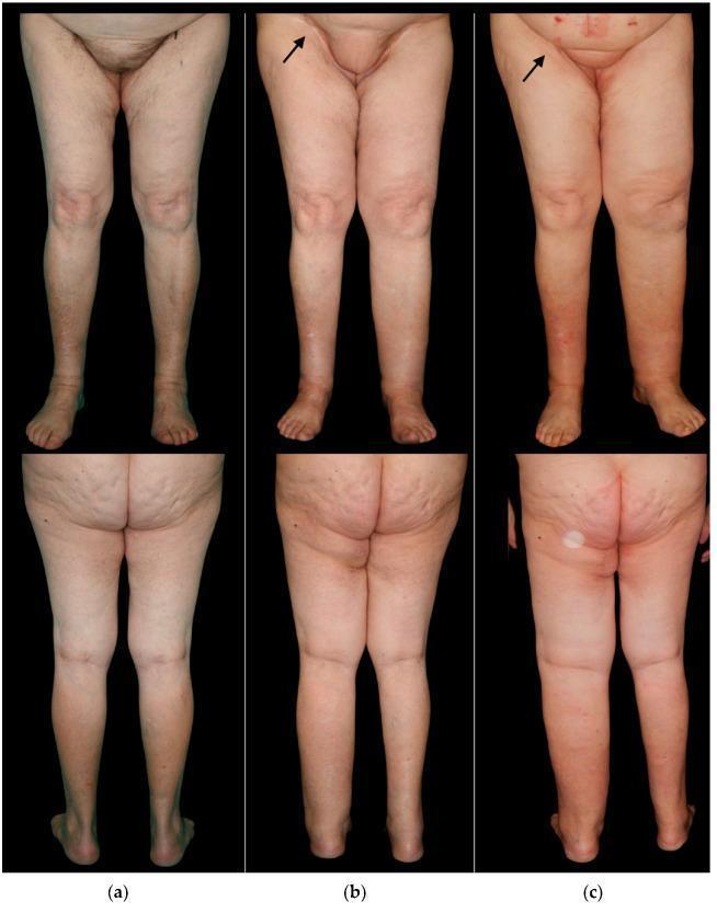
Clinical photographs in frontal and posterior views documenting the long-term progression of lower limb lymphedema in a patient following bilateral groin dissection for vulvar cancer. Panel (a) shows the preoperative baseline with symmetrical limb contours. Panel (b) illustrates the status one year post-surgery, featuring a superficial circumflex iliac artery perforator (SCIP) flap skin island in the right groin (marked with an arrow); the right (treated) leg shows mild swelling, while the left (untreated) leg exhibits moderate lymphedema. Panel (c) shows the 5-year follow-up, demonstrating progressive, severe lymphedema of the left lower limb characterized by significant volumetric increase, loss of anatomical definition at the ankle and knee, and a reddish skin hue indicative of chronic stasis. The right limb remains stable with mild swelling. This timeline demonstrates the efficacy of lymphatic reconstruction (SCIP flap) in mitigating secondary lymphedema compared to the contralateral untreated side. Target educational concepts include postoperative complications of lymphadenectomy, lymphatic flap reconstruction, and clinical staging of chronic lymphedema.
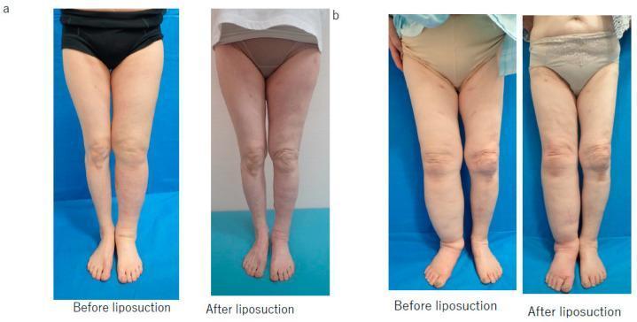
This clinical comparison chart presents two sets of 'before' and 'after' clinical photographs of lower extremity secondary lymphedema treated with suction-assisted lipectomy (liposuction). Panel (a) illustrates a successful clinical outcome in a 71-year-old woman with left lower limb lymphedema. The 'before' image shows significant edema-related enlargement of the left calf and ankle with loss of anatomical definition. The 'after' image, taken following 2000 mL fat removal and a dual compression regimen (stocking and bandage), shows a marked reduction in limb circumference and restoration of leg contour. Panel (b) depicts an unsuccessful result in a 78-year-old woman with right lower limb lymphedema. The 'before' image demonstrates substantial swelling throughout the right leg, particularly distal to the knee. The 'after' image, following 1800 mL fat removal and bandage-only compression, shows persistent limb hypertrophy and lack of significant volume reduction. The series serves as a clinical case study comparing the efficacy of different post-operative compression strategies in managing chronic lymphedema after lymphaticovenous anastomosis failure.
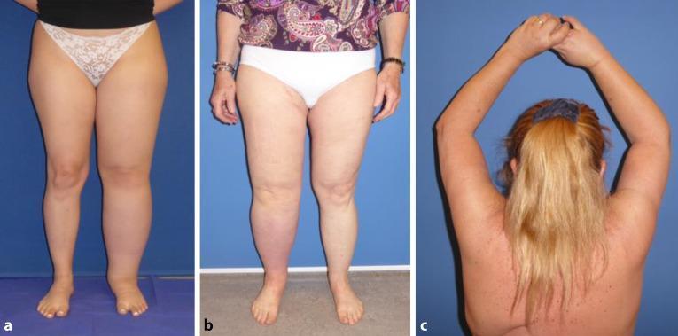
This composite clinical photograph displays three examples of lymphedema, a condition characterized by impaired lymphatic drainage and resultant tissue swelling. Panel A illustrates idiopathic primary lymphedema in the left lower extremity of a 19-year-old female; the affected leg shows significant circumferential enlargement and loss of normal anatomical contours, particularly around the ankle and calf. Panel B shows secondary lymphedema in the right lower extremity of a 62-year-old female following uterine cancer treatment and lymph node dissection. This limb exhibits marked edema and distinctive reddish-purple skin discoloration (erythema) on the lower leg, suggesting chronic inflammatory changes or circulatory stasis. Panel C shows secondary lymphedema in the right upper extremity of a 58-year-old female following mastectomy and axillary lymph node dissection. The right arm demonstrates increased girth compared to the left, visible from the posterior view with arms elevated. These images serve as educational examples of both primary (idiopathic) and secondary (iatrogenic/cancer-related) lymphedema, highlighting characteristic asymmetric swelling and associated skin changes.
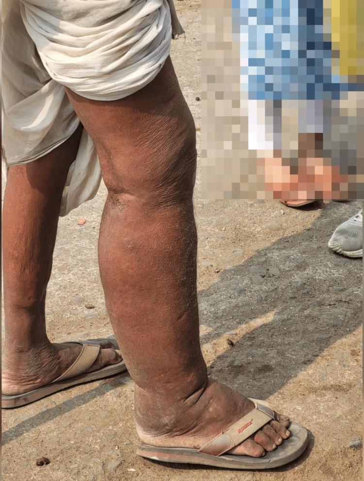
Clinical photograph of a 58-year-old male presenting with asymmetric lower extremity swelling, characteristic of chronic lymphatic filariasis (elephantiasis). The image shows a comparative view of the legs where the right lower limb exhibits advanced stage lymphedema. Key findings include massive non-pitting edema extending from the thigh to the foot, resulting in a distorted, cylindrical limb shape. The skin on the affected right leg is significantly thickened, displaying hyperkeratosis, prominent skin folds, and a rough, brawny texture (pachydermia) compared to the relatively normal appearance of the left leg. The foot shows significant swelling with obscured anatomical contours and secondary skin changes. This visual demonstrates the chronic obstructive stage of a filarial infection, where lymphostasis leads to tissue fibrosis and permanent dermal thickening. The clinical context suggests a history of recurrent adenolymphangitis in an endemic region, illustrating the progression from early pitting edema to late-stage irreversible lymphedema.
cervical cancer staging diagram FIGO
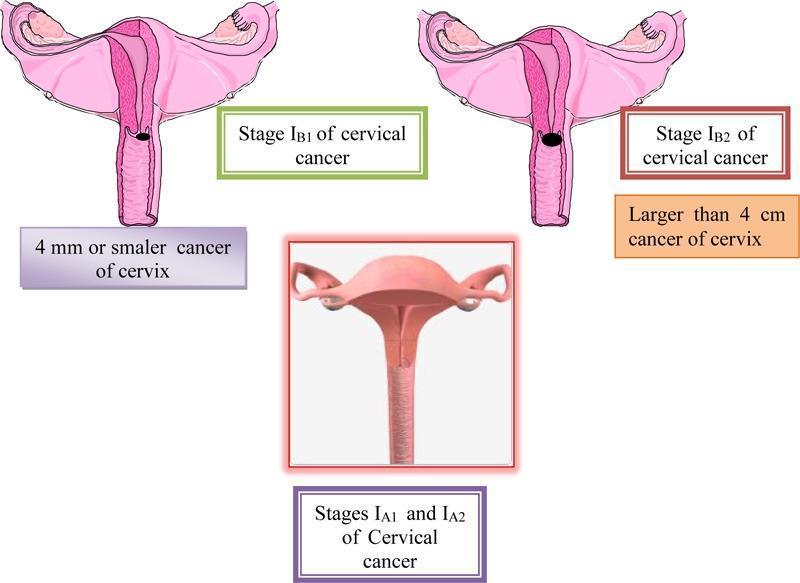
A multi-panel medical illustration and anatomical diagram depicting the FIGO staging of cervical cancer (Stages IA and IB). The central bottom panel displays a 3D-style anatomical diagram representing Stages IA1 and IA2, where the cervix appears macroscopically normal, indicating microinvasive disease that is not visible to the naked eye. The top left panel illustrates Stage IB1, showing a cross-section of the uterus and cervix with a small, localized dark lesion on the cervical canal; it is annotated as a cancer of 4 mm or smaller. The top right panel illustrates Stage IB2, showing a significantly larger, darker, and more prominent exophytic/ulcerative lesion on the cervix, annotated as being larger than 4 cm. All panels show relevant reproductive anatomy including the uterus, fallopian tubes, and ovaries. This educational graphic serves to distinguish clinical stages of cervical carcinoma based on tumor size and macroscopic visibility, relevant for gynecologic oncology and diagnostic pathology training.
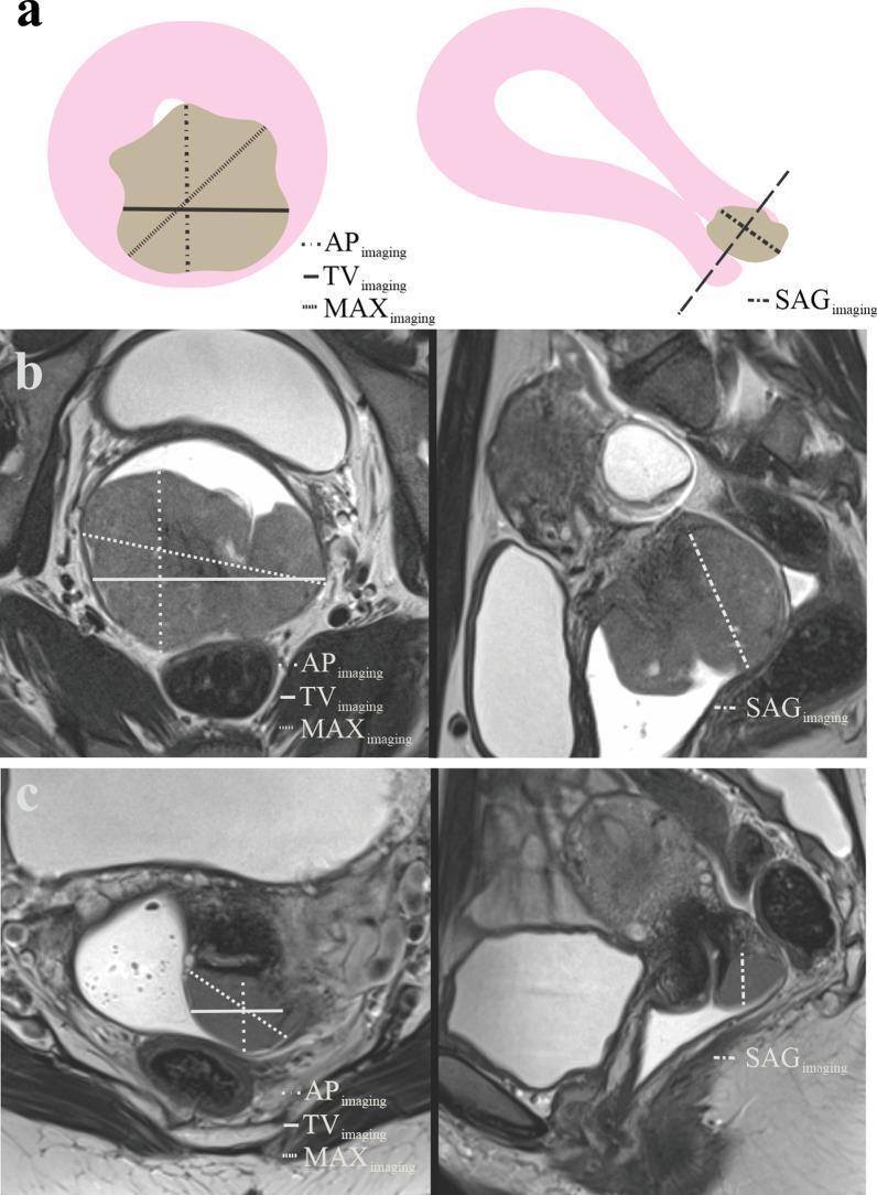
This educational image details the radiological assessment and measurement protocols for cervical cancer using MRI.
Kegel exercise pelvic floor contraction diagram
PMID: 36001098
PMID: 32367126
PMID: 39177709
PMID: 25198150
PMID: 39765163
PMID: 32601784
PMID: 40463868
complete decongestive therapy lymphedema bandaging compression
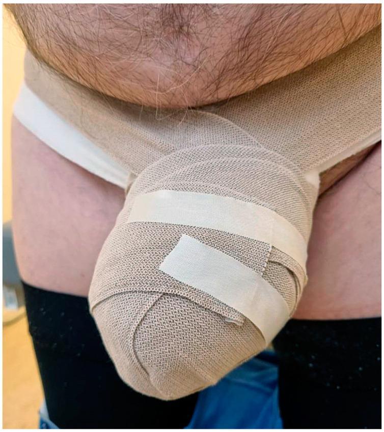
A clinical photograph of the male pelvic region demonstrating the application of low-stretch compression bandages for the management of secondary genital lymphedema. The tan, woven, short-stretch bandages are meticulously wrapped around an enlarged penis and scrotum, creating a bulbous, reinforced mass secured with white adhesive medical tape. The bandaging extends around the waist to provide a stable anchor. The patient is wearing white briefs as a base layer, and dark-colored garments (likely compression stockings or trousers) are visible on the upper thighs. This image illustrates a key component of Complete Decongestive Therapy (CDT) used to reduce edema volume, manage skin tension, and prevent complications like cellulitis in patients with lymphatic obstruction following oncological treatment, such as rectal cancer surgery. The focus is on the specialized bandaging technique required for complex anatomical regions where standard compression garments may be inadequate.
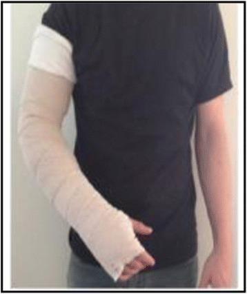
This clinical photograph demonstrates the application of multi-layer compression bandaging (CB) on the right upper extremity of an individual. The bandage system is applied over a protective white tubular sleeve or stockinette used to protect the skin and distal joints. The compression wrap is composed of non-elastic, short-stretch bandage material applied in a spiral or herringbone pattern to provide high working pressure and low resting pressure. The coverage extends from the metacarpophalangeal joints of the hand, through the wrist and forearm, and up to the mid-humerus, just below the deltoid insertion. The fingers and thumb are left exposed to monitor neurovascular status and allow for functional movement. This type of compression intervention is a core component of Complete Decongestive Therapy (CDT) for the management of breast cancer-related lymphedema or chronic edema. The image illustrates the specific technique for night-time self-management or clinician-assisted bandaging to maintain limb volume reduction.
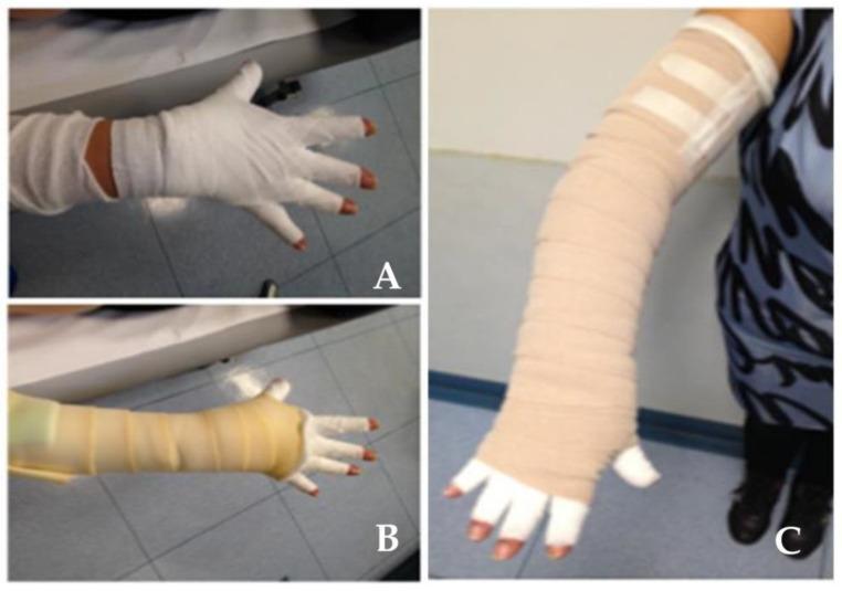
This clinical photograph illustrates three stages (A, B, and C) of applying multi-layer compression bandaging for the management of lymphedema in the upper limb. Panel A shows the initial layer on the hand and wrist, utilizing a white, conforming cotton-based material that covers the palm, dorsum, and individual fingers while leaving the distal fingertips exposed for neurovascular monitoring. Panel B demonstrates the progression, where a secondary, tan-colored short-stretch compression bandage is applied over the wrist and proximal forearm, overlapping the initial hand wrap. Panel C depicts the completed compression garment extending from the hand to the proximal upper arm, just below the shoulder. The final layer consists of tan, textured short-stretch bandages applied in a spiral or herringbone pattern to provide high working pressure and low resting pressure, facilitating lymphatic drainage. The proximal end is secured with white adhesive medical tape. This visual illustrates the distal-to-proximal gradient compression technique essential in Complete Decongestive Therapy (CDT).
female pelvic organs sagittal anatomy uterus bladder rectum
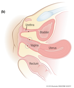
Summary : This is a labeled anatomical illustration showing a sagittal (side) cross-section of the female pelvis, highlighting the spatial relationships between the urethra, bladder, vagina, uterus, and rectum.
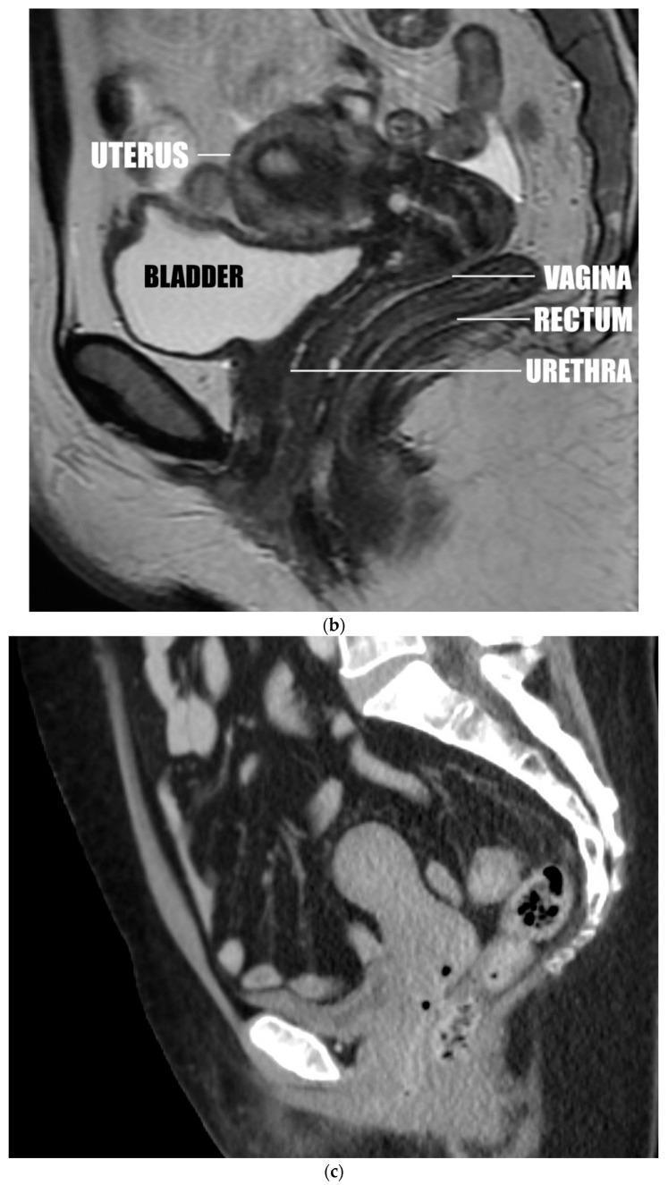
This composite educational graphic features diagnostic images in the sagittal plane demonstrating various uterine positions and pelvic floor anatomy. Image (b) is a T2-weighted sagittal MRI of the female pelvis showing an anteverted uterus. Key anatomical structures are labeled, illustrating the uterus positioned superior to the bladder, with the vagina situated inferior to the uterus and posterior to the bladder. The rectum is shown posterior to the vagina, while the urethra is visible anterior and slightly inferior to the vaginal canal. Image (c) is a sagittal CT reconstruction depicting an anteverted uterus that is separated from the bladder. The series serves as an educational tool for identifying uterine version and insertion types (anterior, apical, or posterior) and understanding their spatial relationships with adjacent pelvic organs. The imaging highlights the clinical significance of uterine orientation in relation to pelvic floor support and conditions such as uterine prolapse.
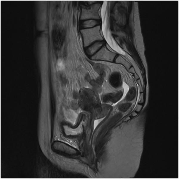
This diagnostic image is a T2-weighted sagittal MRI of the female pelvis, demonstrating a case of post-radiation cervical necrosis. The image provides a lateral view of the pelvic organs, including the uterus, cervix, bladder, and rectum. The primary finding is located at the cervix, which exhibits a highly heterogeneous signal intensity with irregular, poorly defined margins. Areas of hyperintensity are visible, suggesting edema or necroinflammatory exudate, while darker areas may represent tissue breakdown or fibrosis. The anatomical relationship between the bladder (anteriorly) and the rectum (posteriorly) is maintained, though the cervical borders appear distorted compared to normal pelvic anatomy. This visual representation is clinically significant for distinguishing late radiation-induced complications from tumor recurrence in oncology patients. The imaging serves as an educational tool for identifying cervical tissue changes, such as inflammation and structural distortion, following pelvic radiotherapy.
radical hysterectomy pelvic autonomic nerve anatomy
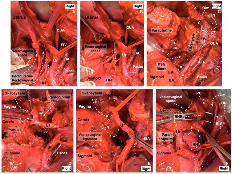
A composite of six clinical photographs (A–F) demonstrating surgical dissection stages of the three parametria during a nerve-sparing radical hysterectomy. The images illustrate the complex pelvic neurovascular and ligamentous anatomy in the right pelvic sidewall. Key structures identified across the panels include the uterus, cervix, vagina, and sigmoid colon. Vascular landmarks include the external iliac artery (EIA) and vein (EIV), internal iliac artery (IIA) and vein (IIV), and the obliterated umbilical artery (OUA). The dissection highlights the ureter (Ur) and its relationship to autonomic nerves, specifically the hypogastric nerve (HN) within the ureterohypogastric fascia, pelvic splanchnic nerve (PSN) fibers, and bladder nerve branches (BNBs). Surgical spaces such as the rectovaginal, Yabuki, and Okabayashi paravaginal spaces are developed to isolate the vesicovaginal ligament and paracervix while preserving the inferior hypogastric plexus. The series demonstrates the precise identification and mobilization of the ureter and paracolpium required to protect pelvic autonomic innervation during oncological resection.
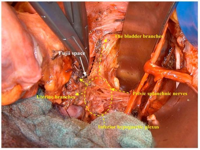
This clinical photograph captures an intraoperative dissection during a nerve-sparing radical hysterectomy, focusing on the pelvic autonomic nervous system. The image demonstrates the Fujii space, a surgical landmark located mediodorsal to the bladder branches and lateral to the vaginal wall. Surgical instruments are used to retract tissue and expose the inferior hypogastric plexus, which appears as a complex network of nerve fibers and connective tissue in the lower central field. Key anatomical structures identified via annotations include the pelvic splanchnic nerves entering the plexus and the emerging uterine and bladder branches of the inferior hypogastric plexus. The visualization highlights the spatial relationship between these neural pathways and the pelvic viscera, emphasizing the surgical technique required to isolate and preserve autonomic function. This clinical imaging is essential for understanding the surgical anatomy of the lateral parametrium and the preservation of pelvic nerves during oncological procedures in gynecologic surgery.
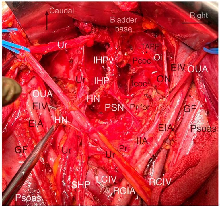
This clinical photograph shows a deep surgical dissection of the pelvic autonomic nervous system and surrounding anatomy during a nerve-sparing radical hysterectomy. The image illustrates the complex relationship between vascular, nervous, and muscular structures within the pelvic sidewall and floor. Key vascular landmarks include the External Iliac Artery (EIA) and Vein (EIV) running along the Psoas muscle, the Internal Iliac Artery (IIA), and the Common Iliac vessels near the Promontorium (Pr). The Pelvic Autonomic Nerve System (PANS) is highlighted centrally, featuring the Superior Hypogastric Plexus (SHP), Hypogastric Nerves (HN), Pelvic Splanchnic Nerves (PSN), and the Inferior Hypogastric Plexus (IHP) with its vesical branches (IHPv) extending toward the bladder base. The Ureter (Ur) is visible traversing the field, providing a critical landmark for surgical planes. Deep muscular structures of the pelvic floor, including the Piriformis (Prifor), Obturator Internus (Oi), and the levator ani components (Pcoc, Icoc), are clearly exposed. This visual serves as an educational resource for identifying anatomical landmarks essential for performing systematic nerve-sparing pelvic surgery.
pelvic floor rehabilitation cervical cancer survivorship clinical guideline 2024 2025
mkdir -p /tmp/workspace/pelvic-floor-rehab-ppt/assets /tmp/workspace/pelvic-floor-rehab-ppt/output && ls -la /tmp/workspace/
cd /tmp/workspace/pelvic-floor-rehab-ppt/assets && python /tmp/skills/shared/scripts/fetch_images.py \ "https://cdn.orris.care/cdss_images/GLGCA_21892_1763336687101_32f67122-95a5-4f6d-8b4b-86bdc65d5dce.png" \ "https://cdn.orris.care/cdss_images/pmc_clinical_VQA_4f3eba852a08414d1c411b65d902ccce640867cadf7d0008bce744477e4368e2.jpg" \ "https://cdn.orris.care/cdss_images/pmc_clinical_VQA_3f4de1b52001f3bb888c2efea727ed93cd5e7d9f9e43ed11f1e68b026517a577.jpg" \ "https://cdn.orris.care/cdss_images/pmc_clinical_VQA_52a333ac4bc359c9c0af18768c77fb2345c4d27439d5a3e8d57151e7f7fce519.jpg" \ "https://cdn.orris.care/cdss_images/pmc_clinical_VQA_5e51b4460ab47709ad13100f283405268a329497b2833ea3f5eb296ace8878cb.jpg" \ "https://cdn.orris.care/cdss_images/pmc_clinical_VQA_329edef3018f086488d2c7c2cb118f0f83a48b6fef502cc80c234a58f4cec45f.jpg" \ "https://cdn.orris.care/cdss_images/pmc_clinical_VQA_0442782d090ab4de84d072b3529a57afd385caa77c8b39560da4a535e4d001e2.jpg" \ "https://cdn.orris.care/cdss_images/pmc_clinical_VQA_0b1ced6dee7cdb4835ca54468d4d6bdc47043159b894032be1546ec05db7394e.jpg" \ "https://cdn.orris.care/cdss_images/pmc_clinical_VQA_0f6bdae723d69486aec56dcf57534eadd94b434749c8714e7cb38ba36ac392d1.jpg" \ > fetch_result.json python3 -c " import json data = json.load(open('fetch_result.json')) import base64 names = ['pelvis_sagittal','pelvic_floor_anatomy','nerve_sparing_anatomy','post_rt_mri','biofeedback','lymphedema_progression','lymphedema_types','cdt_bandaging','figo_staging'] for i,item in enumerate(data): if item.get('base64'): b64 = item['base64'].split(',',1)[1] if ',' in item['base64'] else item['base64'] ext = 'png' if 'png' in item['base64'][:30] else 'jpg' fname = f'{names[i]}.{ext}' with open(fname,'wb') as f: f.write(base64.b64decode(b64)) print('saved', fname) else: print('FAILED', names[i], item.get('error')) " ls -la /tmp/workspace/pelvic-floor-rehab-ppt/assets
~/pelvic-floor-rehab-ppt/build_deck.py
#!/usr/bin/env python3
"""
Build: Rebuilding the Base - Pelvic Floor Rehabilitation in Cervical Cancer
Survivorship. ~90-minute CME-style deck for medical staff.
"""
from pptx import Presentation
from pptx.util import Inches, Pt, Emu
from pptx.dml.color import RGBColor
from pptx.enum.text import PP_ALIGN, MSO_ANCHOR
from pptx.enum.shapes import MSO_SHAPE
from pptx.oxml.ns import qn
import os

ASSETS = "/tmp/workspace/pelvic-floor-rehab-ppt/assets"
OUT = "/tmp/workspace/pelvic-floor-rehab-ppt/output/Rebuilding_the_Base_Pelvic_Floor_Rehab_Cervical_Cancer.pptx"

# ---------- Palette ----------
NAVY = RGBColor(0x0B, 0x2E, 0x4E)
TEAL = RGBColor(0x0E, 0x7C, 0x86)
GOLD = RGBColor(0xC9, 0x9A, 0x3D)
LIGHT = RGBColor(0xF4, 0xF7, 0xF8)
WHITE = RGBColor(0xFF, 0xFF, 0xFF)
DARK_TXT = RGBColor(0x22, 0x2A, 0x30)
GREY = RGBColor(0x6B, 0x74, 0x7C)
ROSE = RGBColor(0xA1, 0x3C, 0x5A)

SW, SH = Inches(13.333), Inches(7.5)

prs = Presentation()
prs.slide_width = SW
prs.slide_height = SH
BLANK = prs.slide_layouts[6]

def add_slide():
    return prs.slides.add_slide(BLANK)

def bg(slide, color=WHITE):
    slide.background.fill.solid()
    slide.background.fill.fore_color.rgb = color

def rect(slide, x, y, w, h, color, line=False):
    shp = slide.shapes.add_shape(MSO_SHAPE.RECTANGLE, x, y, w, h)
    shp.fill.solid()
    shp.fill.fore_color.rgb = color
    if line:
        shp.line.color.rgb = color
    else:
        shp.line.fill.background()
    shp.shadow.inherit = False
    return shp

def textbox(slide, x, y, w, h, text, size=18, color=DARK_TXT, bold=False,
            align=PP_ALIGN.LEFT, font="Calibri", italic=False, anchor=None,
            line_spacing=1.0):
    tb = slide.shapes.add_textbox(x, y, w, h)
    tf = tb.text_frame
    tf.word_wrap = True
    if anchor:
        tf.vertical_anchor = anchor
    lines = text.split("\n")
    for i, ln in enumerate(lines):
        p = tf.paragraphs[0] if i == 0 else tf.add_paragraph()
        p.text = ln
        p.alignment = align
        p.line_spacing = line_spacing
        for r in p.runs:
            r.font.size = Pt(size)
            r.font.color.rgb = color
            r.font.bold = bold
            r.font.italic = italic
            r.font.name = font
    return tb

def footer(slide, page_no, section=""):
    rect(slide, 0, SH - Inches(0.32), SW, Inches(0.32), NAVY)
    textbox(slide, Inches(0.3), SH - Inches(0.34), Inches(8), Inches(0.3),
            "Rebuilding the Base | Pelvic Floor Rehabilitation in Cervical Cancer Survivorship",
            size=9, color=WHITE)
    textbox(slide, SW - Inches(1.2), SH - Inches(0.34), Inches(0.9), Inches(0.3),
            str(page_no), size=9, color=WHITE, align=PP_ALIGN.RIGHT)

def kicker_title(slide, kicker, title, title_size=30, kicker_color=GOLD):
    rect(slide, 0, 0, SW, Inches(1.35), NAVY)
    rect(slide, 0, Inches(1.35), SW, Inches(0.06), GOLD)
    textbox(slide, Inches(0.55), Inches(0.15), Inches(11), Inches(0.35), kicker.upper(),
            size=13, color=kicker_color, bold=True)
    textbox(slide, Inches(0.55), Inches(0.48), Inches(12.2), Inches(0.85), title,
            size=title_size, color=WHITE, bold=True)

def bullets(slide, x, y, w, h, items, size=16, color=DARK_TXT, gap=8, bullet_color=TEAL):
    tb = slide.shapes.add_textbox(x, y, w, h)
    tf = tb.text_frame
    tf.word_wrap = True
    first = True
    for item in items:
        if isinstance(item, tuple):
            text, level = item
        else:
            text, level = item, 0
        p = tf.paragraphs[0] if first else tf.add_paragraph()
        first = False
        p.space_after = Pt(gap)
        p.level = min(level, 4)
        marker = {0: "\u25AA  ", 1: "\u2013  ", 2: "\u2022  "}.get(level, "\u2022  ")
        p.text = marker + text
        for r in p.runs:
            r.font.size = Pt(size - level * 1.5)
            r.font.color.rgb = color if level == 0 else GREY
            r.font.name = "Calibri"
            r.font.bold = (level == 0)
    return tb

def pic_framed(slide, path, x, y, w, h, caption=None, cap_size=11):
    pic = slide.shapes.add_picture(path, x, y, height=h)
    # constrain width if too wide
    if pic.width > w:
        ratio = w / pic.width
        pic.width = w
        pic.height = Emu(int(pic.height * ratio))
        pic.left = x
        pic.top = y
    # center horizontally in box
    pic.left = int(x + (w - pic.width) / 2)
    box_bottom = y + h
    line = slide.shapes.add_connector(1, pic.left, box_bottom + Inches(0.03),
                                       pic.left + pic.width, box_bottom + Inches(0.03))
    line.line.color.rgb = GOLD
    line.line.width = Pt(1.5)
    if caption:
        textbox(slide, x, box_bottom + Inches(0.08), w, Inches(0.6), caption,
                size=cap_size, color=GREY, italic=True, align=PP_ALIGN.CENTER)
    return pic

def section_divider(slide_no, num, title, subtitle):
    s = add_slide(); bg(s, NAVY)
    rect(s, 0, Inches(3.3), SW, Inches(0.08), GOLD)
    textbox(s, Inches(0.9), Inches(2.0), Inches(3), Inches(1.2), f"{num:02d}",
            size=72, color=GOLD, bold=True)
    textbox(s, Inches(2.6), Inches(2.15), Inches(10), Inches(1.1), title,
            size=34, color=WHITE, bold=True)
    textbox(s, Inches(2.6), Inches(3.55), Inches(9.8), Inches(1.0), subtitle,
            size=16, color=RGBColor(0xCF, 0xDE, 0xE5))
    footer(s, slide_no)
    return s

def content_slide(slide_no, kicker, title, items, size=16, extra=None, title_size=28):
    s = add_slide(); bg(s)
    kicker_title(s, kicker, title, title_size=title_size)
    bullets(s, Inches(0.6), Inches(1.65), Inches(12.1), Inches(5.2), items, size=size)
    if extra:
        extra(s)
    footer(s, slide_no)
    return s

def two_col_slide(slide_no, kicker, title, left_items, right_items, left_head="", right_head="",
                   size=15):
    s = add_slide(); bg(s)
    kicker_title(s, kicker, title)
    colw = Inches(5.9)
    lx, rx = Inches(0.55), Inches(6.85)
    y0 = Inches(1.6)
    if left_head:
        rect(s, lx, y0, colw, Inches(0.45), TEAL)
        textbox(s, lx+Inches(0.15), y0+Inches(0.03), colw-Inches(0.3), Inches(0.4), left_head,
                size=15, color=WHITE, bold=True)
        y0b = y0 + Inches(0.55)
    else:
        y0b = y0
    if right_head:
        rect(s, rx, y0, colw, Inches(0.45), ROSE)
        textbox(s, rx+Inches(0.15), y0+Inches(0.03), colw-Inches(0.3), Inches(0.4), right_head,
                size=15, color=WHITE, bold=True)
    bullets(s, lx, y0b, colw, Inches(5.0), left_items, size=size)
    bullets(s, rx, y0b, colw, Inches(5.0), right_items, size=size)
    footer(s, slide_no)
    return s

def image_slide(slide_no, kicker, title, img_path, caption, items=None, img_w=Inches(6.0)):
    s = add_slide(); bg(s)
    kicker_title(s, kicker, title)
    if items:
        bullets(s, Inches(0.55), Inches(1.65), Inches(6.1), Inches(5.1), items, size=15)
        pic_framed(s, img_path, Inches(6.95), Inches(1.7), img_w, Inches(4.4), caption)
    else:
        pic_framed(s, img_path, Inches(3.2), Inches(1.55), Inches(7.0), Inches(4.85), caption)
    footer(s, slide_no)
    return s

def table_slide(slide_no, kicker, title, headers, rows, col_widths=None, size=13, title_size=26):
    s = add_slide(); bg(s)
    kicker_title(s, kicker, title, title_size=title_size)
    n_rows = len(rows) + 1
    n_cols = len(headers)
    left, top = Inches(0.5), Inches(1.6)
    width, height = Inches(12.35), Inches(0.5 * n_rows) if 0.5*n_rows < 5.4 else Inches(5.4)
    gtbl = s.shapes.add_table(n_rows, n_cols, left, top, width, height).table
    if col_widths:
        for i, w in enumerate(col_widths):
            gtbl.columns[i].width = w
    for j, h in enumerate(headers):
        c = gtbl.cell(0, j)
        c.text = h
        c.fill.solid(); c.fill.fore_color.rgb = NAVY
        p = c.text_frame.paragraphs[0]
        p.font.size = Pt(size+1); p.font.bold = True; p.font.color.rgb = WHITE
    for i, row in enumerate(rows, start=1):
        for j, val in enumerate(row):
            c = gtbl.cell(i, j)
            c.text = str(val)
            c.fill.solid()
            c.fill.fore_color.rgb = LIGHT if i % 2 == 0 else WHITE
            p = c.text_frame.paragraphs[0]
            p.font.size = Pt(size); p.font.color.rgb = DARK_TXT
    footer(s, slide_no)
    return s

def quote_slide(slide_no, kicker, title, quote, source):
    s = add_slide(); bg(s, NAVY)
    kicker_title(s, kicker, title)
    rect(s, Inches(0.9), Inches(2.6), Inches(0.1), Inches(2.6), GOLD)
    textbox(s, Inches(1.3), Inches(2.3), Inches(11.0), Inches(2.8), quote, size=22, color=WHITE,
            italic=True, line_spacing=1.2)
    textbox(s, Inches(1.3), Inches(5.3), Inches(11.0), Inches(0.5), source, size=14, color=GOLD)
    footer(s, slide_no)
    return s

page = 0
def N():
    global page
    page += 1
    return page

# ============================================================ TITLE
s = add_slide(); bg(s, NAVY)
rect(s, 0, 0, SW, Inches(0.12), GOLD)
rect(s, 0, SH-Inches(0.12), SW, Inches(0.12), GOLD)
textbox(s, Inches(0.9), Inches(1.6), Inches(11.5), Inches(0.5), "PELVIC FLOOR REHABILITATION SERIES",
        size=15, color=GOLD, bold=True)
textbox(s, Inches(0.9), Inches(2.1), Inches(11.5), Inches(1.7), "Rebuilding the Base",
        size=50, color=WHITE, bold=True)
textbox(s, Inches(0.9), Inches(3.35), Inches(11.5), Inches(1.1),
        "Pelvic Floor Rehabilitation in Cervical Cancer Survivorship",
        size=26, color=RGBColor(0xCF,0xDE,0xE5))
rect(s, Inches(0.9), Inches(4.55), Inches(2.6), Inches(0.05), GOLD)
textbox(s, Inches(0.9), Inches(4.8), Inches(11), Inches(0.5),
        "An Evidence-Based Update for Medical Staff", size=17, color=WHITE, italic=True)
textbox(s, Inches(0.9), Inches(6.5), Inches(11), Inches(0.6),
        "Multidisciplinary In-Service | Approx. 90 minutes", size=13, color=GREY)
N()

# ============================================================ AGENDA
items = [
 "Welcome, learning objectives & why this matters now (10 min)",
 "Pathophysiology: how treatment injures the pelvic floor (15 min)",
 "Clinical manifestations across body systems (12 min)",
 "Assessment, screening tools & the MDT pathway (12 min)",
 "Evidence-based rehabilitation interventions - the core evidence base (25 min)",
 "Evidence summary, guideline alignment & practice gaps (8 min)",
 "Case studies & building a service pathway (10 min)",
 "Key takeaways, resources & discussion / Q&A (8 min)",
]
content_slide(N(), "Session Roadmap", "Agenda for the Next 90 Minutes", items, size=17)

# Learning objectives
items = [
 "Describe how surgery, radiotherapy and chemotherapy for cervical cancer damage pelvic floor structures, nerves and lymphatics.",
 "Recognise the prevalence and spectrum of urinary, bowel, sexual, lymphatic and musculoskeletal pelvic floor dysfunction (PFD) in survivors.",
 "Apply validated screening questions and know when/how to refer for specialist pelvic floor assessment.",
 "Summarise the evidence for pelvic floor muscle training, biofeedback, vaginal dilation, manual therapy and lymphoedema management.",
 "Identify at least one practice change to integrate pelvic floor rehabilitation into routine survivorship care in your own service.",
]
content_slide(N(), "Learning Objectives", "By the End of This Session You Will Be Able To...", items, size=17)

# ============================================================ SECTION 1
section_divider(N(), 1, "The Survivorship Challenge",
                 "Why pelvic floor health deserves a dedicated place in cervical cancer follow-up")

items = [
 "Cervical cancer is the 4th most common cancer in women worldwide; with earlier detection and chemoradiation advances, 5-year survival now exceeds 65-70% in many settings.",
 "A growing survivor population means late and long-term treatment effects are now a mainstream survivorship-care issue, not a rare complication.",
 "Standard curative treatments - radical hysterectomy with pelvic/para-aortic lymphadenectomy, external beam radiotherapy, brachytherapy and concurrent chemotherapy - all directly traverse pelvic floor structures.",
 "Pelvic floor dysfunction (PFD) is under-recognised: patients rarely volunteer urinary, bowel, sexual or lymphatic symptoms unless directly asked.",
 "\u201cSurviving\u201d cancer and \u201cliving well\u201d after cancer are not the same outcome - function and quality of life must be actively rehabilitated.",
]
content_slide(N(), "Setting the Scene", "Cervical Cancer Survivorship in 2026", items, size=16)

items = [
 "Prevalence of female sexual dysfunction (FSD) after treatment: 45.0% (95% CI 21.8-68.3%)",
 "Prevalence of urinary incontinence (UI): 34.1% (95% CI 25.5-42.7%)",
 "Prevalence of faecal incontinence (FI): 11.1% (95% CI 4.8-17.3%)",
 "Lower-limb lymphoedema incidence after gynaecological cancer treatment: 7.4-55.9% (cervical cancer range)",
 "These are pooled estimates from systematic review / meta-analysis data across heterogeneous populations and treatment types - true burden in unscreened populations is likely higher.",
]
def extra_src(s):
    textbox(s, Inches(0.6), Inches(6.75), Inches(12), Inches(0.5),
            "Source: Shan X et al. Int Urogynecol J. 2023 (PMID 36001098); Decorte T et al. Front Oncol. 2025 (PMID 40463868)",
            size=11, color=GREY, italic=True)
content_slide(N(), "The Numbers", "The Burden of Pelvic Floor Dysfunction After Cervical Cancer",
              items, size=16, extra=extra_src)

# ============================================================ SECTION 2 - Pathophysiology
section_divider(N(), 2, "Pathophysiology",
                 "How surgery, radiotherapy and chemotherapy injure the pelvic floor unit")

image_slide(N(), "Foundations", "Pelvic Floor Anatomy: The Structures at Risk",
            f"{ASSETS}/pelvic_floor_anatomy.jpg",
            "Coronal schematic of the levator ani, puborectalis and anal/urogenital sphincter complex.",
            items=[
                "The pelvic floor is a musculofascial sling (levator ani complex: puborectalis, pubococcygeus, iliococcygeus) plus the urethral and anal sphincters.",
                "It supports the bladder, uterus/vaginal vault and rectum, and coordinates continence, sexual function and postural/core stability.",
                "It shares fascia, vasculature and autonomic innervation with the structures removed or irradiated during cervical cancer treatment - injury is rarely isolated to one system.",
            ])

image_slide(N(), "Surgical Injury", "Radical Hysterectomy & Lymphadenectomy",
            f"{ASSETS}/nerve_sparing_anatomy.jpg",
            "Intraoperative dissection showing the pelvic autonomic nerve system (hypogastric nerve, pelvic splanchnic nerves, inferior hypogastric plexus) at risk during radical hysterectomy.",
            items=[
                "Radical (Type C) hysterectomy removes the parametrium/paracolpium, placing the inferior hypogastric plexus, pelvic splanchnic nerves and bladder/uterine branches at direct risk.",
                "Even with nerve-sparing technique, partial denervation of bladder, rectum and vagina is common, producing voiding dysfunction, constipation and reduced vaginal lubrication/sensation.",
                "Pelvic and para-aortic lymphadenectomy interrupts lymphatic drainage of the lower limb - the principal driver of secondary lower-limb lymphoedema (LLL).",
                "Vaginal cuff shortening and adhesions after surgery further reduce vaginal calibre and pelvic floor mobility.",
            ], img_w=Inches(5.6))

items = [
 "Acute phase (weeks): mucosal inflammation, oedema, epithelial denudation - cystitis, proctitis, vaginal mucositis.",
 "Chronic phase (months-years): progressive microvascular injury, obliterative endarteritis and myofibroblast activation drive fibrosis of pelvic connective tissue, vaginal wall and pelvic floor muscle.",
 "Fibrosis reduces vaginal elasticity/length (radiation-induced vaginal stenosis), muscle compliance and bladder/rectal wall distensibility.",
 "Brachytherapy delivers a very high local dose to the vaginal vault and cervix - the single biggest driver of vaginal stenosis and dyspareunia.",
 "Radiation can also injure lumbosacral plexus and pelvic autonomic nerves, and damage residual lymphatic channels, compounding surgical lymphatic injury.",
]
content_slide(N(), "Radiation Injury", "The Fibrosis Cascade: Acute to Late Radiation Effects", items, size=15.5)

image_slide(N(), "Radiation Injury", "Imaging Correlate: Post-Radiation Tissue Change",
            f"{ASSETS}/post_rt_mri.jpg",
            "T2-weighted sagittal MRI showing heterogeneous, distorted cervical signal after pelvic radiotherapy - correlating with clinical fibrosis and stenosis.",
            items=[
                "Post-treatment imaging frequently shows disrupted tissue planes, oedema and fibrotic change at the cervix/vaginal vault.",
                "These structural changes correlate clinically with reduced vaginal length/calibre, dyspareunia and difficulty with speculum or bimanual examination.",
                "Understanding the imaging correlate helps clinicians validate patient-reported symptoms that are otherwise invisible on routine exam.",
            ], img_w=Inches(5.0))

two_col_slide(N(), "Systemic Contributors", "Chemotherapy, Menopause & the Whole-Person Picture",
    left_items=[
        "Platinum-based chemotherapy: peripheral neuropathy affecting pelvic floor sensory/motor input and bladder sensation.",
        "Treatment-induced ovarian failure -> abrupt surgical/radiation menopause -> vaginal atrophy, reduced lubrication, dyspareunia.",
        "Fatigue, deconditioning and reduced physical activity during treatment weaken global core and pelvic floor musculature.",
    ],
    right_items=[
        "Chronic pelvic pain and myofascial overactivity can develop as a protective/guarding response to surgical and radiation pain.",
        "Body image change, fear of recurrence and relationship strain compound physiological sexual dysfunction.",
        "Net effect: PFD in survivors is multifactorial - anatomical, neurological, vascular, hormonal AND psychological.",
    ],
    left_head="Systemic / Hormonal", right_head="Psychosocial")

items = [
 ("Bladder", 0), ("Urgency, frequency, stress/urge incontinence, incomplete emptying, recurrent UTI", 1),
 ("Bowel", 0), ("Faecal urgency/incontinence, radiation proctitis, chronic constipation, tenesmus", 1),
 ("Sexual / Vaginal", 0), ("Dyspareunia, vaginal stenosis and shortening, reduced lubrication, altered sensation, loss of desire", 1),
 ("Lymphatic", 0), ("Lower-limb and/or genital lymphoedema, heaviness, recurrent cellulitis", 1),
 ("Musculoskeletal", 0), ("Pelvic floor over-activity or under-activity, myofascial pain, low back/hip pain, reduced core control", 1),
]
content_slide(N(), "Integrating the Model", "One Injury, Five Overlapping Domains of Dysfunction", items, size=16)

# ============================================================ SECTION 3 - Clinical manifestations
section_divider(N(), 3, "Clinical Manifestations",
                 "Recognising the full spectrum of pelvic floor dysfunction in survivors")

two_col_slide(N(), "Genitourinary", "Urinary & Bowel Dysfunction",
    left_items=[
        "Stress urinary incontinence (denervation of urethral sphincter support)",
        "Urgency/urge incontinence (detrusor overactivity post-radiation)",
        "Voiding dysfunction / incomplete emptying, risk of chronic retention",
        "Recurrent urinary tract infection",
    ],
    right_items=[
        "Faecal incontinence / urgency",
        "Radiation proctitis: bleeding, tenesmus, mucus discharge",
        "Chronic constipation from pelvic nerve injury and opioid use",
        "Anal sphincter weakness after prolonged straining or fistula",
    ],
    left_head="Bladder", right_head="Bowel")

items = [
 "Vaginal stenosis and shortening - most common and most under-reported late effect of pelvic radiotherapy.",
 "Dyspareunia (introital and deep) from fibrosis, dryness and reduced elasticity.",
 "Reduced lubrication and altered arousal/sensation from autonomic nerve injury and hormonal loss.",
 "Body-image change, loss of desire, relationship and intimacy concerns - present in the majority of survivors who are sexually active.",
 "Prevalence of any female sexual dysfunction after cervical cancer treatment: ~45% pooled (range up to 68% in some cohorts).",
]
content_slide(N(), "Sexual Health", "Sexual Dysfunction: The Most Prevalent, Least Discussed Effect", items, size=16)

image_slide(N(), "Lymphatic System", "Lower-Limb Lymphoedema (LLL): What It Looks Like",
            f"{ASSETS}/lymphedema_types.jpg",
            "Secondary lower-limb lymphoedema (panel B) after gynaecological cancer treatment and lymph-node dissection, showing asymmetric swelling and skin change.",
            items=[
                "Incidence after cervical cancer treatment: 7.4-55.9% depending on extent of lymphadenectomy, radiotherapy field and BMI.",
                "Risk factors: number of lymph nodes removed, adjuvant pelvic radiotherapy, BMI > 25 kg/m\u00b2 - NOT surgical technique per se.",
                "Onset can be delayed months to years after treatment - a common reason it is missed at short-term follow-up.",
                "May be unilateral or bilateral, and can involve the genitals as well as the lower limb.",
            ], img_w=Inches(5.4))

image_slide(N(), "Lymphatic System", "Progression If Unmanaged",
            f"{ASSETS}/lymphedema_progression.jpg",
            "Longitudinal example: mild swelling at 1 year progressing to severe, fibrotic lymphoedema at 5 years without intervention.",
            items=[
                "Lymphoedema is progressive and largely irreversible once fibrotic (\u201cbrawny\u201d) skin change sets in.",
                "Early detection and early complete decongestive therapy dramatically change the long-term trajectory.",
                "This is a strong argument for baseline limb measurement and structured surveillance in cervical cancer follow-up clinics.",
            ], img_w=Inches(5.0))

items = [
 "Chronic pelvic pain from myofascial guarding, scar tissue, adhesions and nerve injury - can outlast the original tissue insult.",
 "Pelvic floor muscle overactivity (a protective/splinting response) can itself cause voiding dysfunction and dyspareunia, distinct from underactivity/weakness.",
 "Low back and hip pain from altered core recruitment and prolonged deconditioning.",
 "Reduced exercise tolerance and fear-avoidance of physical activity, compounding lymphoedema and cardiometabolic risk.",
]
content_slide(N(), "Musculoskeletal", "Chronic Pelvic Pain & Myofascial Dysfunction", items, size=16)

items = [
 "Anxiety, depression and body-image disturbance are common and bidirectionally linked with pelvic floor symptoms.",
 "Symptoms are frequently under-reported: patients assume they are a normal or unavoidable part of \u201csurvival\u201d, or feel embarrassed to raise them.",
 "Untreated PFD is strongly associated with reduced health-related quality of life, relationship strain and reduced return to work/social participation.",
 "Clinician take-home: proactive, structured enquiry is required - patients will very rarely raise these symptoms unprompted.",
]
content_slide(N(), "Whole-Person Impact", "Quality of Life & the Psychosocial Dimension", items, size=16)

# ============================================================ SECTION 4 - Assessment
section_divider(N(), 4, "Assessment & Screening",
                 "Building routine pelvic floor enquiry into survivorship follow-up")

items = [
 "Every follow-up visit after cervical cancer treatment should include brief screening for urinary, bowel, sexual and lymphatic symptoms - not only disease surveillance.",
 "Ask directly and normalise the conversation: \u201cMany women notice changes in bladder, bowel, intimacy or leg swelling after this treatment - have you noticed any of these?\u201d",
 "Red flags for urgent/expedited referral: new-onset heavy bleeding, fistula symptoms (urine/stool per vagina), rapidly progressive limb swelling with pain/redness (rule out DVT/cellulitis), suspected recurrence.",
 "Pre-treatment (prehabilitation) baseline assessment is increasingly recommended - identify and treat pre-existing PFD before it is compounded by therapy.",
]
content_slide(N(), "Principles", "Screening: Ask Proactively, Every Visit", items, size=16)

headers = ["Domain", "Validated Tool", "What it Captures"]
rows = [
 ["Pelvic floor / urogenital symptoms", "PFDI-20 (Pelvic Floor Distress Inventory)", "Bladder, bowel, prolapse symptom distress"],
 ["Sexual function", "FSFI (Female Sexual Function Index)", "Desire, arousal, lubrication, orgasm, satisfaction, pain"],
 ["Cervical-cancer-specific QoL", "EORTC QLQ-CX24", "Symptom experience, body image, sexual/vaginal function"],
 ["Urinary incontinence", "ICIQ-SF", "Frequency, severity and impact of leakage"],
 ["Lymphoedema", "Limb circumference / volumetry, ISL staging", "Objective staging (0-III) of lymphoedema severity"],
]
table_slide(N(), "Tools", "Validated Screening & Outcome Instruments", headers, rows, size=13)

two_col_slide(N(), "Physical Assessment", "What a Pelvic Floor Physical Assessment Involves",
    left_items=[
        "Focused history: onset, bother, impact on function/relationships",
        "Visual inspection: scarring, atrophy, prolapse, stenosis",
        "Digital vaginal/rectal exam: modified Oxford Scale (0-5) for pelvic floor muscle strength",
        "Assessment for over-activity (guarding) vs under-activity (weakness) - management differs",
    ],
    right_items=[
        "Surface EMG or manometric biofeedback for objective muscle activity",
        "Vaginal length/calibre assessment (with consent, trauma-informed approach)",
        "Limb circumference measurement / bioimpedance for lymphoedema staging",
        "Functional and psychosocial screening (mood, body image, relationship impact)",
    ],
    left_head="Core Elements", right_head="Objective Measures")

items = [
 "Gynaecologic oncology - disease surveillance, medical management, referral hub",
 "Pelvic floor physiotherapist - muscle training, biofeedback, manual therapy, dilator coaching",
 "Specialist lymphoedema therapist - complete decongestive therapy, compression fitting",
 "Sexual health / psychosexual counsellor or psychologist - desire, body image, relationship work",
 "Continence nurse specialist / stomal therapist where relevant",
 "Primary care / survivorship nurse navigator - ongoing screening, coordination, patient advocacy",
]
content_slide(N(), "Team-Based Care", "The Multidisciplinary Pelvic Health Team", items, size=16)

# ============================================================ SECTION 5 - Evidence based interventions (core)
section_divider(N(), 5, "Evidence-Based Rehabilitation",
                 "What actually works - and how strong is the evidence?")

items = [
 "Prehabilitation (pre-treatment): baseline pelvic floor education/exercise before radiotherapy begins",
 "During treatment: symptom-titrated pelvic floor exercise, early dilator introduction where appropriate, patient education",
 "Early survivorship (0-12 months): structured pelvic floor muscle training, biofeedback, dilator programme, lymphoedema surveillance",
 "Extended survivorship (>12 months): maintenance exercise, management of established stenosis/lymphoedema/pain, psychosexual support",
 "This is a continuum, not a one-off referral - rehabilitation should be revisited at each survivorship milestone.",
]
content_slide(N(), "A Framework", "The Rehabilitation Timeline: Prehab to Late Survivorship", items, size=16)

quote_slide(N(), "Feasibility Evidence", "Does Prehabilitation Work?",
    "\u201cPre-rehabilitation teaching of pelvic floor muscle exercises before radiation therapy might be a protective factor for preserving pelvic floor muscle strength and preventing incontinence one month after treatment... a feasible intervention.\u201d",
    "Sacomori C et al. Int Urogynecol J. 2020 (PMID 32601784) - pilot study, n=49 cervical cancer patients, stages I-III")

image_slide(N(), "Core Intervention", "Pelvic Floor Muscle Training (PFMT)",
            f"{ASSETS}/biofeedback.jpg",
            "Pressure biofeedback (left) vs verbal-cue training (right) for teaching correct pelvic floor muscle contraction.",
            items=[
                "Structured PFMT (e.g. Kegel-based programmes: 8-12 sustained contractions, several sets/day) is the foundation of conservative pelvic floor rehabilitation.",
                "Systematic review evidence (Brennen 2020): PFMT + counselling + core/yoga exercise gave moderate-level evidence of benefit for sexual function (SMD -0.96, 95% CI -1.22 to -0.70) and quality of life (SMD 0.63, 95% CI 0.38 to 0.88) in cervical cancer survivors.",
                "Correct technique matters: differentiate over-active (needs down-training/relaxation) from under-active (needs strengthening) pelvic floor before prescribing exercise.",
            ], img_w=Inches(5.4))

items = [
 "Biofeedback (pressure, EMG or manometric) gives patients real-time visual/auditory confirmation of correct pelvic floor contraction - improves learning and adherence versus verbal instruction alone.",
 "Electrical stimulation can help patients who cannot yet initiate a voluntary contraction (e.g. after nerve injury).",
 "Cyr et al. 2024 systematic review (gynaecological cancer): active PFM therapy with or without electrostimulation improved bladder outcomes; evidence for bowel outcomes remains limited.",
 "Devan Moy & Simarro Gonzalez 2025 (systematic review of reviews): pelvic floor exercise, with or without biofeedback, PLUS patient education carries a high degree of recommendation for sexual dysfunction after gynaecological cancer.",
]
content_slide(N(), "Core Intervention", "Biofeedback & Electrostimulation: Strengthening the Evidence", items, size=15.5)

items = [
 "Multimodal (combined) therapy consistently outperforms single-modality treatment: active PFM training + structured education/counselling is the most effective combination identified across the current evidence base.",
 "Cyr et al. 2024 (20 studies, 11 RCTs, gynaecological cancer): combined PFM (active > passive) + education most effective for vaginal, pelvic floor, sexual and pelvic floor muscle function outcomes.",
 "Dosage, adherence and clinical supervision were repeatedly identified as key moderators of effect - unsupervised handouts alone are unlikely to be sufficient.",
 "Practical implication: pair every exercise prescription with structured teaching, a clear rationale, and a follow-up check of technique.",
]
content_slide(N(), "Core Intervention", "Multimodal Therapy: More Than the Sum of Its Parts", items, size=16)

items = [
 "Rationale: regular dilation (or resumption of intercourse) after pelvic radiotherapy is widely recommended to reduce vaginal stenosis and preserve function for both intimacy and gynaecological follow-up examination.",
 "Cochrane review (Miles & Johnson, 2014 update): no RCT evidence to date proves routine dilation prevents stenosis or improves quality of life; several observational studies suggest frequent dilator use is associated with less self-reported stenosis, but reverse causation (healthier vaginas comply more) cannot be excluded.",
 "Brennen 2020 systematic review: dilator therapy reduced vaginal complications with low-level evidence (OR 0.37, 95% CI 0.17-0.80).",
 "Devan Moy 2025: vaginal dilators received only a moderate recommendation due to inconsistent protocols and risk of bias in the underlying studies.",
 "Clinical consensus (Bakker/Delphi, 2014) still supports offering structured dilator programmes with clear education, given plausible benefit, low harm and patient acceptability when introduced sensitively.",
]
content_slide(N(), "Core Intervention - Under Debate", "Vaginal Dilator Therapy: Strong Tradition, Mixed Trial Evidence",
              items, size=14.5)

items = [
 "Manual therapy (myofascial release, scar mobilisation, trigger-point release) targets fibrotic tissue restriction and pelvic floor overactivity contributing to pain and dyspareunia.",
 "Devan Moy 2025: manual techniques received a moderate recommendation - plausible benefit, but limited high-quality comparative trial data specific to gynaecological cancer survivors.",
 "Often combined with relaxation-based pelvic floor down-training in patients whose primary problem is muscle guarding rather than weakness.",
 "Requires specific training - referral to a pelvic health physiotherapist is appropriate rather than generic \u201cKegel advice\u201d in these patients.",
]
content_slide(N(), "Core Intervention", "Manual Therapy for Pelvic Pain & Fibrosis", items, size=16)

image_slide(N(), "Lymphoedema Management", "Complete Decongestive Therapy (CDT)",
            f"{ASSETS}/cdt_bandaging.jpg",
            "Multi-layer short-stretch compression bandaging, a core technique of Complete Decongestive Therapy (illustrative upper-limb example; principle is identical for lower-limb/genital lymphoedema).",
            items=[
                "CDT = manual lymphatic drainage + multi-layer compression bandaging + remedial exercise + skin care, followed by lifelong compression garment maintenance.",
                "Early referral to a certified lymphoedema therapist improves long-term limb volume outcomes and reduces cellulitis risk.",
                "Weight management (BMI target <25 kg/m\u00b2) and graded exercise reduce both incidence and severity of lower-limb lymphoedema.",
                "Baseline (pre-treatment) limb measurement enables early objective detection of change.",
            ], img_w=Inches(5.2))

items = [
 "Psychosexual counselling and structured education, delivered early and normalised as part of routine care, is rated as a high-recommendation intervention across systematic reviews.",
 "NCCN Survivorship Guidelines (v2.2024) endorse pelvic physical therapy for sexual dysfunction associated with pelvic floor dysfunction, and vaginal dilators/moisturisers as options for dyspareunia.",
 "Bakker/Delphi consensus (2014): recommend clear rationale-based education, gradual introduction, and clinician-patient rapport as key facilitators of dilator/rehabilitation adherence.",
 "Telerehabilitation (supervised digital group PFMT sessions) is emerging as a feasible option to extend access - ongoing RCTs (e.g. NCT06786091) are testing digital pelvic floor exercise programmes after cervical cancer treatment.",
]
content_slide(N(), "Enablers", "Education, Counselling & Extending Access", items, size=15.5)

# ============================================================ SECTION 6 - Evidence summary
section_divider(N(), 6, "Evidence Summary",
                 "Grading the evidence base and identifying gaps for future research")

headers = ["Intervention", "Key Outcome", "Evidence Level", "Recommendation"]
rows = [
 ["Pelvic floor muscle training (+ counselling/core exercise)", "Sexual function SMD -0.96; QoL SMD 0.63", "Moderate (SR, Brennen 2020)", "Strong"],
 ["PFMT + biofeedback +/- e-stim", "Improved bladder outcomes; sexual/PFM function", "Moderate (SR, Cyr 2024)", "High"],
 ["Combined PFM + education (multimodal)", "Best overall vaginal/sexual/PFM outcomes", "Moderate (SR, Cyr 2024)", "High"],
 ["Vaginal dilator therapy", "Reduced self-reported stenosis (obs. data)", "Low / no RCT proof (Cochrane 2014)", "Moderate"],
 ["Manual therapy for pelvic pain", "Plausible benefit for pain/dyspareunia", "Low (limited trials)", "Moderate"],
 ["Complete decongestive therapy (lymphoedema)", "Reduced limb volume, fewer complications", "Established in oncology lymphoedema generally", "Strong"],
 ["Pre-rehabilitation before radiotherapy", "Feasible; may preserve PFM strength", "Low (single pilot study)", "Emerging / promising"],
]
table_slide(N(), "The Evidence at a Glance", "Summary Table: Interventions, Evidence & Recommendation Strength",
            headers, rows, size=11.5, title_size=24)

items = [
 "Most trials are small, single-centre, and heterogeneous in outcome measures, making meta-analysis difficult (as seen across every systematic review cited today).",
 "High-quality RCTs specifically testing vaginal dilator protocols are still lacking, despite decades of clinical use - the Cochrane review found zero eligible RCTs in both its 2010 and 2014 updates.",
 "Bowel dysfunction interventions remain the least studied domain relative to bladder and sexual outcomes.",
 "Long-term (>5 year) outcome data and cost-effectiveness data for structured pelvic floor rehabilitation programmes are sparse.",
 "Take-home for practice: absence of definitive RCT proof for a specific technique (e.g. dilators) does not mean \u201cdo nothing\u201d - it means individualised, shared decision-making using the best available, moderate-level evidence.",
]
content_slide(N(), "Honest Appraisal", "Where the Evidence Base Still Falls Short", items, size=15.5)

# ============================================================ SECTION 7 - Cases & implementation
section_divider(N(), 7, "Bringing It to the Bedside",
                 "Case studies and building a pelvic floor rehabilitation pathway")

items = [
 "42-year-old woman, FIGO stage IB2, treated with radical hysterectomy + pelvic lymphadenectomy.",
 "6 weeks post-op: reports occasional urinary leakage on coughing and \u201cheaviness\u201d in the left leg by evening.",
 "Discussion prompts: What would you screen for at this visit? Which validated tool would you use? Who would you refer to first, and why?",
 "Evidence-based plan: modified Oxford scale assessment, baseline limb circumference, referral to pelvic floor physiotherapist for individualised PFMT +/- biofeedback, education on lymphoedema self-monitoring.",
]
content_slide(N(), "Case Study 1", "Post-Surgical Patient at 6-Week Follow-Up", items, size=16)

items = [
 "38-year-old woman, FIGO stage IIB, treated with concurrent chemoradiation and brachytherapy, now 4 months post-treatment.",
 "Reports severe dyspareunia, has stopped attempting intercourse, and has not resumed vaginal dilator use since finishing treatment 'because it hurt too much'.",
 "Discussion prompts: How would you reframe the conversation about dilator use? What does the evidence tell us about setting expectations? Who else should be involved?",
 "Evidence-based plan: psychosexual counselling referral, review dilator technique/size with pelvic health physiotherapist, consider vaginal moisturiser/lubricant and topical oestrogen per gynae-oncology guidance, address pain-related fear-avoidance before re-escalating dilation frequency.",
]
content_slide(N(), "Case Study 2", "Post-Chemoradiation Patient with Vaginal Stenosis", items, size=15.5)

two_col_slide(N(), "Implementation", "Building a Pelvic Floor Rehabilitation Pathway in Your Service",
    left_items=[
        "No pelvic health physiotherapy resource locally identified",
        "Staff discomfort/lack of training in asking about sexual, bladder, bowel symptoms",
        "Patient embarrassment and low awareness that these symptoms are treatable",
        "Fragmented follow-up between oncology, gynaecology, physiotherapy and psychology",
    ],
    right_items=[
        "Build a simple 4-item screening checklist into every survivorship visit template",
        "Identify (or train) at least one pelvic health physiotherapy contact for referral",
        "Standardise patient education materials and normalise the conversation from day one of consent/counselling",
        "Establish a clear referral pathway and track outcomes (PFDI-20 / FSFI) over time",
    ],
    left_head="Common Barriers", right_head="Practical Solutions")

items = [
 "Cervical cancer survivorship care must include structured, proactive assessment of bladder, bowel, sexual and lymphatic function - not only surveillance for recurrence.",
 "Pelvic floor dysfunction is highly prevalent (sexual dysfunction ~45%, urinary incontinence ~34%, lymphoedema up to ~56%) yet consistently under-reported by patients.",
 "The strongest evidence supports multimodal pelvic floor muscle training combined with structured education and, where indicated, biofeedback.",
 "Vaginal dilator therapy remains standard practice and is reasonable to recommend, but should be framed honestly: strong clinical consensus, weaker RCT proof.",
 "Early referral - ideally starting before or during radiotherapy - to a multidisciplinary pelvic health team improves the trajectory of recovery.",
]
content_slide(N(), "Key Takeaways", "What to Carry Back to Your Clinical Practice", items, size=15.5)

items = [
 "Ask about bladder, bowel, sexual and limb symptoms at every survivorship visit - normalise the conversation.",
 "Know your local (or regional) pelvic health physiotherapy and lymphoedema therapy referral pathway.",
 "Use a validated screening tool (PFDI-20, FSFI, ICIQ-SF or EORTC QLQ-CX24) rather than relying on incidental disclosure.",
 "Discuss prehabilitation and early pelvic floor exercise with patients before radiotherapy starts, where feasible.",
 "Document baseline limb measurements to enable early detection of lymphoedema.",
]
content_slide(N(), "Call to Action", "Five Things You Can Do Starting This Week", items, size=17)

# References
refs = [
 "Shan X, Qian M, Wang L, Liu X. Prevalence of pelvic floor dysfunction and sexual dysfunction in cervical cancer survivors: a systematic review and meta-analysis. Int Urogynecol J. 2023. PMID 36001098.",
 "Brennen R, Lin KY, Denehy L, Frawley HC. The effect of pelvic floor muscle interventions on pelvic floor dysfunction after gynecological cancer treatment: a systematic review. Phys Ther. 2020. PMID 32367126.",
 "Cyr MP, Jones T, Brennen R, Colombage U, Frawley HC. Effectiveness of pelvic floor muscle and education-based therapies in females treated for gynecological cancer: a systematic review. Curr Oncol Rep. 2024. PMID 39177709.",
 "Devan Moy N, Simarro Gonzalez MC. Efficacy of pelvic rehabilitation in managing sexual dysfunctions secondary to gynecological cancer: systematic review of reviews. Rehabilitacion. 2025. PMID 39765163.",
 "Miles T, Johnson N. Vaginal dilator therapy for women receiving pelvic radiotherapy. Cochrane Database Syst Rev. 2014. PMID 25198150.",
 "Sacomori C, Araya-Castro P, Diaz-Guerrero P, et al. Pre-rehabilitation of the pelvic floor before radiation therapy for cervical cancer: a pilot study. Int Urogynecol J. 2020. PMID 32601784.",
 "Decorte T, Cerckel M, Kheir GB, et al. Risk factors for lower limb lymphedema after gynecological cancer treatment: a systematic review. Front Oncol. 2025. PMID 40463868.",
 "Bakker RM, ter Kuile MM, Vermeer WM, et al. Sexual rehabilitation after pelvic radiotherapy and vaginal dilator use: consensus using the Delphi method. Int J Gynecol Cancer. 2014. PMID 25248115.",
 "National Comprehensive Cancer Network (NCCN). NCCN Guidelines Insights: Survivorship, Version 2.2024. J Natl Compr Canc Netw. 2024.",
]
s = add_slide(); bg(s)
kicker_title(s, "References", "Key Evidence Cited in This Presentation", title_size=26)
bullets(s, Inches(0.55), Inches(1.6), Inches(12.2), Inches(5.3), refs, size=12.5, gap=9)
footer(s, N())

# Thank you / Q&A
s = add_slide(); bg(s, NAVY)
rect(s, 0, Inches(3.3), SW, Inches(0.08), GOLD)
textbox(s, Inches(0.9), Inches(2.3), Inches(11.5), Inches(1.0), "Thank You",
        size=44, color=WHITE, bold=True)
textbox(s, Inches(0.9), Inches(3.5), Inches(11.5), Inches(0.7),
        "Discussion & Questions", size=22, color=GOLD)
textbox(s, Inches(0.9), Inches(4.6), Inches(11), Inches(1.2),
        "Rebuilding the base means restoring function, dignity and quality of life -\nnot only treating the cancer.",
        size=16, color=RGBColor(0xCF,0xDE,0xE5), italic=True, line_spacing=1.3)
footer(s, N())

os.makedirs(os.path.dirname(OUT), exist_ok=True)
prs.save(OUT)
print("SAVED:", OUT)
print("TOTAL SLIDES:", len(prs.slides.__iter__.__self__._sldIdLst))
cd /tmp/workspace/pelvic-floor-rehab-ppt && python3 build_deck.py
cd /tmp/workspace/pelvic-floor-rehab-ppt && python3 -c " from pptx import Presentation prs = Presentation('output/Rebuilding_the_Base_Pelvic_Floor_Rehab_Cervical_Cancer.pptx') print('Slides:', len(prs.slides)) for i, slide in enumerate(prs.slides, 1): n_pics = sum(1 for sh in slide.shapes if sh.shape_type == 13) n_tbl = sum(1 for sh in slide.shapes if sh.has_table) title = '' for sh in slide.shapes: if sh.has_text_frame and sh.text_frame.text.strip(): title = sh.text_frame.text.strip().split(chr(10))[0] break print(f'{i:2d} | pics={n_pics} tbl={n_tbl} | {title[:70]}') " 2>&1 | head -60 ls -la /tmp/workspace/pelvic-floor-rehab-ppt/output/
Rebuilding the Base Pelvic Floor Rehab Cervical Cancer
Presentation · PPTX
- Title & Agenda (3 slides) - roadmap and learning objectives
- Section 1 - The Survivorship Challenge - epidemiology and burden data (FSD ~45%, UI ~34%, FI ~11%, lower-limb lymphoedema up to 56%)
- Section 2 - Pathophysiology - pelvic floor anatomy, radical hysterectomy/nerve injury, radiation fibrosis cascade, chemotherapy/menopause effects, with anatomy and MRI images
- Section 3 - Clinical Manifestations - bladder, bowel, sexual, lymphatic, musculoskeletal and psychosocial domains, with lymphoedema clinical photos
- Section 4 - Assessment - screening principles, validated tools (PFDI-20, FSFI, EORTC QLQ-CX24, ICIQ-SF), physical exam, MDT roles
- Section 5 - Evidence-Based Interventions (core, ~25 min) - prehabilitation, PFMT, biofeedback/e-stim, multimodal therapy, vaginal dilator controversy, manual therapy, complete decongestive therapy, education/telerehab
- Section 6 - Evidence Summary - a graded evidence table across all interventions plus an honest gaps/limitations slide
- Section 7 - Cases & Implementation - two clinical vignettes, barriers/solutions for building a pathway
- Close - key takeaways, 5 action items, full reference list (9 PubMed-sourced citations), thank-you/discussion slide
PLEASE REVIEW ALL SLIDES , PLEASE INCLUDE REQUIRED SLIDES CONTENTS AND PICTURES FOR THE WEBINAR TOPICS OF Rebuilding the Base: Pelvic Floor Rehabilitation in Cervical Cancer Survivorship
cd /tmp/workspace/attachments/3c1c57be-156a-4b40-b28f-32b885769c3b && python3 -c " from pptx import Presentation prs = Presentation('Healing_Through_Pelvic_Floor_Physical_Therapy_Webinar (1).pptx') print('TOTAL SLIDES:', len(prs.slides)) for i, slide in enumerate(prs.slides, 1): print(f'--- SLIDE {i} ---') for sh in slide.shapes: if sh.has_text_frame and sh.text_frame.text.strip(): print(sh.text_frame.text.strip()) if sh.shape_type == 13: print('[IMAGE]', sh.name) if sh.has_table: tbl = sh.table for row in tbl.rows: print(' | '.join(c.text for c in row.cells)) print() "
~/pelvic-floor-rehab-ppt
pelvic floor three muscle layers superficial deep diagram
diaphragm pelvic floor breathing coordination inhale exhale diagram
cat cow yoga stretch pelvic exercise
child pose happy baby stretch pelvic mobility exercise
abdominal scar tissue mobilization massage technique
obturator internus piriformis pelvic wall muscle anatomy
genitourinary syndrome of menopause vaginal atrophy diagram
physical therapist pelvic floor patient consultation clinical assessment
pelvic floor muscle layers levator ani puborectalis coccygeus diagram
vaginal dilator set graduated sizes
Educational medical cross-section diagram of the female pelvic floor showing three muscle layers labeled clearly: Layer 1 superficial perineal pouch (bulbospongiosus, ischiocavernosus, superficial transverse perineal, external anal sphincter), Layer 2 urogenital diaphragm / deep perineal compartment (deep transverse perineal, external urethral sphincter), Layer 3 pelvic diaphragm (levator ani group - pubococcygeus, puborectalis, iliococcygeus, coccygeus). Clean anatomical textbook illustration style, labeled, side-by-side layered view
Anatomical side-view diagram of a woman's torso showing the diaphragm and pelvic floor working together during breathing. Panel one labeled INHALATION shows diaphragm descending downward and pelvic floor muscles lengthening and relaxing. Panel two labeled EXHALATION shows diaphragm rising upward and pelvic floor gently lifting and contracting. Simple clean medical education illustration with arrows showing movement direction
Simple clean line-art fitness illustration collage showing six gentle pelvic floor stretches performed by a woman: happy baby pose, butterfly stretch, cat-cow stretch, child's pose, deep squat, cobra stretch. Arranged in a grid, minimalist medical education style
cd /tmp/workspace/pelvic-floor-rehab-ppt/assets && python /tmp/skills/shared/scripts/fetch_images.py \ "https://cdn.orris.care/cdss_images/pmc_clinical_VQA_2a9abe32c1e65f2fd6ed6a226defd6932921276fd9e019e456ad18809c522d4a.jpg" \ "https://cdn.orris.care/cdss_images/pmc_clinical_VQA_6b4f114c7af1fcdf75d4876e0db4aab82f51e44742a9a6b480fb6a6329af718a.jpg" \ "https://cdn.orris.care/cdss_images/pmc_clinical_VQA_520973deea4ce7c06ef41677c9ede3507f2d41f1e1f8597e28c7b32b104ac859.jpg" \ > fetch_result2.json python3 -c " import json, base64 data = json.load(open('fetch_result2.json')) names = ['gsm_schirmer_test','general_strengthening_exercise','levator_ani_closure_diagram'] for i,item in enumerate(data): if item.get('base64'): b64 = item['base64'].split(',',1)[1] if ',' in item['base64'] else item['base64'] fname = f'{names[i]}.jpg' with open(fname,'wb') as f: f.write(base64.b64decode(b64)) print('saved', fname) else: print('FAILED', names[i], item.get('error')) " ls -la /tmp/workspace/pelvic-floor-rehab-ppt/assets
cd /tmp/workspace/pelvic-floor-rehab-ppt/assets && python /tmp/skills/shared/scripts/fetch_images.py \ "https://cdn.orris.care/image-gen/490d1b047c1e4d18b0dcfbc189e98466.png" \ "https://cdn.orris.care/image-gen/5e4cbdd720664f19a55d4b5c1f5e3d05.png" \ "https://cdn.orris.care/image-gen/f88de47b916a4e4bbf9156b1340494da.png" \ > fetch_result3.json python3 -c " import json, base64 data = json.load(open('fetch_result3.json')) names = ['pelvic_floor_3layers_diagram','breathing_pf_coordination_diagram','pelvic_girdle_stretches_collage'] for i,item in enumerate(data): if item.get('base64'): b64 = item['base64'].split(',',1)[1] if ',' in item['base64'] else item['base64'] fname = f'{names[i]}.png' with open(fname,'wb') as f: f.write(base64.b64decode(b64)) print('saved', fname) else: print('FAILED', names[i], item.get('error')) " ls -la /tmp/workspace/pelvic-floor-rehab-ppt/assets
Rebuilding the Base Pelvic Floor Rehab Cervical Cancer
Presentation · PPTX
- Title & Agenda (3 slides) - roadmap and learning objectives
- Section 1 - The Survivorship Challenge - epidemiology and burden data (FSD ~45%, UI ~34%, FI ~11%, lower-limb lymphoedema up to 56%)
- Section 2 - Pathophysiology - pelvic floor anatomy, radical hysterectomy/nerve injury, radiation fibrosis cascade, chemotherapy/menopause effects, with anatomy and MRI images
- Section 3 - Clinical Manifestations - bladder, bowel, sexual, lymphatic, musculoskeletal and psychosocial domains, with lymphoedema clinical photos
- Section 4 - Assessment - screening principles, validated tools (PFDI-20, FSFI, EORTC QLQ-CX24, ICIQ-SF), physical exam, MDT roles
- Section 5 - Evidence-Based Interventions (core, ~25 min) - prehabilitation, PFMT, biofeedback/e-stim, multimodal therapy, vaginal dilator controversy, manual therapy, complete decongestive therapy, education/telerehab
- Section 6 - Evidence Summary - a graded evidence table across all interventions plus an honest gaps/limitations slide
- Section 7 - Cases & Implementation - two clinical vignettes, barriers/solutions for building a pathway
- Close - key takeaways, 5 action items, full reference list (9 PubMed-sourced citations), thank-you/discussion slide
An error occurred (ValidationException) when calling the InvokeCodeInterpreter operation: Code interpreter session 01M0VS0M399G0DFJ9N2XF654N2 is not active
We encountered an error while processing your request. Please try again or reach out to us for help.
An error occurred (ValidationException) when calling the InvokeCodeInterpreter operation: Code interpreter session 01M0VS0M399G0DFJ9N2XF654N2 is not active
We encountered an error while processing your request. Please try again or reach out to us for help.
PLEASE REVIEW ALL SLIDES , PLEASE INCLUDE REQUIRED SLIDES CONTENTS AND PICTURES FOR THE WEBINAR TOPICS OF Rebuilding the Base: Pelvic Floor Rehabilitation in Cervical Cancer Survivorship
~/pelvic-floor-rehab-ppt/assets
~/pelvic-floor-rehab-ppt
An error occurred (ValidationException) when calling the InvokeCodeInterpreter operation: Code interpreter session 01M0VS0M399G0DFJ9N2XF654N2 is not active
We encountered an error while processing your request. Please try again or reach out to us for help.