
Absent nasal bone in fetus
Reading File
Absent nasal bone on fetal ultrasound is a well-known soft marker for Down syndrome (trisomy 21), first described by Cicero et al. in 2001. It is assessed during the 11-13+6 week first-trimester scan. Let me pull the authoritative textbook content and recent evidence together.
Searching sources...
Finding Sources
Searching PubMed
"nasal bone" AND fetus AND "Down syndrome"
Reading File
Reading File
Searching PubMed
"nasal bone" AND prenatal AND trisomy
Finding Sources
Reading File
Reading File
Reading File
Finding Sources
Reading File
Reading File
Now let me fetch the key ultrasound images from the textbook to include in the answer.
Loading Image
Loading Image
Here is a thorough, textbook-based summary of absent nasal bone in the fetus.
Absent Nasal Bone in the Fetus
Background / Pathophysiology
Absent nasal bone (NB) on prenatal ultrasound reflects delayed or absent ossification of the fetal nasal bone. The flat nasal bridge is a well-recognized feature of Down syndrome (trisomy 21), and histopathologic studies confirm that NB ossification is absent in about one-quarter of Down syndrome fetuses evaluated between 15-40 weeks' gestation (Stempfle et al.), and absent in one-third by radiographic-pathologic analysis (Tuxen et al.). - Creasy & Resnik's Maternal-Fetal Medicine, p. 649
When and How It Is Assessed
First Trimester (11-13+6 weeks)
Assessment is done in the midsagittal plane with the transducer at ~45 degrees to the profile.
Technical criteria (Fetal Medicine Foundation):
- Image magnified so each caliper movement = 0.1 mm change
- Midsagittal view of fetal profile (tip of nose, 3rd/4th ventricles visible; orbit and zygoma NOT seen)
- Transducer parallel to the direction of the nose, tilted side-to-side to confirm NB is separate from overlying skin
How to call "absent":
When a normal NB is present, three echogenic lines are visible - the NB and overlying skin form an "equal sign" (=). The NB echogenicity is greater than (or at least equal to) the overlying skin. If the NB echo is less echogenic than the skin edge and thinner than the overlying skin, it is not yet ossified and is classified as absent.
Note: The echogenic skin edge can be mistaken for the nasal bone. Careful tilting technique is essential.
It takes an average of 80 studies for a clinician to become proficient in NB assessment.
Ultrasound Images
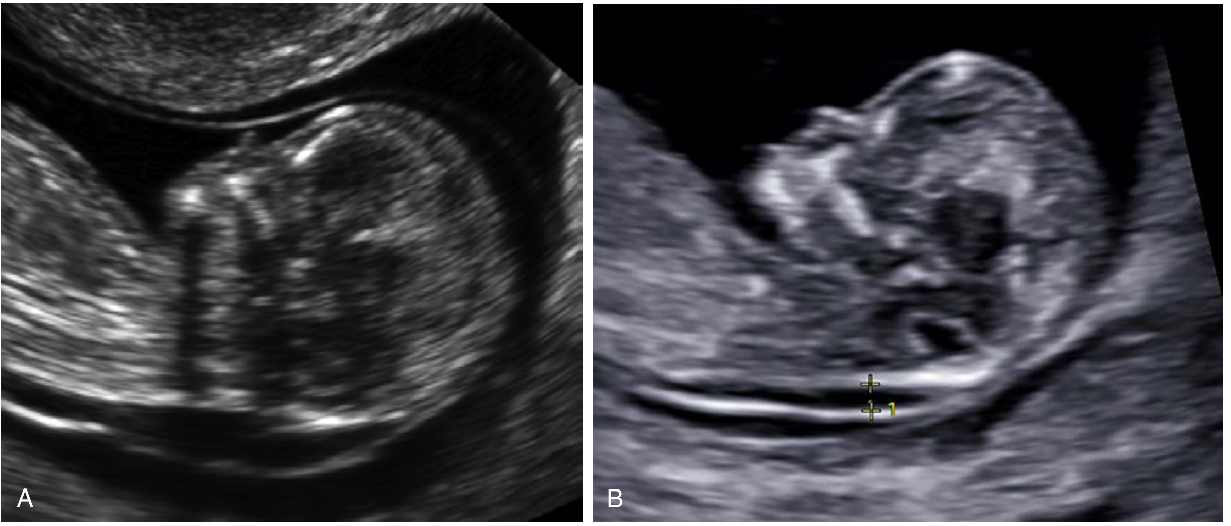
Fig 29.22 - Absent nasal bone in two different fetuses with trisomy 21. (A) Absent NB with enlarged NT. (B) Absent NB; the echogenic skin edge could be mistaken for the NB. - Creasy & Resnik's MFM
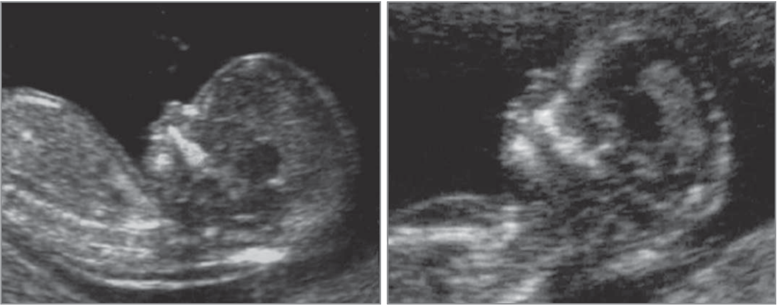
Fig 30.5 - First-trimester ultrasound: euploid fetus (left, NB present) vs. trisomy 21 fetus (right, NB absent). - Creasy & Resnik's MFM
Performance as a Screening Marker
First Trimester
| Parameter | Value |
|---|---|
| Absent NB in trisomy 21 fetuses | 73% |
| Absent NB in euploid fetuses | 0.5% |
| Sensitivity (NB alone for trisomy 21) | 65% |
| False-positive rate | 0.8% |
| Positive predictive value | ~54% (1 in 2 fetuses with absent NB had T21) |
| Likelihood ratio increase for trisomy 21 | 87-fold |
| Negative likelihood ratio | 0.35 |
Sonek et al. found that combining NB + NT + maternal age gives a 93% detection rate for Down syndrome at a 5% false-positive rate; adding serum biochemistry can push detection to 97% in high-risk populations.
Adding NB to the standard combined first-trimester screen (NT + biochemistry) reduces the false-positive rate from 5% to 2.5% while maintaining 90% detection.
Second Trimester
- Hypoplastic or absent NB occurs in up to 60% of Down syndrome pregnancies imaged in the second trimester
- Complete absence: ~37% of affected cases
- Hypoplasia: ~50% of affected cases
- In normal pregnancies: absence in 0.9%, hypoplasia in 2.4%
- May be the best single ultrasound marker for second-trimester risk assessment when performed by experienced operators
Second-trimester markers associated with Down syndrome (Box 30.2) include: brachycephaly, increased nuchal thickness, CHD, hyperechoic bowel, shortened femur/humerus, renal pyelectasis, duodenal atresia, hypoplastic 5th digit midphalanx, echogenic intracardiac focus, sandal gap foot, and short or absent nasal bone.
Ethnic Variation
This is a critical confounder. Absent NB is more common in Afro-Caribbean and southern Asian fetuses, even without aneuploidy:
| Ethnicity | Absent NB in euploid pregnancies | LR for Trisomy 21 |
|---|---|---|
| White/Caucasian | 2.5% | 31.3 |
| Afro-Caribbean | 9.0% | 8.8 |
| Southern Asian | 5.0% | 14.2 |
The NB is also more likely to be absent with increasing NT thickness in euploid pregnancies.
Proposed Screening Strategy (Two-Stage Approach)
Nikolaides et al. proposed using NB as a contingency (second-stage) marker rather than a universal first-tier screen:
- Risk ≥ 1:100 after NT + biochemistry → offer CVS directly
- Risk < 1:1000 → reassurance, no further testing
- Risk 1:101 to 1:1000 → perform NB assessment (applies to ~15% of pregnancies)
This achieves 90% DS detection at 2.5% false-positive rate without requiring universal NB assessment by all operators.
Clinical Management When Absent NB Is Found
- Isolated absent NB in first trimester (NT < 95th percentile): Low likelihood of chromosomal microarray (CMA) abnormality; not routinely an indication for invasive testing alone
- Isolated absent NB in euploid fetus without other second-trimester anomalies: Usually associated with good neonatal outcome
- Absent NB + increased NT or other markers: Significantly increases aneuploidy risk; discuss invasive diagnostic testing (CVS/amniocentesis) and/or cell-free DNA (cfDNA)
- Credentialed providers only: The NTQR and Fetal Medicine Foundation offer certification for NB assessment
Other Aneuploidies
While trisomy 21 is the primary association, absent NB has also been observed in other chromosomal anomalies. Recent chromosomal microarray studies (PMID 35506821, 38766695) show that even in second-trimester isolated absent/hypoplastic NB, pathogenic copy number variants can occasionally be found, supporting the value of CMA when combined with other risk factors.
Summary Points
- Absent NB is a first- and second-trimester soft marker strongly associated with trisomy 21
- It is assessed in the midsagittal plane at 45 degrees; the NB must be less echogenic than the overlying skin to call it absent
- Sensitivity for trisomy 21: ~65-73% (first trimester), ~60% (second trimester); false-positive rate: <1%
- Ethnic variation significantly alters the likelihood ratio - always adjust for race
- Best used as a contingency marker in intermediate-risk patients, not universal first-tier screening
- Isolated absent NB with normal NT and no other anomalies has a generally favorable prognosis
- Combined screening (NT + biochemistry + NB) achieves ~93-97% DS detection
Source: Creasy & Resnik's Maternal-Fetal Medicine: Principles and Practice, Chapters 18, 29, 30
Fetal medicine counselling
Asking for Preferences
What aspect of fetal medicine counselling are you looking for?
Hypoplastic left heart syndrome in fetus
Hypoplastic left heart syndrome (HLHS) is one of the most severe congenital heart defects, characterized by underdevelopment of the left-sided cardiac structures - left ventricle, mitral valve, aortic valve, and ascending aorta. It is duct-dependent and lethal without intervention. Let me pull the full textbook content.
Searching sources...
Reading File
Finding Sources
Searching PubMed
"hypoplastic left heart syndrome" AND prenatal AND fetal
Reading File
Reading File
Finding Sources
Reading File
Reading File
Reading File
Reading File
Reading File
Loading Image
Loading Image
Reading File
Finding Sources
Reading File
I now have comprehensive content to write a thorough answer.
Hypoplastic Left Heart Syndrome (HLHS) in the Fetus
Definition
HLHS is a heterogeneous spectrum of congenital heart defects characterized by severe underdevelopment of all left-sided cardiac structures - left ventricle (LV), mitral valve, aortic valve, and ascending aorta - resulting in inability to sustain systemic cardiac output. It is one of the most common and most feared fetal cardiac diagnoses. Without intervention, HLHS is invariably fatal and accounts for 25% of early cardiac deaths in neonates. - Schwartz's Principles of Surgery, 11e, p. 801
Prevalence: 0.2 per 1000 live births; occurs twice as often in males as females.
Anatomy and Subtypes
HLHS is classified into four anatomic subtypes based on valvular morphology:
| Subtype | Aortic Valve | Mitral Valve |
|---|---|---|
| A | Stenosis | Stenosis |
| B | Atresia | Atresia ← most classic |
| C | Atresia | Stenosis |
| D | Stenosis | Atresia |
Aortic atresia tends to produce more severe ascending aorta hypoplasia than aortic stenosis. The ascending aorta may be as small as < 2 mm in diameter.
Additional features:
- Coarctation shelf present in 80% of patients
- Large ductus arteriosus; main pulmonary artery dilated
- Segmental pulmonary arteries are small (reduced intrauterine pulmonary blood flow)
- Left atrium typically smaller than normal; leftward displacement of septum primum
- Interatrial communication (foramen ovale) is nearly always present but is often restrictive
Pathophysiology
HLHS likely results from altered intracardiac blood flow during development. When aortic outflow is obstructed (atretic or stenotic valve), the LV is deprived of the normal high-pressure systolic stimulus for growth, and downstream structures (ascending aorta) receive only low-pressure diastolic retrograde flow via the ductus, causing progressive hypoplasia.
Postnatal circulation in HLHS:
- All pulmonary, systemic, and coronary blood flow comes from the RV via the ductus
- Pulmonary venous return enters the left atrium but cannot cross the stenotic/atretic mitral valve → shunted right-to-left across the foramen ovale → volume loads the RV
- As pulmonary vascular resistance falls postnatally, RV output is preferentially directed to the lungs → systemic hypoperfusion + coronary ischemia
- Ductal closure = incompatible with life
Prenatal Diagnosis (Fetal Echocardiography)
Key Diagnostic Views
Four-Chamber View (4CV) - always abnormal:
- Markedly abnormal 4CV at midgestation
- No inflow into the LV (no mitral valve / mitral atresia)
- Severely hypoplastic LV with intact ventricular septum (usually)
- The RV dominates the heart
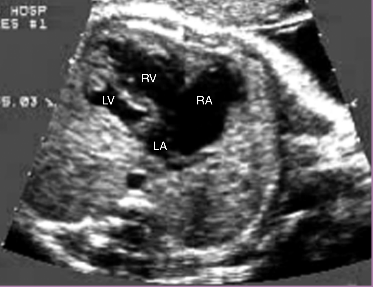
Fig 23.61 - Four-chamber view: HLHS with mitral atresia and VSD. The VSD allows some LV filling, explaining the relatively preserved (though still hypoplastic) LV size. LA = left atrium; RA = right atrium; RV = right ventricle; LV = left (hypoplastic) ventricle. - Creasy & Resnik's MFM
Aortic Arch View:
- Aortic arch fills retrograde from the ductus arteriosus
- Color flow Doppler confirms pulsatile retrograde flow into the transverse arch
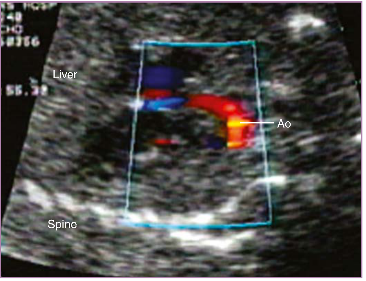
Fig 23.64 - Sagittal color Doppler: Retrograde flow into the transverse aortic arch (Ao) in a fetus with HLHS - the hallmark of duct-dependent systemic circulation. - Creasy & Resnik's MFM
Critical Additional Assessment: The Foramen Ovale
The foramen ovale must be carefully evaluated with 2D and color flow Doppler. A restrictive foramen ovale is a critical negative prognostic finding:
- Restriction to left-to-right flow → pulmonary venous congestion → left atrial hypertension → dilated LA and pulmonary veins
- Can lead to irreversible pulmonary vascular disease
- May develop as late as the third trimester
- May not be confirmed by 2D alone; requires spectral and color flow Doppler ± pulmonary vein assessment
- Fetuses with restrictive/intact atrial septum may benefit from percutaneous enlargement of the foramen ovale during the second trimester to prevent pulmonary vascular disease
Tricuspid valve and RV function must also be assessed - tricuspid regurgitation and RV dysfunction are negative prognostic markers.
Progressive/Evolving HLHS
Some cases do not begin as full HLHS:
- Severe aortic stenosis present by 18-20 weeks may evolve into HLHS by term - the LV initially dilates (dilated cardiomyopathy pattern), then becomes hypoplastic
- Signs of evolving HLHS: dilated poorly functioning LV, endocardial fibroelastosis (bright LV endocardium), mitral regurgitation, L-to-R foramen ovale flow, retrograde transverse arch flow
Associated Abnormalities
- Increased risk for aneuploidy (chromosomal anomalies)
- Increased risk for central nervous system abnormalities (congenital or acquired)
- Associated VSD (if present, LV may retain relatively normal size due to R-to-L shunting)
Karyotyping / chromosomal microarray should be offered to all parents with a fetal HLHS diagnosis.
Antenatal Management
Delivery Planning
- Deliver at a tertiary care center experienced with complex congenital heart defects
- Fetuses with HLHS + restrictive/intact foramen ovale: consider planned cesarean delivery to allow controlled environment for immediate neonatal intervention
- Prostaglandin E1 (PGE1) must be ready to initiate at delivery to maintain ductal patency
Fetal Intervention (Selected Cases)
Three conditions are currently treated with prenatal cardiac intervention:
| Indication | Procedure | Goal |
|---|---|---|
| Restrictive atrial septum in HLHS | Atrial septostomy | Prevent irreversible pulmonary vascular disease |
| Aortic stenosis with evolving HLHS | Balloon aortic valvuloplasty | Promote LV growth → biventricular circulation |
| Pulmonary atresia with hypoplastic right heart | Balloon pulmonary valvuloplasty | Promote RV growth |
Fetal Aortic Valvuloplasty (FAV) - Results:
- In 108 fetuses (2002-2018): >83% technically successful
- Biventricular circulation achieved in 33-38% of technically successful cases
- Overall intraprocedural complications: 48.1% (fetal bradycardia, pericardial effusion, pleural effusion, balloon rupture)
- Fetal death within 2 days: 16.7%
- Survival at 1 year: 80% ± 4%; at 5 years: 75% ± 5%
Postnatal Management: Three-Stage Palliation
All infants with HLHS require staged palliative surgery. Cardiac transplantation is an alternative for select cases.
Stage 1 - Norwood Procedure (within first week of life)
Described by Norwood et al. in 1983, modified to the current three-stage approach. Creates a single outflow vessel (neoaorta) arising from the RV:
- Pulmonary root connected to native ascending aorta
- Hypoplastic arch augmented with homograft patch
- Pulmonary blood flow via either:
- Modified Blalock-Taussig shunt (mBTS): innominate artery → right pulmonary artery
- Sano shunt (RV-PA conduit): 5-6 mm Gore-tex graft directly from RV to PA
- Wide atrial septectomy to prevent pulmonary venous hypertension
Sano vs. mBTS (SVR Trial, NIH-sponsored RCT):
- Transplantation-free survival at 12 months: Sano 74% vs. mBTS 64% (P = .01)
- Sano group had more unintended interventions and complications
- RV size/function at 14 months: similar between groups
Stage 2 - Glenn Procedure (~6 months)
- Bidirectional cavopulmonary anastomosis
- Superior vena cava connected to right pulmonary artery
Stage 3 - Fontan Procedure (~2-3 years)
- Inferior vena cava connected to pulmonary artery (total cavopulmonary connection)
- Separates pulmonary and systemic circuits
Prognosis and Outcomes
- With modern surgical management, Norwood survival exceeds 90% at experienced centers
- Outcomes for HLHS remain significantly worse than for other complex CHD
- Predictors of poor outcome:
- Low birth weight
- Significant tricuspid regurgitation
- Restrictive interatrial communication
- Poor RV function
- Extracardiac or chromosomal anomalies
- Significant neurodevelopmental morbidity remains a concern (CNS injury from chronic hypoxemia + surgical insults)
Counselling Points for HLHS
When HLHS is diagnosed prenatally, structured multidisciplinary counselling must cover:
- What HLHS is - underdeveloped left heart, duct-dependent systemic circulation
- Natural history without treatment - invariably fatal
- Three management options:
- Three-stage palliative surgery (Norwood → Glenn → Fontan)
- Cardiac transplantation (limited by organ availability; 24% die waiting)
- Comfort/palliative care only
- Surgical risks and outcomes - >90% Norwood survival at expert centers, but lifetime morbidity remains
- Neurodevelopmental concerns - higher rates of cognitive, motor, and behavioral difficulties
- Risk of aneuploidy - offer chromosomal testing
- Foramen ovale restriction - if present, worsens prognosis and changes delivery planning
- Delivery planning - need for tertiary center, PGE1 readiness, possible cesarean for restrictive FO
- Quality of life considerations - Fontan circulation is palliation, not cure; long-term Fontan complications (protein-losing enteropathy, arrhythmia, hepatic disease, lymphatic issues)
- Recurrence risk - familial congenital heart defects
A 2025 systematic review (Zeleznik et al., PMID 41458986) highlights the importance of ethical frameworks and multidisciplinary team involvement when counselling families facing this diagnosis.
Sources: Creasy & Resnik's Maternal-Fetal Medicine: Principles and Practice (Chapters 23, 30, 34); Schwartz's Principles of Surgery, 11e (Chapter 20)
Hypoplastic nasal bone at 14 weeks scan
Hypoplastic (short) nasal bone at 14 weeks is a soft marker for aneuploidy, particularly trisomy 21. It differs from "absent" nasal bone - the bone is present but shorter than expected for gestational age. Let me pull the full textbook content.
Searching sources...
Finding Sources
Searching PubMed
"nasal bone" AND hypoplastic AND "first trimester" AND trisomy
Finding Sources
Reading Article
PMID: 34171388
Reading File
Reading File
Your credits are exhausted. You can purchase a top-up or subscribe to a plan to continue using Orris. If you're already subscribed, your daily credits will reset tomorrow.
This is a shared conversation. Sign in to Orris to start your own chat.