
Vericose vein
Reading File
Now let me search the medical library for varicose veins content in parallel.
Finding Sources
Finding Sources
Searching PubMed
"varicose veins"[MeSH Terms] AND treatment
Good. Now let me read the key sections from Bailey & Love, Harrison's, and Schwartz's in parallel.
Reading File
Reading File
Reading File
Let me read more from Bailey & Love (complications, investigations, treatment) and fetch the varicose veins image.
Reading File
Loading Image
Now let me read the complications and treatment sections from Bailey & Love.
Reading File
Reading File
Let me read the complications, CEAP classification table, and treatment section from Harrison's and Bailey & Love.
Reading File
Reading File
I now have comprehensive information from multiple authoritative textbooks. Let me compile the full answer.
Varicose Veins
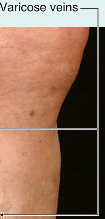
Clinical photograph demonstrating varicose veins of the lower limb - Gray's Anatomy for Students
Definition
Varicose veins are dilated, bulging, tortuous superficial veins measuring at least 3 mm in diameter. They must be distinguished from:
-
Reticular veins - dilated intradermal veins, 1-3 mm, appearing blue-green, do NOT protrude from the skin surface
-
Telangiectasias (spider veins) - <1 mm diameter, near skin surface, forming blue/purple/red linear or spider-web patterns
-
Harrison's Principles of Internal Medicine 22E, p.2223
Epidemiology
-
Adult prevalence: 30-50% for visible varicose veins
-
Gender: Higher prevalence in women (most studies)
-
Age: Prevalence rises sharply with age - from ~11.5% (age 18-24) to ~55.7% (age 55-64) in the Edinburgh Vein Study
-
Risk factors: Pregnancy, family history, increasing BMI, tall stature, advancing age, ethnicity
-
Bailey & Love's Short Practice of Surgery 28th Ed, p.1052
Classification
Primary vs. Secondary
| Type | Cause |
|---|---|
| Primary | Intrinsic defect of saphenous vein valves, weak vein wall, high intraluminal pressure; ~50% have family history |
| Secondary | Venous hypertension from deep-venous insufficiency, DVT post-thrombotic syndrome, incompetent perforating veins, arteriovenous fistulas |
CEAP Classification (Clinical, Etiologic, Anatomic, Pathophysiologic)
| Grade | Description |
|---|---|
| C0 | No visible/palpable signs |
| C1 | Telangiectasias or reticular veins |
| C2 | Varicose veins |
| C2r | Recurrent varicose veins |
| C3 | Edema |
| C4a | Pigmentation or eczema |
| C4b | Lipodermatosclerosis or atrophie blanche |
| C4c | Corona phlebectatica |
| C5 | Healed venous ulcer |
| C6 | Active venous ulcer |
| C6r | Recurrent active venous ulcer |
- Harrison's Principles of Internal Medicine 22E, Table 293-1
Anatomy Relevant to Varicose Veins
The great saphenous vein (GSV) - the longest vein in the body - runs from the medial foot, anterior to the medial malleolus, along the medial calf and thigh, draining into the common femoral vein. The small saphenous vein (SSV) runs from the dorsolateral foot, posterior to the lateral malleolus, along the posterolateral calf, draining into the popliteal vein.
- GSV incompetence: ~60% of cases - medial thigh and calf varicosities
- SSV incompetence: ~20% of cases - posterolateral calf varicosities
- Anterolateral thigh/calf: suggests non-saphenous or tributary source
Symptoms
-
Aching, heaviness, throbbing, burning, "bursting" sensation
-
Pruritus (itching)
-
Ankle swelling
-
Symptoms worsen with prolonged standing/sitting and throughout the day
-
Symptoms relieved by leg elevation or compression hosiery
-
Symptoms are independent of the degree of reflux or visible size of varices
-
Significant quality-of-life deficit documented in studies
-
Bailey & Love's 28th Ed, p.1052; Schwartz's Principles of Surgery 11th Ed
Complications
| Complication | Notes |
|---|---|
| Superficial thrombophlebitis | Painful, cord-like inflammation |
| Bleeding | From attenuated vein clusters; can be dramatic |
| Chronic venous insufficiency | Venous hypertension, extravasation of fluid/blood |
| Lipodermatosclerosis | Fibrotic, woody induration of subcutaneous tissue |
| Hyperpigmentation | Hemosiderin deposition |
| Venous eczema | Stasis dermatitis |
| Venous ulceration | Typically above medial malleolus (gaiter area) |
Clinical Examination & Special Tests
Brodie-Trendelenburg Test - Distinguishes primary (superficial insufficiency) from secondary (deep venous insufficiency) varicose veins:
- Elevate leg supine, empty veins
- Apply tourniquet to proximal thigh, ask patient to stand
- Rapid filling within 30s = deep venous insufficiency + incompetent perforators (secondary)
- Filling only after tourniquet removal = primary superficial incompetence
Perthes Test - Assesses deep venous obstruction:
- Tourniquet on mid-thigh while standing
- Patient walks 5 minutes
- Collapse of superficial veins = patent deep system
- Further distension = deep venous obstruction
- Harrison's 22E, p.2224
Investigations
Duplex Ultrasound (gold standard):
- Combines B-mode imaging + spectral Doppler
- Detects venous obstruction AND venous reflux in superficial + deep veins
- Reflux: reversal of flow >0.5 sec during Valsalva or distal compression/release
- Patient stands facing examiner, leg externally rotated
- Groin scan: identifies "Mickey Mouse" sign - GSV medial to common femoral artery at saphenofemoral junction (SFJ)
- "Saphenous eye" view - transverse cross-section of GSV in fascial compartment
MR/CT/Conventional Venography - reserved for suspected IVC/iliofemoral pathology (post-DVT, May-Thurner syndrome, extrinsic tumour compression)
Treatment
Conservative Measures
- Leg elevation, avoid prolonged standing
- Graduated elastic compression stockings (20-30, 30-40, or 40-50 mmHg)
- Trial of compression hosiery to confirm venous origin of symptoms
Interventional Indications
- Symptoms unrelieved despite compression
- Lipodermatosclerosis
- Venous ulcer
1. Sclerotherapy
- Liquid sclerotherapy: for telangiectasias and small varicosities
- Foam sclerotherapy: for larger veins; a multicenter RCT showed significant symptom relief vs placebo
- Sclerosants: hypertonic saline (11.7-23.4%), sodium tetradecyl sulfate (0.125-1%), polidocanol (0.5-1.0%)
- Mechanism: destroys venous endothelium
- Compression bandages applied for 3-5 days post-injection, then elastic stockings for ≥2 weeks
- Complications: local hyperpigmentation, allergic reaction, superficial thrombophlebitis, DVT, skin necrosis
2. Endovenous Laser Ablation (EVLA)
- Laser fibre inserted into vein, wavelength typically 1470 nm
- Thermal energy generated ablates the vein wall
- Tumescent perivenous anaesthesia is administered first
- Better for very large veins (>15 mm diameter)
- Less expensive consumables
3. Radiofrequency Ablation (RFA)
- Catheter-based (e.g., ClosureFast™); automated treatment cycle
- Simpler to use, single-button press; shorter learning curve
- Marginally less post-procedural pain and bruising than EVLA
- Both EVLA and RFA: equally effective, faster recovery than open surgical stripping
4. Phlebectomy (Ambulatory/Hook Phlebectomy)
- Surgical avulsion of varicosities through small stab incisions
- Can be performed concomitantly with endothermal ablation or as a staged procedure
- Done under tumescent anaesthesia
5. Surgical Stripping (Historical Gold Standard)
- High tie and stripping of GSV/SSV
- Now largely replaced by endovenous techniques due to equivalent efficacy but slower recovery
6. Nonthermal, Nonsclerosant Techniques (Newer)
-
Mechanical endovenous ablation (MOCA) combining catheter injury + sclerosant
-
Cyanoacrylate glue - proprietary adhesive formulations showing promising early results
-
Schwartz's Principles of Surgery 11th Ed, p. (block 11); Bailey & Love 28th Ed; Harrison's 22E, p.2224-2225
Recent Evidence
A 2024 meta-analysis (PMID: 38316290) comparing endovenous radiofrequency ablation vs laser ablation found both techniques to have equivalent efficacy for lower extremity varicose veins, supporting current practice of choosing between them based on operator experience and vein characteristics.
This is a shared conversation. Sign in to Orris to start your own chat.