
Explain in easy way... episcleritis
Reading File
Now let me search the medical library and web in parallel.
Finding Sources
Searching the Web
episcleritis simple nodular causes symptoms treatment
Reading File
Reading File
Reading File
Reading File
Loading Image
Here is a clear, easy explanation of episcleritis:
Episcleritis - A Simple Explanation
What is it?
Think of the eye like a layered cake. The white part of your eye is the sclera. Just above it is a thin, clear tissue called the episclera. When that thin layer gets inflamed (irritated and swollen), it's called episcleritis.
It looks like a red eye, but it is NOT the same as pink eye (conjunctivitis). It's generally a benign (harmless), self-limiting condition - meaning it goes away on its own.
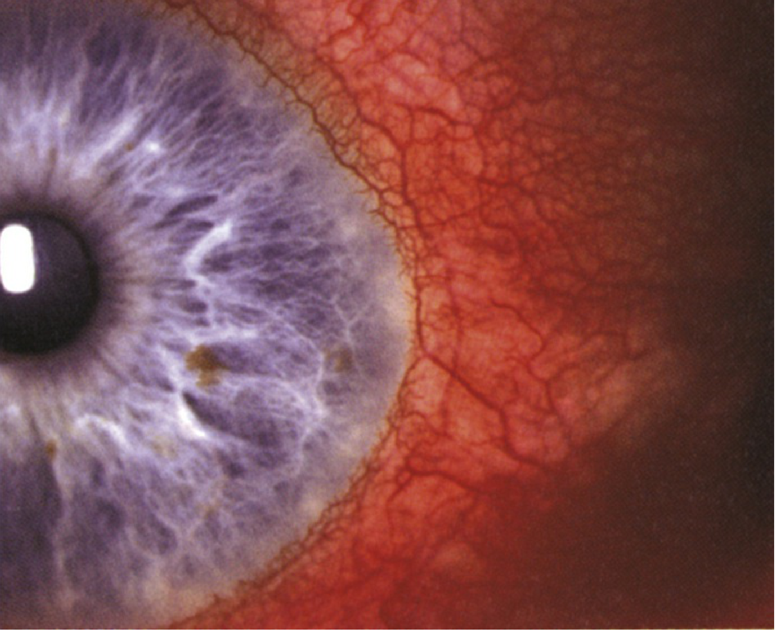
Two Types
| Feature | Simple Episcleritis (75%) | Nodular Episcleritis (25%) |
|---|---|---|
| Appearance | Redness only, no lump | Redness + a small raised nodule |
| Onset | Sudden, peaks in 24 hrs | Slower onset |
| Duration | Few days to 3 weeks | Longer course |
| Pain | Often mild or absent | More painful |
| Systemic disease link | Less common | More commonly linked |
Simple episcleritis can be either sectoral (just one patch of redness) or diffuse (whole white of the eye is red).
Who Gets It?
- Most common in middle-aged women
- Can be bilateral (both eyes) in over half of cases
- Recurs in about 60% of patients, but episodes tend to become less frequent over time
- Rare in children
What Causes It?
- Most often: No known cause (idiopathic) - about 60% of cases have no underlying disease
- Sometimes linked to systemic conditions:
- Rheumatoid arthritis
- Crohn's disease / Inflammatory bowel disease
- Gout
- Herpes zoster (shingles)
- Lupus (SLE)
- Rosacea
- Rarely: infections (syphilis, Lyme disease), medications (topiramate, pamidronate)
Symptoms
- Red or pink eye (sectoral patch or the whole white)
- Mild discomfort or grittiness - up to 50% feel no pain at all
- Mild watering
- Sometimes mild photophobia (light sensitivity)
- No discharge (this helps rule out conjunctivitis)
- Vision is always normal - episcleritis does NOT affect vision
How is it Diagnosed?
The key clinical test is the phenylephrine 2.5% drop test:
- Put the drop in the eye, wait 10-15 minutes
- In episcleritis: the red vessels blanch (go white) because the vessels are superficial
- In scleritis (deeper, more serious): vessels do NOT blanch - they stay red/purple
Another clue: the engorged episcleral vessels can be gently moved with a cotton swab - they're not fixed like in scleritis.
Key Difference: Episcleritis vs. Scleritis
| Feature | Episcleritis | Scleritis |
|---|---|---|
| Depth | Superficial (episclera) | Deep (sclera itself) |
| Pain | Mild or none | Severe, deep, boring pain |
| Colour | Bright red | Violaceous (bluish-purple) hue |
| Phenylephrine test | Vessels blanch | Vessels do NOT blanch |
| Vessels move with swab? | Yes | No |
| Vision affected? | No | Can be |
| Systemic disease link | Sometimes | Frequently |
| Danger level | Benign | Can be sight-threatening |
Treatment
Treatment depends on severity:
- Mild cases: No treatment needed - just cool compresses or refrigerated artificial tears (lubricating eye drops)
- Moderate cases:
- Topical NSAIDs (e.g., diclofenac 0.1% four times a day)
- Mild topical steroids (e.g., fluorometholone 0.1% four times a day)
- Severe or recurrent cases:
- Oral NSAIDs such as ibuprofen 200-600 mg three times a day for 10-14 days (take with food)
- Naproxen or flurbiprofen are alternatives
- If systemic disease found: Treat the underlying cause with a rheumatologist
Prognosis
Excellent. Episcleritis is not dangerous and does not cause permanent eye damage or vision loss. Most episodes last a few days to 3 weeks. It can recur but tends to become less frequent over time.
Key memory tip: Episcleritis = superficial, benign, bright red, blanches with phenylephrine, mild pain or none, good prognosis. Scleritis = deep, potentially serious, violaceous/purple, does NOT blanch, severe pain, worse prognosis.
Sources: Kanski's Clinical Ophthalmology 10th Ed.; Wills Eye Manual
This is a shared conversation. Sign in to Orris to start your own chat.