
Hsts ammonia
Reading File
Quick answer: Ammonia (NH3) is a nitrogenous waste product that is toxic to the brain. It is normally converted to urea in the liver (urea cycle) and excreted by the kidneys. Elevated blood ammonia (hyperammonemia) causes hepatic encephalopathy, among other conditions. The kidneys also produce ammonia to aid in acid-base balance.
Searching sources...
Finding Sources
Finding Sources
I see the user typed "Hsts ammonia" - this appears to be a query about ammonia in the context of HSTs (likely "Homeostasis" or could be a specific acronym). Let me clarify this is likely about "HSTs" = possibly "Hepatic Steatosis", "Hyperammonemia", or a lab context. The query might be about Henry's Clinical Diagnosis (lab methods) - let me read the key sources in parallel.
Reading File
Reading File
Reading File
I notice "Hsts ammonia" likely means the user is asking about HSTs (possibly "Hepatic Steatosis" or more likely "Hyperammonemia Syndromes" / urea cycle disorders). Let me also fetch the urea cycle diagram and get the lab reference ranges, while reading the neurological section too.
Loading Image
Reading File
Reading File
I now have comprehensive information about ammonia from multiple authoritative textbooks. Let me compose a thorough answer. Note that "Hsts" is most likely a typo or autocorrect for "HSTs" - possibly meaning "tests" (i.e., the user is asking about ammonia tests/lab values), or it could be "hosts" or simply a garbled prefix. Given the lab-focused results and context, I'll provide a complete clinical overview of ammonia covering biochemistry, metabolism, lab measurement, and clinical significance.
Ammonia - Clinical Overview
What Is Ammonia?
Ammonia (NH3/NH4+) is a nitrogenous compound derived primarily from amino acid and nucleic acid catabolism. It is toxic - particularly to the brain - and must be rapidly detoxified. At physiological pH (~7.4), the vast majority (~98.3%) exists as the ionized form NH4+, with only ~1.7% as the free NH3 form. Small changes in pH cause exponential shifts in the NH3:NH4+ ratio, which has important implications for transport and toxicity.
- Henry's Clinical Diagnosis and Management by Laboratory Methods, p. 528
Urea Cycle (Krebs-Henseleit Cycle)
Ammonia is detoxified exclusively in the liver via the urea cycle. The liver uniquely possesses all the required enzymes:
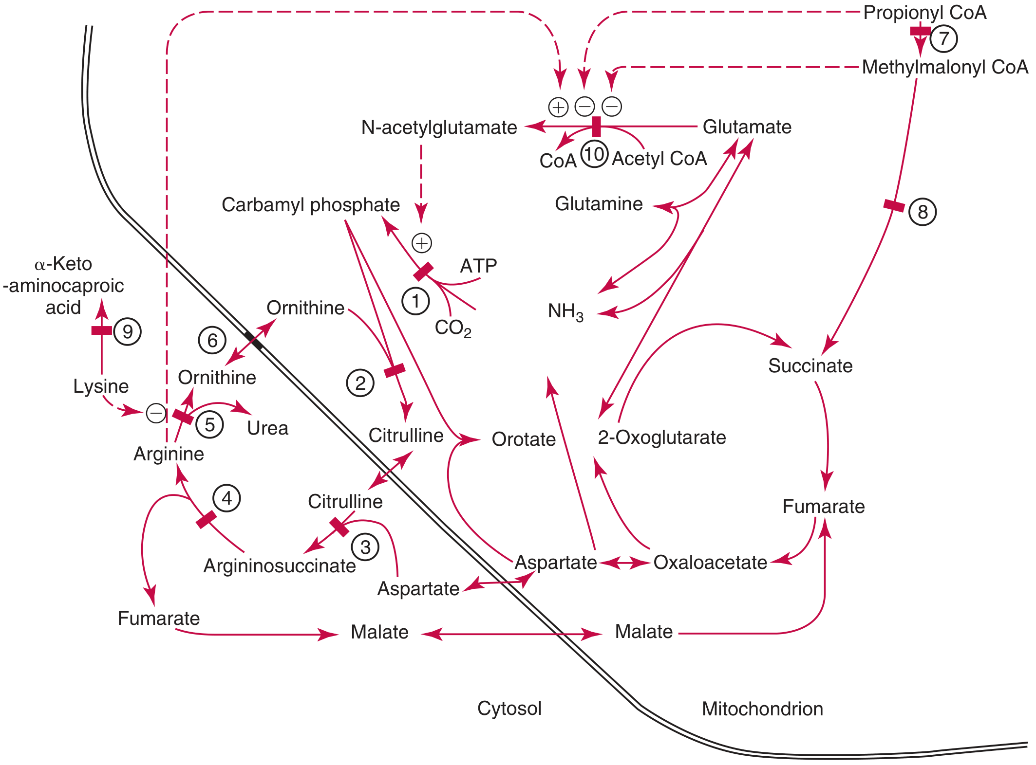
Key steps (mitochondria -> cytosol):
- NH3 + CO2 + ATP -> Carbamoyl phosphate (enzyme: CPS-1 - rate-limiting step)
- Carbamoyl phosphate + Ornithine -> Citrulline (enzyme: OCT - unique to liver)
- Citrulline + Aspartate -> Argininosuccinate (enzyme: ASS)
- Argininosuccinate -> Arginine + Fumarate (enzyme: ASL)
- Arginine -> Ornithine + Urea (enzyme: Arginase) - Urea is then renally excreted
Congenital deficiency of any of these enzymes causes hyperammonemia (urea cycle disorders). The numbered defect sites visible in the diagram correspond to: (1) CPS-1, (2) OCT, (3) ASS, (4) ASL, (5) Arginase, (6) mitochondrial ornithine transport, (7) propionyl-CoA carboxylase, (8) methylmalonyl-CoA mutase, (9) L-lysine dehydrogenase, and (10) N-acetylglutamate synthetase.
- Tietz Textbook of Laboratory Medicine, 7th Edition, p. 1977-1978
Renal Ammonia Metabolism
The kidney is a net producer of ammonia (renal vein ammonia > arterial ammonia). This is critical for acid-base homeostasis:
-
Primary site: Proximal tubule (60-70% of renal ammoniagenesis under basal conditions; 70-80% during metabolic acidosis)
-
Key enzyme: Phosphate-dependent glutaminase (PDG/KGA), which converts glutamine -> glutamate + NH4+
-
Further processing: Glutamate dehydrogenase (GDH) converts glutamate -> alpha-ketoglutarate + NH4+
-
NH3 chemistry: NH4+ can substitute for K+ at K+-transport sites due to similar biophysical properties in aqueous solution
-
Ammonia is selectively transported to the urine (for acid excretion) or to the renal vein; integrated transport occurs in the proximal tubule, thick ascending limb (TAL), and collecting duct
-
Brenner and Rector's The Kidney, 2-Volume Set
Causes of Hyperammonemia
| Category | Examples |
|---|---|
| Liver disease | Cirrhosis, fulminant hepatic failure, Reye syndrome |
| Portosystemic shunting | Intrahepatic (cirrhosis) or surgical shunts |
| Urea cycle enzyme defects | CPS-1 deficiency, OTC deficiency, citrullinemia, etc. |
| Organic acidemias | Propionic acidemia, methylmalonic acidemia |
| Drugs | Valproate, salicylates |
| Renal failure | Increased urea diffuses into GI tract -> bacterial conversion to NH3 |
| GI bleeding | Bacterial metabolism of protein in blood |
| Excess dietary protein / constipation | Increased gut ammonia production |
- Tietz Textbook of Laboratory Medicine, 7th Edition, p. 1978
Ammonia Toxicity: Mechanisms in the CNS
Ammonia causes hepatic encephalopathy through several mechanisms:
- GABA depletion: Ammonia reacts with glutamic acid to form glutamine (reversing glutaminase). This depletes glutamate, which is the precursor for GABA synthesis, leading to reduced inhibitory neurotransmission.
- Astrocyte toxicity: Ammonia is directly toxic to astrocytes.
- Glutamine accumulation: Acts as an osmolyte contributing to cerebral edema.
- Synergy with inflammation and oxidative stress: Complex interaction that amplifies neurotoxicity.
Key clinical note: Arterial ammonia correlates better with encephalopathy severity than venous levels. Lowering ammonia invariably reduces encephalopathy. In acute liver failure, ammonia >200 µmol/L (340 µg/dL) is associated with cerebral edema and poor prognosis.
- Henry's Clinical Diagnosis and Management by Laboratory Methods, p. 528-529
Laboratory Measurement
Method: Enzymatic assay using glutamate dehydrogenase (GLDH):
- NH3 + alpha-ketoglutarate + NADPH -> Glutamate + NADP+
- Ammonia concentration is determined by the decrease in absorbance at 340 nm (as NADPH -> NADP+)
Reference interval (enzymatic method):
- 15 to 45 µg/dL (11 to 32 µmol/L)
Key interferences and pre-analytical issues:
- Hemolysis - RBCs contain ~3x the ammonia of plasma; falsely elevates results
- Cigarette smoke - contains ammonia; keep samples away from smoke
- ALT up to 2400 U/L does not interfere
- Sample stability: 3 hours at 2-4°C, or 24 hours at -20°C
- Fasting venous plasma sample is preferred
Clinical utility:
-
Useful in differential diagnosis of encephalopathy (to determine if hepatic origin)
-
Especially helpful in diagnosing Reye syndrome and inherited urea cycle disorders
-
NOT useful for monitoring encephalopathy severity in known chronic liver disease (poor correlation at that stage; ~70% of cirrhotic patients without encephalopathy already have elevated levels)
-
Henry's Clinical Diagnosis and Management by Laboratory Methods, p. 529; Tietz Textbook of Laboratory Medicine, 7th Edition, p. 1978-1979
This is a shared conversation. Sign in to Orris to start your own chat.