
Diabetic nephropathy Notes pathology by robbins mbbs 6 marks in flow chart and easy way
Diabetic Nephropathy - Pathology Notes (Robbins)
Flow Chart Format | 6 Marks | MBBS
OVERVIEW BOX
DIABETES (Type 1 or Type 2)
         ↓
    Hyperglycemia (chronic, uncontrolled)
         ↓
    KIDNEY DAMAGE (prime target)
         ↓
  Three major categories of lesions:
  1. GLOMERULAR LESIONS (most important)
  2. VASCULAR LESIONS
  3. PYELONEPHRITIS / PAPILLARY NECROSIS
FLOW CHART 1 - GLOMERULAR LESIONS (Most Important)
HYPERGLYCEMIA
      │
      ▼
┌─────────────────────────────────────────────────────────┐
│         THREE GLOMERULAR LESIONS                        │
│                                                         │
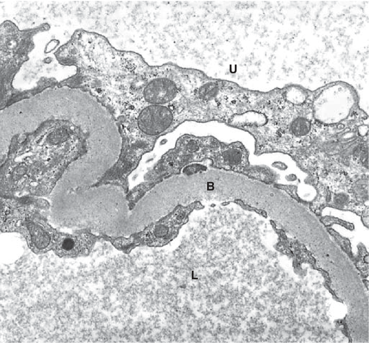
│  ① Capillary Basement Membrane (GBM) Thickening        │
│      ↓ earliest change                                  │
│      Detectable on ELECTRON MICROSCOPY (EM)             │
│      Occurs within few years of onset of DM             │
│      Seen BEFORE any change in renal function           │
│                                                         │
│  ② Diffuse Mesangial Sclerosis                          │
│      ↓                                                  │
│      ↑ Mesangial matrix + mesangial cell proliferation  │
│      + Basement membrane thickening                     │
│      Common in DM > 10 years / older patients /         │
│         hypertensive patients                           │
│      When SEVERE → NEPHROTIC SYNDROME                   │
│      (Proteinuria + Hypoalbuminemia + Edema)            │
│                                                         │
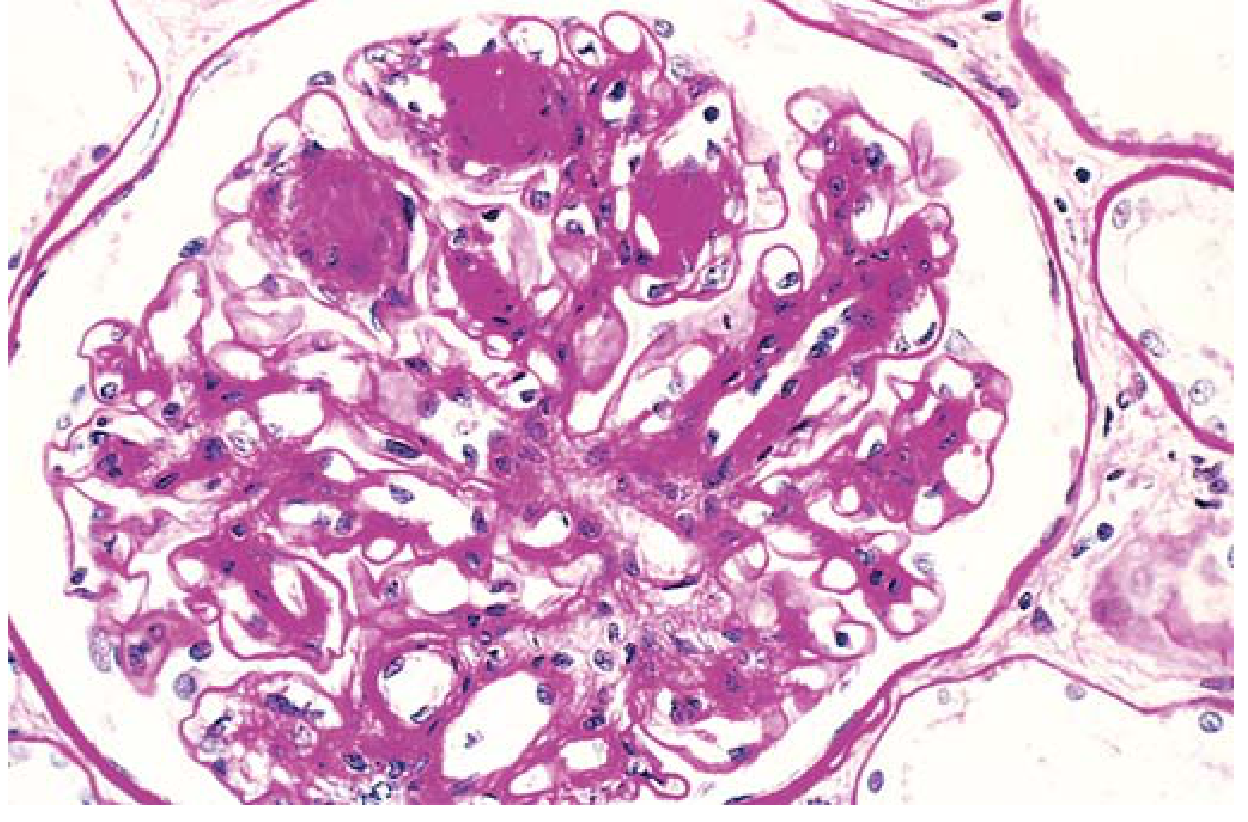
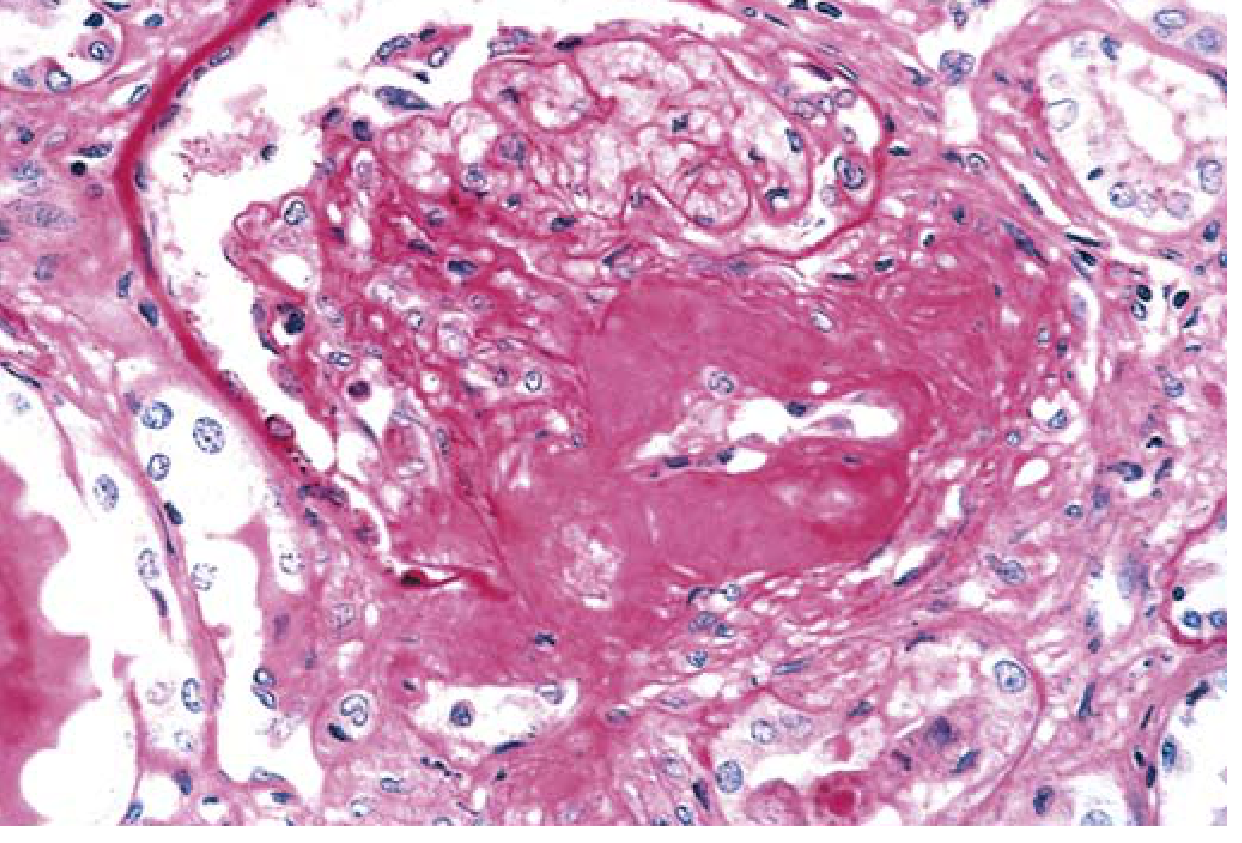
│  ③ Nodular Glomerulosclerosis                           │
│      = KIMMELSTIEL-WILSON (K-W) LESION ★               │
│      ↓                                                  │
│      Ball-like laminated matrix deposits                │
│      Located in PERIPHERY of glomerulus                 │
│      Seen in ~15%-30% of long-term diabetics            │
│      PATHOGNOMONIC of Diabetes                          │
│      (Unlike diffuse form, nodular = virtually          │
│       diagnostic of DM)                                 │
│      → Major contributor to renal dysfunction           │
└─────────────────────────────────────────────────────────┘
FLOW CHART 2 - VASCULAR LESIONS
DIABETES
    ↓
Macrovascular disease (atherosclerosis)
    +
Renal Arteriolosclerosis
    ↓
HYALINE ARTERIOLOSCLEROSIS
    │
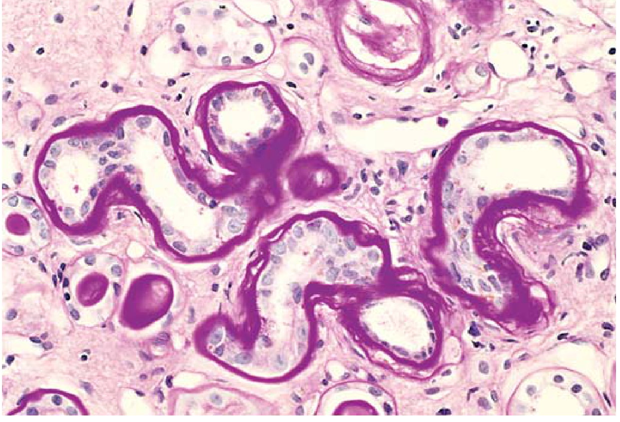
    ├── Affects AFFERENT arteriole (also in non-DM hypertension)
    │
    └── Affects EFFERENT arteriole ← UNIQUE TO DIABETES ★
                ↓
    Vascular compromise + Glomerulosclerosis
                ↓
    Diffuse ischemia → Kidney scarring
                ↓
    Finely granular cortical surface = NEPHROSCLEROSIS
FLOW CHART 3 - PYELONEPHRITIS / PAPILLARY NECROSIS
DIABETES → Impaired immunity + Vascular compromise
    ↓
PYELONEPHRITIS (more severe in diabetics)
    │
    ├── Acute Pyelonephritis
    │
    ├── Chronic Pyelonephritis
    │
    └── NECROTIZING PAPILLITIS (special pattern)
            ↓
        Necrosis of renal papillae
        (particularly dangerous in DM)
PROGRESSION FLOW (Clinical Stages)
Early DM
    ↓
GBM thickening (EM only, no symptoms)
    ↓
Microalbuminuria (30-300 mg/day) — earliest clinical sign
    ↓
Proteinuria (frank, >300 mg/day)
    ↓
Nephrotic Syndrome (severe diffuse/nodular glomerulosclerosis)
    ↓
↓ GFR → Chronic Kidney Disease (CKD)
    ↓
End-Stage Renal Disease (ESRD) - up to 40% of both T1DM & T2DM
MEMORY TABLE - Quick Revision
| Feature | Details |
|---|---|
| Earliest lesion | GBM thickening (EM) |
| Earliest clinical sign | Microalbuminuria |
| Pathognomonic lesion | Nodular glomerulosclerosis (K-W lesion) |
| K-W lesion morphology | Ball-like laminated matrix, periphery of glomerulus |
| Unique vascular finding | Efferent arteriole hyalinosis |
| Frequency of K-W lesion | 15%-30% of long-term DM |
| End result of vascular disease | Nephrosclerosis (granular cortex) |
| Special infection pattern | Necrotizing papillitis |
HISTOLOGY IMAGES FROM ROBBINS
ONE-LINE EXAM TRICKS
- "K-W lesion" = NODULAR glomerulosclerosis = ONLY in DM = pathognomonic
- Diffuse form = more common, NOT pathognomonic
- Efferent arteriole hyalinosis = unique to DM (afferent is seen in hypertension too)
- EM is needed to detect earliest lesion (GBM thickening)
- Necrotizing papillitis = special pyelonephritis pattern in DM
Source: Robbins & Kumar Basic Pathology, Chapter 18 (Endocrine Pancreas), pp. 751-752
Create a summary Word document of these pathology notes for study
ls -la /tmp/workspace/ 2>/dev/null || mkdir -p /tmp/workspace && echo "created"
mkdir -p /tmp/workspace/diabetic-nephropathy && python3 << 'EOF' from docx import Document from docx.shared import Pt, Inches, RGBColor from docx.enum.text import WD_ALIGN_PARAGRAPH from docx.oxml.ns import qn from docx.oxml import OxmlElement import requests import os doc = Document() # ── Page margins ───────────────────────────────────────────────────────────── from docx.shared import Cm section = doc.sections[0] section.top_margin = Cm(2) section.bottom_margin = Cm(2) section.left_margin = Cm(2.5) section.right_margin = Cm(2.5) # ── Default normal style ────────────────────────────────────────────────────── normal = doc.styles["Normal"] normal.font.name = "Arial" normal.font.size = Pt(11) # Helper: add shaded box paragraph def add_shaded_para(doc, text, bg_hex="E8F4FD", bold=False, size=11): p = doc.add_paragraph() p.paragraph_format.space_before = Pt(2) p.paragraph_format.space_after = Pt(2) pPr = p._p.get_or_add_pPr() shd = OxmlElement("w:shd") shd.set(qn("w:val"), "clear") shd.set(qn("w:color"), "auto") shd.set(qn("w:fill"), bg_hex) pPr.append(shd) run = p.add_run(text) run.bold = bold run.font.size = Pt(size) run.font.name = "Arial" return p # Helper: bullet def add_bullet(doc, text, level=0, bold_part=None): p = doc.add_paragraph(style="List Bullet") p.paragraph_format.left_indent = Inches(0.3 * (level + 1)) p.paragraph_format.space_after = Pt(2) if bold_part and bold_part in text: idx = text.index(bold_part) before = text[:idx] after = text[idx + len(bold_part):] if before: r = p.add_run(before); r.font.name = "Arial"; r.font.size = Pt(11) rb = p.add_run(bold_part); rb.bold = True; rb.font.name = "Arial"; rb.font.size = Pt(11) if after: r = p.add_run(after); r.font.name = "Arial"; r.font.size = Pt(11) else: r = p.add_run(text); r.font.name = "Arial"; r.font.size = Pt(11) return p # ── TITLE ───────────────────────────────────────────────────────────────────── title = doc.add_heading("Diabetic Nephropathy", level=1) title.alignment = WD_ALIGN_PARAGRAPH.CENTER for run in title.runs: run.font.color.rgb = RGBColor(0x1A, 0x53, 0x7E) run.font.size = Pt(22) sub = doc.add_paragraph("Pathology Notes | Robbins & Kumar Basic Pathology | MBBS 6 Marks") sub.alignment = WD_ALIGN_PARAGRAPH.CENTER sub.runs[0].font.color.rgb = RGBColor(0x55, 0x55, 0x55) sub.runs[0].font.size = Pt(11) sub.runs[0].italic = True doc.add_paragraph() # spacer # ── OVERVIEW ───────────────────────────────────────────────────────────────── h2 = doc.add_heading("Overview", level=2) for run in h2.runs: run.font.color.rgb = RGBColor(0x1A, 0x53, 0x7E) add_shaded_para(doc, "The kidneys are prime targets of diabetes. Diabetic nephropathy is the leading cause of chronic kidney failure in the United States. " "Up to 40% of both Type 1 and Type 2 diabetics develop end-stage renal disease.", bg_hex="EAF4FB") doc.add_paragraph() # ── MAIN LESIONS OVERVIEW ───────────────────────────────────────────────────── h2 = doc.add_heading("Three Major Categories of Lesions", level=2) for run in h2.runs: run.font.color.rgb = RGBColor(0x1A, 0x53, 0x7E) cats = [ ("1. Glomerular Lesions", "Most important"), ("2. Renal Vascular Lesions", "Principally arteriolosclerosis"), ("3. Pyelonephritis", "Including necrotizing papillitis"), ] for title_txt, detail in cats: p = doc.add_paragraph() p.paragraph_format.left_indent = Inches(0.3) p.paragraph_format.space_after = Pt(3) r1 = p.add_run(f" {title_txt} ") r1.bold = True; r1.font.name = "Arial"; r1.font.size = Pt(11) r2 = p.add_run(f"— {detail}") r2.font.name = "Arial"; r2.font.size = Pt(11) r2.font.color.rgb = RGBColor(0x44, 0x44, 0x44) doc.add_paragraph() # ── SECTION 1: GLOMERULAR LESIONS ───────────────────────────────────────────── h2 = doc.add_heading("1. Glomerular Lesions (Most Important)", level=2) for run in h2.runs: run.font.color.rgb = RGBColor(0xC0, 0x39, 0x2B) # 1a GBM h3 = doc.add_heading("A. Capillary Basement Membrane (GBM) Thickening", level=3) for run in h3.runs: run.font.color.rgb = RGBColor(0x1F, 0x61, 0x8D) add_bullet(doc, "Earliest structural lesion of diabetic nephropathy", bold_part="Earliest structural lesion") add_bullet(doc, "Detected by Electron Microscopy (EM) — within a few years of DM onset", bold_part="Electron Microscopy (EM)") add_bullet(doc, "Precedes any change in renal function") add_bullet(doc, "GBM thickened along its entire length") add_shaded_para(doc, "KEY POINT: Earliest lesion — seen on EM before symptoms appear", bg_hex="FEF9E7", bold=True) doc.add_paragraph() # 1b Diffuse h3 = doc.add_heading("B. Diffuse Mesangial Sclerosis", level=3) for run in h3.runs: run.font.color.rgb = RGBColor(0x1F, 0x61, 0x8D) add_bullet(doc, "Increased mesangial matrix + mesangial cell proliferation", bold_part="Increased mesangial matrix") add_bullet(doc, "Associated with basement membrane thickening") add_bullet(doc, "Found in most patients with DM > 10 years duration", bold_part="DM > 10 years") add_bullet(doc, "More common in older patients and those with hypertension") add_bullet(doc, "When severe → Nephrotic Syndrome:", bold_part="Nephrotic Syndrome") add_bullet(doc, "Proteinuria + Hypoalbuminemia + Edema", level=1) add_bullet(doc, "More common but NOT pathognomonic of diabetes", bold_part="NOT pathognomonic") doc.add_paragraph() # 1c Nodular h3 = doc.add_heading("C. Nodular Glomerulosclerosis (Kimmelstiel-Wilson Lesion) ★", level=3) for run in h3.runs: run.font.color.rgb = RGBColor(0x1F, 0x61, 0x8D) add_shaded_para(doc, "PATHOGNOMONIC of Diabetes — virtually diagnostic", bg_hex="FDEDEC", bold=True) add_bullet(doc, "Ball-like deposits of laminated matrix material", bold_part="Ball-like deposits of laminated matrix") add_bullet(doc, "Located in the PERIPHERY of the glomerulus", bold_part="PERIPHERY") add_bullet(doc, "Seen in ~15%–30% of long-term diabetics", bold_part="15%–30%") add_bullet(doc, "Major contributor to renal dysfunction") add_bullet(doc, "In contrast to diffuse form — nodular = PATHOGNOMONIC", bold_part="PATHOGNOMONIC") doc.add_paragraph() # ── SECTION 2: VASCULAR LESIONS ─────────────────────────────────────────────── h2 = doc.add_heading("2. Renal Vascular Lesions", level=2) for run in h2.runs: run.font.color.rgb = RGBColor(0xC0, 0x39, 0x2B) add_bullet(doc, "Renal atherosclerosis — part of macrovascular disease in DM") add_bullet(doc, "Hyaline arteriolosclerosis — most important vascular change") h3 = doc.add_heading("Hyaline Arteriolosclerosis — Key Points:", level=3) for run in h3.runs: run.font.color.rgb = RGBColor(0x1F, 0x61, 0x8D) add_bullet(doc, "Affects AFFERENT arteriole (also seen in non-DM hypertension)", bold_part="AFFERENT arteriole") add_bullet(doc, "Affects EFFERENT arteriole — UNIQUE TO DIABETES ★", bold_part="EFFERENT arteriole — UNIQUE TO DIABETES ★") add_bullet(doc, "Vascular compromise + Glomerulosclerosis → Diffuse ischemia") add_bullet(doc, "Result: Diffuse kidney scarring → Finely granular cortical surface", bold_part="Finely granular cortical surface") add_bullet(doc, "End appearance = NEPHROSCLEROSIS", bold_part="NEPHROSCLEROSIS") doc.add_paragraph() # ── SECTION 3: PYELONEPHRITIS ───────────────────────────────────────────────── h2 = doc.add_heading("3. Pyelonephritis / Papillary Necrosis", level=2) for run in h2.runs: run.font.color.rgb = RGBColor(0xC0, 0x39, 0x2B) add_bullet(doc, "Acute or chronic inflammation — begins in interstitium, spreads to tubules") add_bullet(doc, "More SEVERE in diabetics than non-diabetics", bold_part="More SEVERE in diabetics") add_bullet(doc, "Special pattern: Necrotizing Papillitis", bold_part="Necrotizing Papillitis") add_bullet(doc, "Necrosis of renal papillae — particularly dangerous in DM", level=1) add_bullet(doc, "Can cause acute renal failure", level=1) doc.add_paragraph() # ── CLINICAL PROGRESSION FLOW ───────────────────────────────────────────────── h2 = doc.add_heading("Clinical Progression (Staging Flow)", level=2) for run in h2.runs: run.font.color.rgb = RGBColor(0x1A, 0x53, 0x7E) stages = [ ("Stage 1 (Early DM)", "GBM thickening — detected only on EM, no clinical signs"), ("Stage 2 (Microalbuminuria)", "Albumin 30–300 mg/day — earliest CLINICAL sign"), ("Stage 3 (Overt Proteinuria)", "Albumin >300 mg/day — frank proteinuria"), ("Stage 4 (Nephrotic Syndrome)", "Diffuse/nodular glomerulosclerosis; GFR declining"), ("Stage 5 (ESRD)", "Up to 40% of T1DM and T2DM; requires dialysis/transplant"), ] for stage, desc in stages: p = doc.add_paragraph() p.paragraph_format.left_indent = Inches(0.3) p.paragraph_format.space_after = Pt(3) r1 = p.add_run(f"{stage}: ") r1.bold = True; r1.font.name = "Arial"; r1.font.size = Pt(11) r1.font.color.rgb = RGBColor(0x1A, 0x53, 0x7E) r2 = p.add_run(desc) r2.font.name = "Arial"; r2.font.size = Pt(11) doc.add_paragraph() # ── QUICK REVISION TABLE ────────────────────────────────────────────────────── h2 = doc.add_heading("Quick Revision Table", level=2) for run in h2.runs: run.font.color.rgb = RGBColor(0x1A, 0x53, 0x7E) table = doc.add_table(rows=1, cols=2) table.style = "Table Grid" # Header row hdr_cells = table.rows[0].cells for cell in hdr_cells: cell._tc.get_or_add_tcPr() shd = OxmlElement("w:shd") shd.set(qn("w:val"), "clear") shd.set(qn("w:color"), "auto") shd.set(qn("w:fill"), "1A537E") cell._tc.tcPr.append(shd) hdr_cells[0].text = "Feature" hdr_cells[1].text = "Key Details" for cell in hdr_cells: for para in cell.paragraphs: for run in para.runs: run.bold = True run.font.color.rgb = RGBColor(0xFF, 0xFF, 0xFF) run.font.name = "Arial" run.font.size = Pt(11) rows_data = [ ("Earliest lesion", "GBM thickening — detected by Electron Microscopy (EM)"), ("Earliest clinical sign", "Microalbuminuria (30–300 mg/day)"), ("Pathognomonic lesion", "Nodular glomerulosclerosis — Kimmelstiel-Wilson (K-W) lesion"), ("K-W lesion morphology", "Ball-like laminated matrix deposits, periphery of glomerulus"), ("K-W lesion frequency", "15%–30% of long-term diabetics"), ("Unique vascular finding", "Efferent arteriole hyalinosis — unique to diabetes"), ("End result of vasculopathy","Nephrosclerosis (finely granular cortical surface)"), ("Special infection pattern", "Necrotizing papillitis"), ("ESRD risk", "Up to 40% of both T1DM and T2DM"), ("Diffuse vs Nodular", "Diffuse = common, NOT pathognomonic; Nodular = PATHOGNOMONIC"), ] fill_colors = ["F2F9FF", "FFFFFF"] for i, (feat, detail) in enumerate(rows_data): row = table.add_row() row.cells[0].text = feat row.cells[1].text = detail for j, cell in enumerate(row.cells): shd = OxmlElement("w:shd") shd.set(qn("w:val"), "clear") shd.set(qn("w:color"), "auto") shd.set(qn("w:fill"), fill_colors[i % 2]) cell._tc.get_or_add_tcPr().append(shd) for para in cell.paragraphs: for run in para.runs: run.font.name = "Arial" run.font.size = Pt(10.5) doc.add_paragraph() # ── EXAM TIPS ───────────────────────────────────────────────────────────────── h2 = doc.add_heading("Exam Memory Tips", level=2) for run in h2.runs: run.font.color.rgb = RGBColor(0x1A, 0x53, 0x7E) tips = [ ('K-W lesion', ' = NODULAR glomerulosclerosis = ONLY in DM = pathognomonic of diabetes'), ('Diffuse form', ' = more common but NOT pathognomonic'), ('Efferent arteriole hyalinosis', ' = UNIQUE to DM (afferent also seen in hypertension)'), ('EM needed', ' to detect earliest lesion (GBM thickening)'), ('Necrotizing papillitis', ' = special pyelonephritis pattern unique to DM'), ('Complications appear', ' approximately 15–20 years after onset of hyperglycemia'), ] for bold_part, rest in tips: p = doc.add_paragraph() p.paragraph_format.left_indent = Inches(0.3) p.paragraph_format.space_after = Pt(3) r = p.add_run("★ ") r.font.color.rgb = RGBColor(0xE6, 0x7E, 0x22) r.font.name = "Arial"; r.font.size = Pt(11) rb = p.add_run(bold_part) rb.bold = True; rb.font.name = "Arial"; rb.font.size = Pt(11) r2 = p.add_run(rest) r2.font.name = "Arial"; r2.font.size = Pt(11) doc.add_paragraph() # ── SOURCE ──────────────────────────────────────────────────────────────────── add_shaded_para(doc, "Source: Robbins & Kumar Basic Pathology, Chapter 18 — Endocrine Pancreas, pp. 751–752", bg_hex="F4F6F7", bold=False) # ── SAVE ───────────────────────────────────────────────────────────────────── out_path = "/tmp/workspace/diabetic-nephropathy/Diabetic_Nephropathy_Pathology_Notes.docx" doc.save(out_path) print(f"Saved: {out_path}") EOF
python3 -c " from docx import Document doc = Document('/tmp/workspace/diabetic-nephropathy/Diabetic_Nephropathy_Pathology_Notes.docx') count = 0 for p in doc.paragraphs: if p.text.strip(): print(f'[{p.style.name}] {p.text[:80]}') count += 1 if count > 30: break print('...') # check table for t in doc.tables: print(f'TABLE: {len(t.rows)} rows x {len(t.columns)} cols') for row in t.rows[:3]: print([c.text for c in row.cells]) "
Diabetic Nephropathy Pathology Notes
Word Document · DOCX
| Section | Content |
|---|---|
| Title + Subtitle | Formatted heading with source citation |
| Overview | Shaded intro box - DM as leading cause of CKD |
| 3 Major Lesion Categories | Clean numbered list with descriptions |
| Glomerular Lesions (A/B/C) | GBM thickening, Diffuse mesangial sclerosis, K-W nodular lesion - each with bullet points and color-coded key boxes |
| Vascular Lesions | Afferent vs efferent arteriole hyalinosis; unique DM finding starred |
| Pyelonephritis | Necrotizing papillitis explained |
| Clinical Staging Flow | 5 stages from early DM to ESRD |
| Quick Revision Table | 10-row color-banded table of must-know facts |
| Exam Memory Tips | 6 starred exam tricks with bold highlights |
| Source | Robbins & Kumar Basic Pathology, pp. 751-752 |