
E tep recurrent inguinal hernia
E-TEP inguinal hernia recurrent
E-TEP extended totally extraperitoneal inguinal hernia repair laparoscopic technique
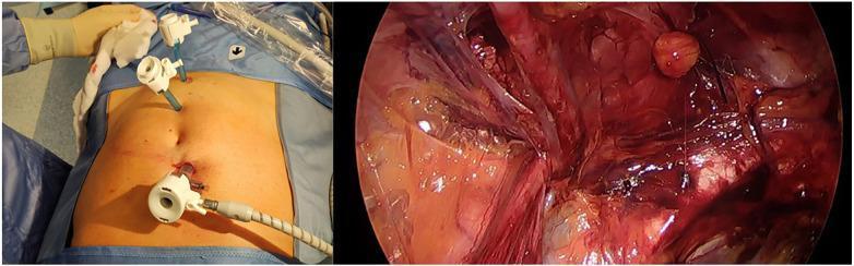
A composite medical image featuring a clinical photograph and a laparoscopic view demonstrating a Totally Extraperitoneal (TEP) inguinal hernia repair procedure. The left panel shows the external abdominal view of a supine patient with surgical draping. Three trocars are visible: one 10 mm optical trocar placed infraumbilically with connected insufflation tubing, and two operative trocars positioned superiorly along the midline. The right panel displays the internal laparoscopic view of the preperitoneal space during inguinal dissection before mesh placement. The surgical field highlights vascularized tissue, anatomical planes, and a prominent reddish-brown structure representing a hernia sac or landmark within the inguinal region. This image illustrates the E-TEP (Enhanced View Totally Extraperitoneal) technique, focusing on trocar disposition to optimize working space and visual access to the inguinal anatomy for hernia management.
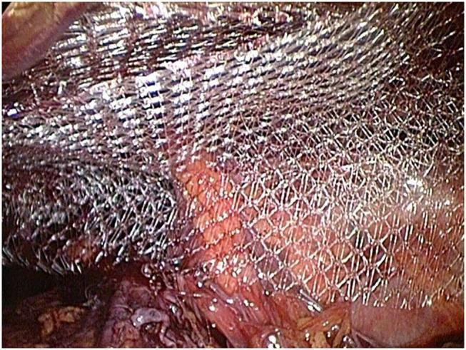
This intraoperative clinical photograph captures a Totally Extraperitoneal (TEP) laparoscopic repair of an inguinal hernia. The central focus is a 15 x 12 cm large-pore monofilament polypropylene surgical mesh (Bard Soft Mesh) being positioned within the preperitoneal space. The mesh exhibits a metallic, silvery-white sheen and a distinct diamond-shaped, porous grid structure designed for optimal tissue integration and pliability. Beneath the semi-transparent mesh, the anatomical landmarks of the inguinal region and Retzius space are visible, specifically showing the reddish-pink vascularized tissue and the underlying musculofascial structures. The image demonstrates the 'double mesh technique' where the prosthetic material is laid over the dissected hernia site without mechanical fixation. This visual serves as a high-level educational example of laparo-endoscopic hernia management, illustrating the proper orientation and coverage of a polypropylene prosthesis over the myopectineal orifice to reinforce the abdominal wall.
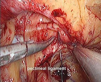
This clinical photograph provides an endoscopic surgical view of the preperitoneal space during a Totally Extraperitoneal (TEP) inguinal hernia repair. The central focus is the pectineal ligament (Cooper's ligament), which is clearly identified by a text label and arrow. The ligament appears as a white, dense fibrous band. Laparoscopic instrumentation is actively engaged in the surgical field: an atraumatic grasping forceps is seen on the left, while a surgical needle and suture material are shown puncturing the pectineal ligament. This step is part of an internal ring suturing technique designed to reduce the size of the hernia defect. The surrounding anatomy shows typical surgical features including minor hemorrhage, adipose tissue, and connective tissue layers within the inguinal region. This image serves as an educational reference for surgical landmarks and laparoscopic suturing techniques in hernia management.
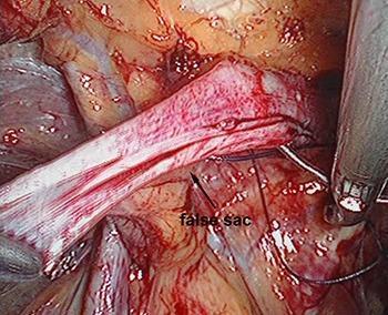
This clinical photograph captures an intraoperative endoscopic view during a Totally Extraperitoneal (TEP) inguinal hernia repair. The central focus is the 'false sac,' identified as a tubular, fibrous structure with a striated, whitish-red texture, indicating vascularization of the peritoneal tissue. A curved surgical suture needle is shown penetrating the base of this sac to secure it to the conjoined tendon or pectineal ligament. Metallic laparoscopic instruments, including grasping forceps, are visible on the periphery, manipulating the surrounding preperitoneal adipose and connective tissues. The image demonstrates the surgical technique of internal ring suturing to reduce the defect size before mesh placement. Key educational concepts include the identification of hernia sac morphology, management of the preperitoneal space, and laparoscopic suturing techniques in abdominal wall reconstruction. The clinical relevance pertains to surgical anatomy and the prevention of hernia recurrence through mechanical reinforcement of the internal ring.
PMID: 41258609
E-TEP for Recurrent Inguinal Hernia
What is E-TEP?
When is E-TEP Used in Recurrent Inguinal Hernia?
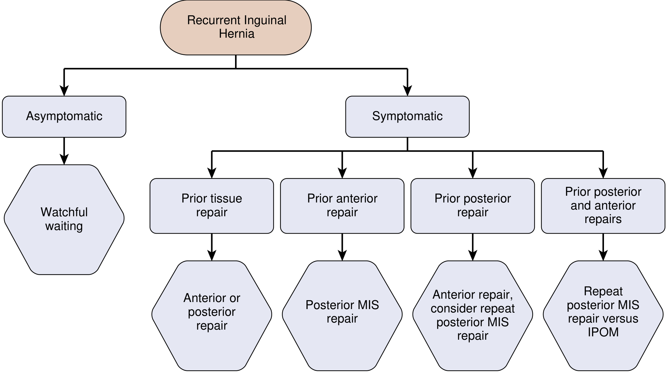
| Prior Repair | Recommended Approach |
|---|---|
| Tissue repair only | Anterior or posterior repair |
| Prior anterior repair (e.g., Lichtenstein) | Posterior MIS (TAPP / TEP / e-TEP) - preferred |
| Prior posterior repair (TAPP/TEP) | Anterior repair; consider repeat posterior MIS |
| Both anterior + posterior repairs | Repeat posterior MIS vs. IPOM |
| Asymptomatic | Watchful waiting |
Advantages of E-TEP/TEP Over TAPP in Recurrence
-
Minimizes intraabdominal adhesions - stays entirely extraperitoneal
-
No peritoneal entry - avoids bowel/visceral injury risk
-
Better mesh positioning - expanded view allows larger mesh with more overlap
-
Simultaneous contralateral inspection - can identify contralateral occult hernia
-
A recent retrospective study found 96% success rate for second laparoscopic repair with low late re-recurrence rates
-
Current Surgical Therapy 14e, p. 711-712
Technical Considerations for E-TEP in Recurrence
Preoperative Planning
- Review all prior operative notes: mesh type, plane used, dates, any infection
- Obtain CT or MRI if occult recurrence or complex anatomy (MRI has highest sensitivity/specificity for occult hernia and mesh neuromas)
- Document MRSA history for antibiotic prophylaxis
Intraoperative Challenges
- Distorted tissue planes + scar tissue make dissection more difficult than primary repair
- Tearing of the peritoneum during preperitoneal dissection is more likely in redo cases
- If peritoneum tears extensively, convert to TAPP - every TEP surgeon should be comfortable with both techniques
- For large inguinoscrotal recurrences, TAPP may be preferred due to better visualization and ability to reduce incarcerated contents
Mesh Considerations
- Always use mesh in recurrent repair - tissue is already weakened from prior repair
- Lightweight polypropylene is preferred: less chronic pain, reduced mesh sensation, better ingrowth
- Prior mesh removal: no clear consensus; complete removal is often difficult and not always necessary
- Only exception to mesh use: active infection (use biologic mesh if still required)
E-TEP vs TEP vs TAPP: Comparative Outcomes
| Parameter | e-TEP | TAPP | TEP |
|---|---|---|---|
| Case selection | Larger (W3) hernias | Irreducible hernias | Smaller hernias |
| Mesh fixation rate | 86.7% | 97.7% | 38.2% |
| Operative time | 87.3 min | 94.1 min | 70.2 min |
| Seroma / SSI / hematoma / recurrence | Comparable across all three | - | - |
The Complex / Multiply Recurrent Patient
- Patients with multiple prior repairs (both open and laparoscopic) are the most challenging
- May have mesh in more than one anatomical plane
- Laparoscopic approach is still recommended first - diagnostic laparoscopy helps survey direct, indirect, and femoral spaces
- If repeat TAPP is not feasible or peritoneum is deficient: IPOM (intraperitoneal onlay mesh) is the last resort using dual-layer mesh with anti-adhesive barrier
- Consider referral to a high-volume hernia center
- Re-recurrence risk increases with each subsequent repair - set realistic patient expectations
E-TEP Technique Highlights (Intraoperative)
- Access: Optical trocar placed below umbilicus, balloon or blunt dissection to enter preperitoneal space
- Extended dissection: Crossing midline to reach contralateral preperitoneal space (the "extended view")
- Hernia sac reduction: Indirect sac dissected off cord structures; internal ring suturing may be used to reduce defect
- Cooper's ligament identification: Mandatory landmark for proper mesh positioning
- Mesh placement: Large mesh (≥15x12 cm) laid over the myopectineal orifice; fixation with tacks or sutures depending on surgeon preference
- No peritoneal closure needed (unlike TAPP)
Summary
- E-TEP is preferred for recurrent inguinal hernia after prior anterior (open) repair, as the posterior plane is undisturbed
- It offers wider mesh coverage and better visualization than standard TEP, with outcomes comparable to TAPP
- Convert to TAPP if peritoneum tears; convert to open if dense adhesions prevent safe laparoscopic dissection
- Always use lightweight polypropylene mesh; optimize all risk factors preoperatively
- For multiply recurrent or complex cases, diagnostic laparoscopy first, IPOM as last resort, and consider specialist referral
What are the risk factors for hernia recurrence after redo repair?
inguinal hernia recurrence risk factors redo repair
inguinal hernia recurrence risk factors laparoscopic repair
PMID: 38717561
Risk Factors for Hernia Recurrence After Redo Repair
1. Nonmodifiable Patient Factors
| Factor | Details |
|---|---|
| Connective tissue disorders | Hernia-prone patients often have aberrant collagen metabolism (e.g., Ehlers-Danlos, Marfan); defective fibrillar collagen impairs mesh integration |
| Hernia type | Direct hernias and sliding hernias have higher recurrence rates than indirect; femoral hernias are notorious for re-recurrence |
| Female sex | Higher recurrence rates reported in females, partly due to higher rates of femoral hernias and smaller pelvis anatomy |
| Number of prior repairs | Re-recurrence risk increases multiplicatively with each subsequent repair |
2. Modifiable Patient Factors
Smoking
- Smoking impairs tissue oxygenation, prolongs inflammatory phase, and delays wound healing
- Increases risk of surgical site infections, hematomas, and seromas - all independent recurrence drivers
- A 2024 meta-analysis of 577,901 patients (Marcolin et al., Hernia) found smoking confers a nearly 3-fold increase in recurrence risk (OR 2.95; 95% CI 2.08-4.18; I² = 0%)
- Nicotine replacement therapy does not fully reverse these risks but can be offered to patients who cannot quit
- Smoking cessation for at least 4 weeks before elective surgery reduces SSI risk in open repairs
Obesity (BMI)
- Increases mechanical strain and tension on the repair construct
- BMI is independently associated with rising SSI rates (risk peaks above BMI 42)
- Obesity impairs wound healing through poor tissue perfusion and immune dysfunction
- Weight loss before redo repair is encouraged, but must be weighed against incarceration risk if delaying surgery
Poor Glycaemic Control / Diabetes
- Hyperglycaemia impairs neutrophil bactericidal activity
- For every 40 mg/dL rise above normoglycaemia: 30% increase in postoperative infection rate
- HbA1c optimization before elective redo repair is standard practice
Immunosuppression
- Corticosteroids and other immunosuppressive agents impair collagen synthesis and wound healing
- Increase surgical site infection risk and secondary mesh failure
Chronic Cough / COPD
- Elevated intra-abdominal pressure from chronic coughing stresses any mesh repair
- Address medically before redo surgery; COPD is listed as a preoperative risk factor in Sabiston (ASA III-IV is an independent predictor)
Postoperative Surgical Site Occurrences (SSOs)
- Seromas, hematomas, wound infections, and wound dehiscence after the redo repair all independently raise re-recurrence risk
- SSOs set off a "vicious cycle of diminishing returns" - wound breakdown leads to mesh exposure, infection, removal, and further tissue weakness
3. Technical Factors
| Technical Error | Consequence |
|---|---|
| Failure to achieve tension-free repair | Tissue repairs (Bassini, McVay, Halsted) carry 50-75% higher recurrence than mesh repairs |
| Inadequate dissection / incomplete sac reduction | Residual hernia sac or missed concurrent direct/indirect component |
| Missed lipoma of the cord | Unresected cord lipoma presents as apparent re-recurrence (pseudo-recurrence) |
| Inadequate mesh size / overlap | Insufficient coverage of myopectineal orifice; mesh must cover all three potential hernia sites (direct, indirect, femoral) |
| Wrong tissue plane | Violating a previously scarred plane instead of finding virgin tissue |
| Surgeon volume / experience | Recurrence rates are inversely related to surgeon volume - low-volume surgeons have significantly higher rates |
| Poor fixation | Mesh migration or folding in early postoperative period before fibrous ingrowth |
Surgeon experience is the most important single variable for both open and laparoscopic approaches. Low-volume surgeons should consider referral of complex recurrent cases to high-volume hernia centres.
4. Mesh Factors
| Factor | Details |
|---|---|
| No mesh used | Tissue-only repairs in redo surgery are almost never appropriate given already weakened tissue |
| Wrong mesh type | Heavy-weight mesh: associated with more chronic pain and mesh contraction; lightweight polypropylene preferred for better tissue integration |
| Mesh infection | Requires removal; bare patch of weakened tissue left behind has very high re-recurrence risk |
| Mesh shrinkage / contraction | All meshes shrink 20-30% post-implantation; undersized mesh or poor overlap leaves defect uncovered |
| Mesh migration | Particularly with non-fixated or improperly placed mesh |
Summary Table
| Category | Key Risk Factors |
|---|---|
| Nonmodifiable | CTD/genetics, direct hernia, femoral hernia, female sex, prior repair count |
| Patient (modifiable) | Smoking, obesity, hyperglycaemia, immunosuppression, COPD/chronic cough |
| Technical | Tension repair, missed sac/lipoma, small mesh, wrong plane, low surgeon volume |
| Mesh | No mesh, infected mesh, shrinkage, migration, heavy-weight mesh |
| Post-op SSO | Infection, seroma, hematoma, wound dehiscence |
Clinical Takeaway
- Stop smoking (minimum 4 weeks, ideally 8+ weeks preoperatively)
- Optimize BMI - weight loss if feasible without undue delay
- Control HbA1c - aim below 8% before elective surgery
- Review prior operative notes - understand exactly what plane and mesh was used
- Approach through unviolated tissue - use posterior (e-TEP/TAPP/TEP) after prior anterior repair
- Use lightweight polypropylene mesh with adequate overlap
- Set realistic patient expectations - re-recurrence rates are higher than primary repair recurrence rates regardless of technique