
What is swelling
swelling definition types causes pathophysiology medical
pitting edema swelling subcutaneous tissue
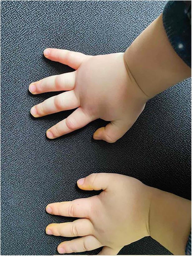
This clinical photograph shows the bilateral hands of a pediatric patient exhibiting significant non-pitting edema. The swelling is most pronounced across the dorsum of the hands, palms, and wrists, causing the skin to appear taut and shiny. The fingers show diffuse enlargement (angioedema) with a loss of normal anatomical definition around the joints. Notably, the skin remains pale and lacks associated erythema, urticaria (hives), or bruising, which are key diagnostic features in distinguishing hereditary angioedema (HAE) from allergic reactions. This image serves as a classic educational representation of peripheral angioedema in a child with HAE Type I, characterized by deep dermal and subcutaneous tissue swelling. The visual evidence illustrates the clinical manifestation of C1 esterase inhibitor deficiency, highlighting the localized, non-inflammatory nature of the edema during an acute attack.

Clinical photograph presenting anterior (left) and lateral (right) views of the male genital region. The images demonstrate significant, diffuse swelling of the penis. The enlargement involves the entire shaft, resulting in a tense, thickened appearance of the subcutaneous tissues. The overlying skin appears smooth and intact without visible ulceration, erythema, or discrete nodules. The lateral view confirms the circumferential nature of the edema. This clinical presentation of diffuse, non-pitting penile swelling can be associated with various conditions, including lymphatic obstruction, vascular malformations, or systemic deposition disorders such as pretibial myxedema (extratecticular thyroid dermopathy). The visual serves as an educational example of localized soft tissue edema in the genital region, highlighting the need for diagnostic differentiation between inflammatory, vascular, and metabolic etiologies.
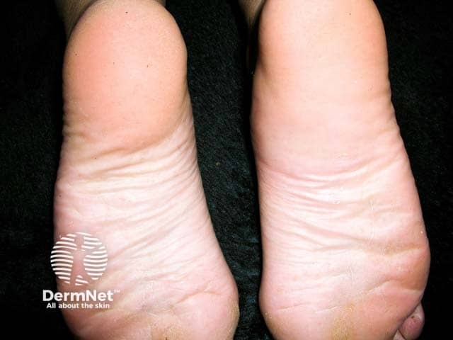
Clinical photography of both feet showing acute non-pitting edema characteristic of angioedema. Modality: clinical photography; technique: color-calibrated digital capture with macro lens, frontal and oblique views. Anatomical context: integumentary system; pedal region, dorsal and plantar aspects. Laterality: bilateral. The swelling predominantly involves the dorsal aspects of the feet with diffuse, soft-tissue enlargement of toes and forefoot; skin remains intact without obvious erythema, warmth, or fluctuance. No vesicles, wheals, or bullae are evident in the image. Subcutaneous tissue appears expanded with smooth contours; no lymphatic stasis or skin crepitus. The edema seems to spare the deeper structures; no obvious necrosis. Observed features are compatible with angioedema, which involves transient, localized swelling of the deeper dermis and subcutaneous tissues. Clinical correlation is necessary to distinguish monosymptomatic angioedema from urticarial edema; systemic symptoms are not captured in this static photograph. Potential etiologies include allergic or nonallergic hypersensitivity reactions, mast cell activation, ACE inhibitor–related angioedema, hereditary C1 esterase inhibitor deficiency, or idiopathic causes. The image highlights diagnostic significance for dermatology and emergency medicine by illustrating typical pedal edema patterns and distribution. This visualization supports differential diagnosis, patient counseling, and monitoring response to therapy such as antihistamines, corticosteroids, or fresh frozen plasma in specific contexts.
Swelling (Edema)
Definition
- Costanzo Physiology 7th Edition, p. 178
- Medical Physiology (Boron & Boulpaep), Box 20-1
How Does Fluid Normally Stay in Balance?
- Hydrostatic pressure (Pc) - pushes fluid OUT of capillaries into tissues
- Oncotic (colloid osmotic) pressure (πc) - pulls fluid BACK into capillaries via plasma proteins
Causes of Swelling / Edema
| Cause | Mechanism | Examples |
|---|---|---|
| Increased capillary hydrostatic pressure | More fluid pushed out of vessels | Heart failure, venous obstruction, prolonged standing |
| Decreased plasma oncotic pressure | Less fluid pulled back into vessels | Nephrotic syndrome (protein lost in urine), liver failure (less protein made), malnutrition |
| Increased capillary permeability | Leaky vessel walls | Burns, inflammation, allergic reactions (histamine release) |
| Impaired lymphatic drainage | Fluid cannot be returned | Lymph node removal/irradiation, parasitic filariasis (elephantiasis) |
- Costanzo Physiology, Table 4.6
Types of Swelling
By distribution:
- Localized - affects one area (e.g., ankle swelling after a sprain, swelling from an insect bite, cellulitis)
- Generalized (Anasarca) - widespread; common in severe heart, kidney, or liver disease
By tissue affected:
| Type | Location | Common Cause |
|---|---|---|
| Peripheral edema | Legs, ankles, feet, hands | Heart failure, venous insufficiency |
| Pulmonary edema | Lungs | Left-sided heart failure |
| Cerebral edema | Brain | Stroke, trauma, infection |
| Ascites | Peritoneal cavity | Liver cirrhosis, portal hypertension |
| Pleural effusion | Pleural space (around lungs) | Heart failure, infection |
| Periorbital edema | Around the eyes | Renal disease, allergy |
| Lymphedema | Extremities | Lymph node removal, filariasis |
By exam finding:
- Pitting edema - finger pressure on swollen tissue leaves a depression (pit), indicating fluid displacement in the interstitium. Classic in heart failure and kidney disease.
- Non-pitting edema - no pit forms; seen in lymphedema and myxedema (thyroid disease).
Where Does Swelling Appear First?
-
Gravity-dependent areas: legs and feet when standing; sacrum (lower back) when bedridden - called dependent edema
-
Loose connective tissue: around the eyes (periorbital edema) - often the first sign of renal disease
-
Generalized: in severe cases, fluid accumulates throughout the body
-
Robbins & Cotran Pathologic Basis of Disease, p. 118
Clinical Consequences
-
Subcutaneous (under-skin) edema: mostly a sign of underlying heart or kidney disease; can impair wound healing
-
Pulmonary edema: impairs gas exchange, causes breathlessness, and increases risk of bacterial infection - can be life-threatening
-
Cerebral edema: the brain swells against the rigid skull, potentially causing brain herniation and death
-
Ascites (abdominal): prone to bacterial infection (peritonitis)
-
Robbins & Cotran Pathologic Basis of Disease, Clinical Features section