
Mammary gland LAQ for 1st year mbbs as per vishram singh
mammary gland anatomy structure diagram
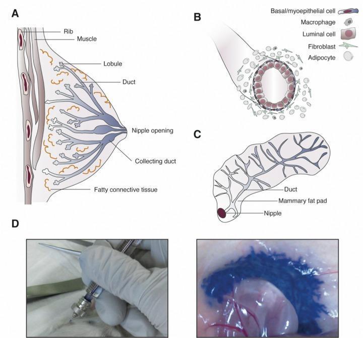
Educational medical graphic detailing mammary gland anatomy and intraductal (ID) injection methodology. Panel A: Cross-sectional anatomical diagram of the human breast, illustrating ribs, pectoral muscle, and the ductolobular system branching from the nipple opening through fatty connective tissue. Panel B: Histological diagram of a mammary duct, highlighting its bilayered epithelium consisting of inner luminal cells and outer basal/myoepithelial cells, surrounded by macrophages, fibroblasts, and adipocytes. Panel C: Schematic of a murine mammary gland, showing the single efferent duct and branching network within the mammary fat pad. Panel D: Clinical photographs demonstrating a preclinical procedure in a mouse model. The left image shows cannulation of the nipple using a short, blunt-ended needle for intraductal delivery. The right image displays the distribution of blue dye within the ductal system, demonstrating successful infusion of ablative reagents. This figure illustrates the translational approach of using the intraductal route for localized delivery of chemotherapeutics or preventive agents in breast cancer research, such as for BRCA1 mutation carriers.
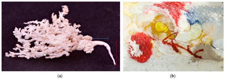
This composite educational image features corrosion casts of mammary gland anatomy. Figure (a) displays a white glandular cast against a black background, illustrating the complex branching architecture of the ductal system. It shows a primary ascendant milk duct (vertical red marker) transitioning into a wider transversal duct (horizontal blue marker) before arborizing into numerous secondary ducts (white arrowhead) and distal clusters representing terminal alveoli (white arrow). The absence of a prominent teat cistern is a notable anatomical variation. Figure (b) demonstrates a cross-sectional view of resin-perfused ducts within preserved tissue. A primary duct is filled with yellow resin, showing the hollow structure of the teat canal, while an adjacent underdeveloped, blind-ended duct is filled with red resin. These casts are used in anatomical research to study ductal morphology, lobation patterns, and developmental variations such as atrophic or rudimentary ducts, providing insight into physiological milk drainage and the potential pathways for disseminated infections.
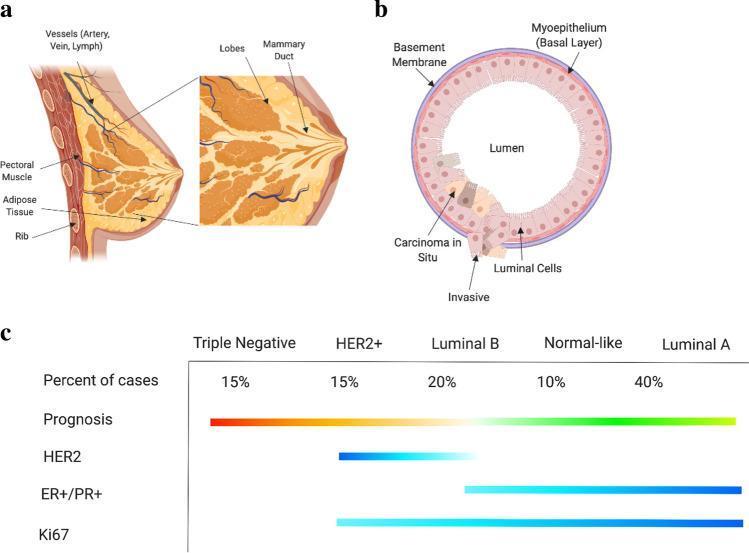
This composite educational infographic illustrates the anatomy of the human breast, its cellular structure, and the molecular subtyping of breast cancer. Panel (a) presents an anatomical diagram of the whole breast in cross-section, labeling the pectoral muscle, ribs, adipose tissue, and the functional unit consisting of lobes and mammary ducts. Panel (b) depicts the cellular anatomy of a mammary duct in cross-section, showing the basement membrane, myoepithelium (basal layer), and luminal cells surrounding a central lumen. It highlights pathological progression from carcinoma in situ to invasive carcinoma, demonstrating the breach of basement membrane integrity. Panel (c) is a comparison chart of breast cancer molecular subtypes: Triple Negative, HER2+, Luminal B, Normal-like, and Luminal A. It correlates these subtypes with clinical and pathological features, including percentage of cases, prognosis (gradient from poor/red to good/green), and the expression levels of key biomarkers such as HER2, ER+/PR+, and Ki67. The visual is designed for medical education regarding oncology subtyping and pathophysiology.
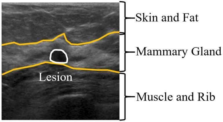
This diagnostic anatomical diagram features a breast ultrasound (BUS) image annotated to illustrate the typical layered structure of breast tissue. From superficial to deep, the three primary layers are identified: 1) the Skin and Fat layer, which presents as a relatively hyperechoic (brighter) and heterogeneous area; 2) the Mammary Gland layer, which is delineated by yellow boundary lines and serves as the primary functional tissue; and 3) the Muscle and Rib layer, characterized by lower echogenicity and deeper anatomical positioning. Within the mammary gland layer, a specific focal finding is highlighted: a rounded, hypoechoic (darker) lesion enclosed by a white circular outline. The diagram demonstrates the importance of anatomical localization for lesion detection in clinical sonography, specifically highlighting that breast pathology typically originates within the mammary gland zone. This educational material is designed for radiology students or medical professionals to improve pattern recognition in B-mode breast ultrasonography.
mammary gland lymphatic drainage breast axillary nodes
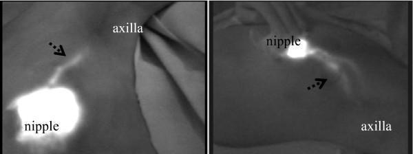
This diagnostic fluorescence image displays two patterns of subcutaneous lymphatic drainage pathways in the human breast, visualized using Indocyanine Green (ICG) fluorescence. Both images demonstrate a high-intensity fluorescent signal at the nipple/subareolar region, representing the injection site. From this origin, linear and slightly tortuous fluorescent streams—indicated by dashed arrows—course towards the axilla (axillary basin). The left image depicts a relatively direct and well-defined pathway, while the right image shows a more diffuse and wider lymphatic channel, potentially illustrating variations in individual anatomy or the merging of multiple lymphatic vessels. The clinical significance of this imagery is the real-time identification of sentinel lymph node drainage pathways, essential for oncological surgical planning and staging. These findings highlight the primary lymphatic flow from the mammary gland towards the axillary lymph nodes, which is a key concept in surgical oncology and breast cancer management.
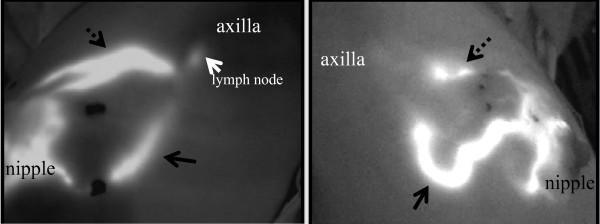
This diagnostic fluorescence image depicts indocyanine green (ICG) lymphography used for identifying lymphatic drainage patterns in the breast. The visualization shows two primary subcutaneous lymphatic pathways originating from the areolar (nipple) region and extending toward the axilla. In the first panel, a direct pathway is visible (dashed arrow), alongside a secondary curvilinear stream (solid arrow) that traverses the lateral aspects of the mammary parenchyma. The fluorescence identifies a distinct 'lymph node' at the mid-axillary destination (white arrow). The second panel demonstrates the convergence of multiple lymphatic vessels into a complex network, highlighting varying vessel thicknesses and signal intensities. This imaging modality is utilized clinically for sentinel lymph node mapping in breast cancer surgery, allowing real-time transcutaneous guidance and anatomical localization of drainage pathways and Berg's level lymph nodes.
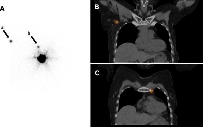
This three-panel composite image displays diagnostic nuclear medicine imaging demonstrating aberrant lymphatic drainage in a patient with ipsilateral breast tumor recurrence (IBTR). Panel A is a planar lymphoscintigraphy image showing a large central area of radiotracer uptake at the injection site. Two secondary foci of uptake are visible: arrow 'a' indicates drainage to the contralateral axilla, and arrow 'b' indicates drainage to the ipsilateral internal mammary chain (IMC). Panels B and C show coronal SPECT/CT fusion images providing anatomical localization for these findings. Panel B demonstrates a 'hot spot' (focal radiotracer uptake) in the right axillary region adjacent to the thoracic wall. Panel C illustrates a 'hot spot' localized to the left internal mammary chain, situated lateral to the sternum. These images illustrate the utility of SPECT/CT in identifying sentinel lymph nodes in non-axillary or contralateral basins, which is clinically significant for staging and surgical planning in recurrent breast cancer cases where normal lymphatic pathways may be surgically disrupted.
Mammary Gland - Long Answer Question (LAQ)
For 1st Year MBBS | As Per Vishram Singh Pattern
INTRODUCTION
SITUATION AND EXTENT
-
Located in the superficial fascia overlying the anterior thoracic wall
-
Extends from the 2nd to 6th rib vertically
-
Extends from the lateral border of the sternum to the mid-axillary line horizontally
-
Overlies mainly the pectoralis major and partly serratus anterior muscles
-
The axillary tail (axillary process / tail of Spence) - the superolateral extension - curves around the lower border of pectoralis major, pierces the deep fascia, and enters the axilla
-
Gray's Anatomy for Students, p. 274
STRUCTURE OF THE BREAST
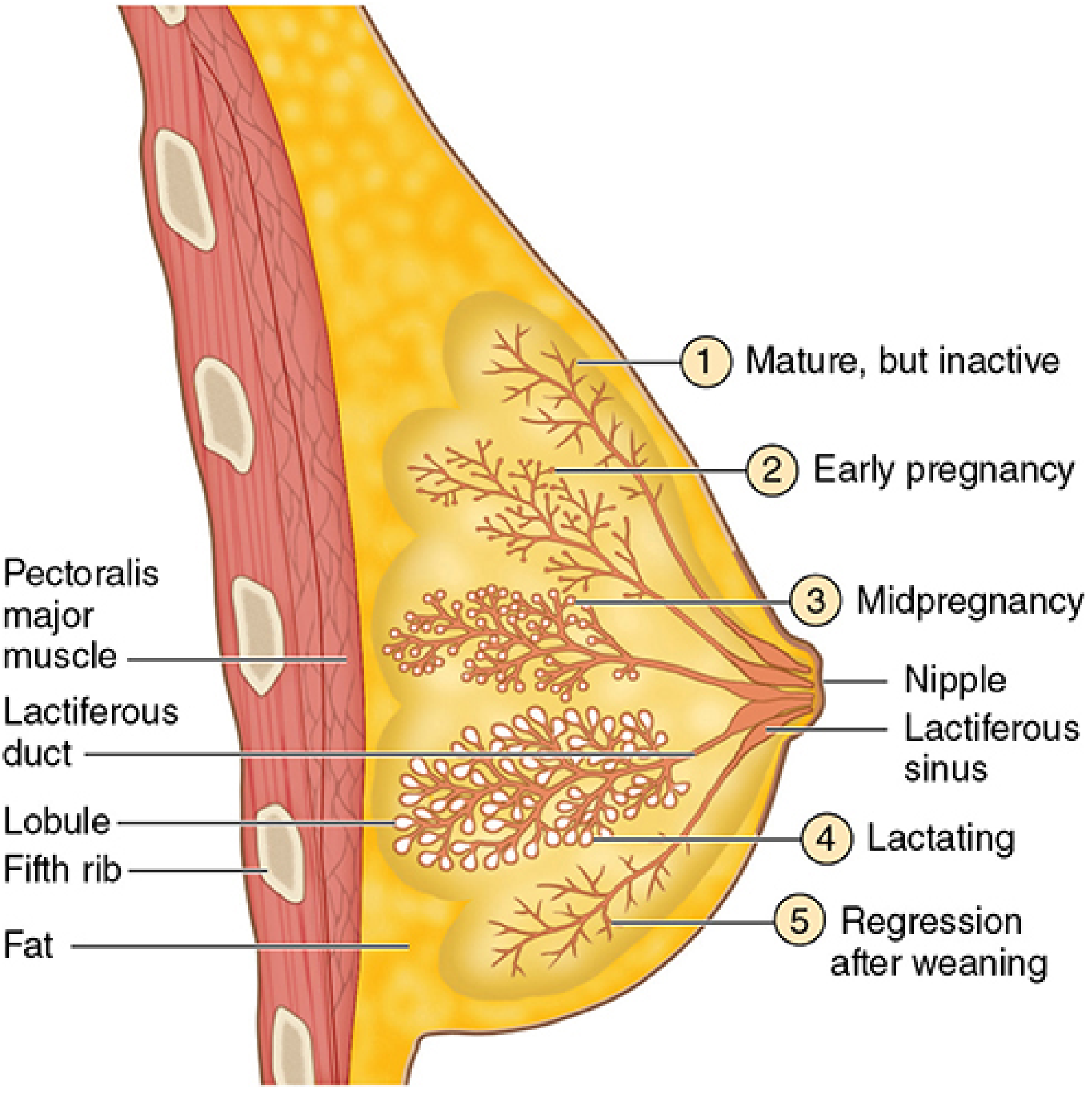
1. Glandular Tissue
- 15-20 lobes arranged radially around the nipple
- Each lobe is a compound tubulo-acinar gland separated by dense fibrous septa and adipose tissue
- Each lobe subdivides into lobules containing terminal duct lobular units (TDLUs) - the functional secretory units
- Each lobe has its own lactiferous duct that opens independently at the nipple
- Just before the opening, each lactiferous duct dilates to form the lactiferous sinus (ampulla), which acts as a reservoir for milk
2. Fibrous (Stromal) Tissue
- Dense fibrous septa separate the lobes
- Suspensory ligaments of Cooper (ligamenta suspensoria) - fibrous bands connecting the overlying dermis to the underlying deep fascia, passing between the lobules
- These ligaments keep the breast elevated
3. Adipose Tissue
- Fat constitutes most of the breast volume in non-pregnant, non-lactating women
- Distributed between and within the lobes
Nipple and Areola
- The nipple is a cylindrical projection at the tip of the breast; it contains numerous smooth muscle fibers running parallel to the lactiferous ducts, which cause erection when stimulated
- The areola is the pigmented circular skin around the nipple; it contains Montgomery's glands (modified sebaceous glands that hypertrophy in pregnancy to lubricate the nipple)
- The areola keratinocytes contain abundant melanin and darken further during pregnancy
- Junqueira's Basic Histology, 17e, p. 1155-1156
RELATIONS
| Surface | Relation |
|---|---|
| Anterior | Skin and superficial fascia |
| Posterior | Pectoralis major (mainly), serratus anterior (laterally), external oblique (inferiorly) |
| Between breast and pectoralis major | Retromammary space - loose areolar tissue allowing free movement; clinically important in retromammary abscess |
BLOOD SUPPLY
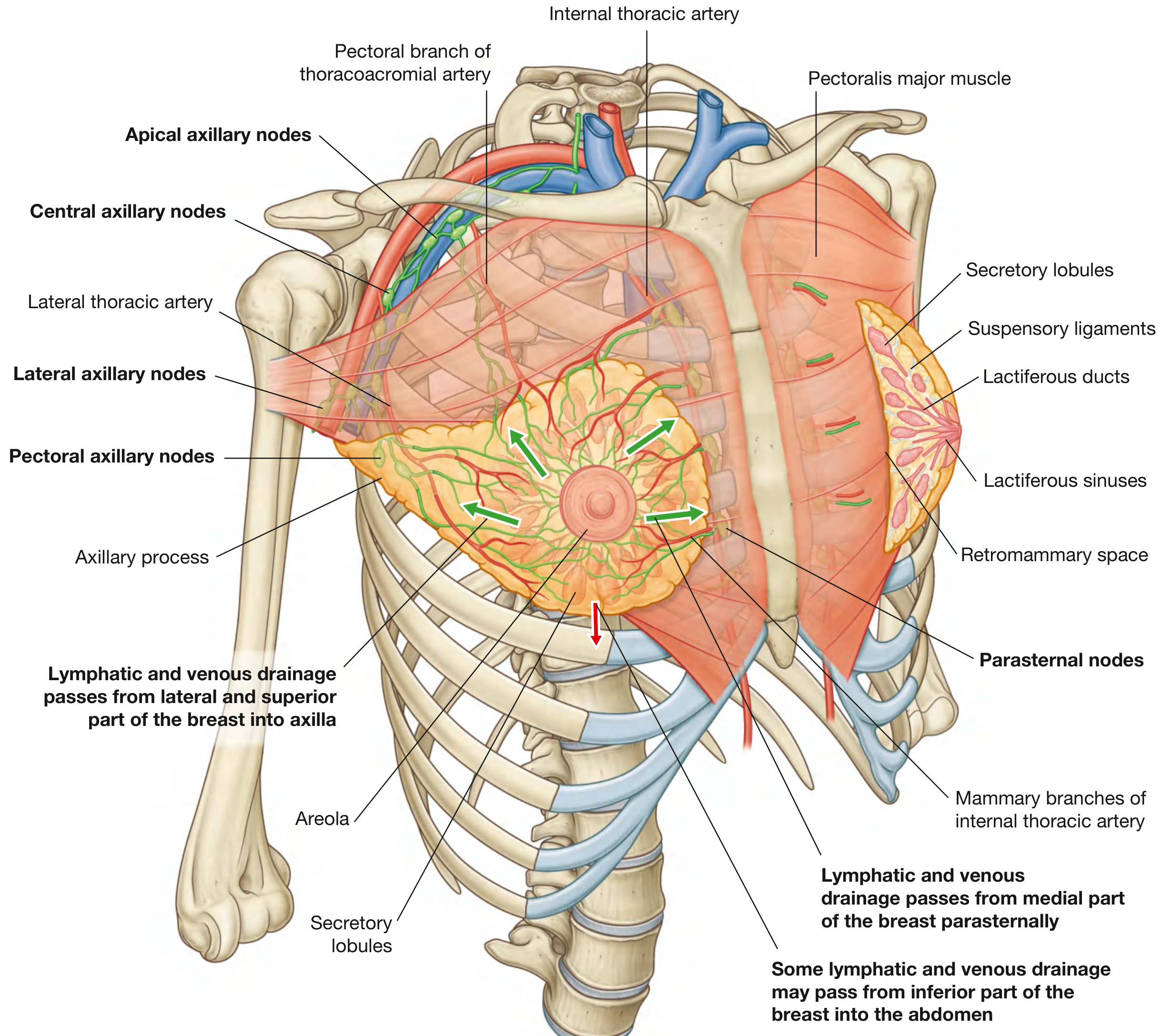
Arteries:
- Internal thoracic (internal mammary) artery - perforating branches (2nd, 3rd, 4th) supply the medial part (largest contribution - ~60%)
- Lateral thoracic artery - branch of axillary artery; supplies lateral part
- Pectoral branches of thoracoacromial artery - branch of axillary artery
- Posterior intercostal arteries (2nd, 3rd, 4th) - lateral branches
- Superior thoracic artery - minor contribution
Veins:
- Internal thoracic vein (medially)
- Axillary vein (laterally)
- Intercostal veins - these communicate with the vertebral venous plexus (of Batson), providing a route for metastatic spread to the vertebral column and brain
- Fischer's Mastery of Surgery, 8th ed.
LYMPHATIC DRAINAGE
Axillary route (75% of drainage):
- Pectoral (anterior) nodes - receive drainage from the lateral and central breast
- Central nodes - receive from pectoral and lateral nodes
- Apical nodes - the final station before the subclavian trunk
Parasternal (internal mammary) route (~25%):
- Drains medial and deep portions into parasternal (internal mammary) nodes along the internal thoracic vessels; these drain into the bronchomediastinal trunk
Minor routes:
- Intercostal nodes near the heads of the ribs (drain into thoracic duct)
- Inferior pathway - some drainage may pass to abdominal lymphatics
- Cross-communication to the opposite breast is possible
NERVE SUPPLY
- Anterior and lateral cutaneous branches of the 2nd to 6th intercostal nerves supply the skin of the breast and the nipple
- The nipple and areola are especially richly supplied with sensory nerve endings
- Sympathetic nerves supply smooth muscle fibers of the nipple and areola (causing erection)
- The 4th intercostal nerve provides the principal sensory supply to the nipple
HISTOLOGY OF THE MAMMARY GLAND
(A) Inactive (Non-pregnant, Non-lactating) State:
- Breast is mostly fat and fibrous connective tissue
- Small ducts with minimal branching; few, small, rudimentary TDLUs
- Lactiferous sinuses lined by stratified cuboidal epithelium
- Lactiferous ducts and terminal ducts lined by simple cuboidal epithelium with myoepithelial cells on the basal surface
- Sparse smooth muscle fibers encircle the larger ducts
(B) During Puberty:
- Under estrogen influence, fat accumulates and ducts elongate
- Lobules develop with branching ducts; alveoli remain small and rudimentary
- In the premenstrual phase, slight edema of connective tissue makes breasts larger
(C) During Pregnancy:
- Under combined action of estrogen, progesterone, prolactin and placental lactogen:
- Extensive proliferation of TDLUs and secretory alveoli
- Alveoli lined by cuboidal epithelium with numerous myoepithelial cells
- Stroma becomes less prominent; infiltrated by lymphocytes and plasma cells
- Fat stores decrease as glandular tissue expands
(D) During Lactation:
- Alveoli greatly enlarged; lumens filled with milk
- Alveolar cells show active secretion (both merocrine and apocrine components)
- Proteins and carbohydrates - released by exocytosis (merocrine)
- Lipids - released by apocrine secretion (fat droplets bud off with cell membrane)
- Myoepithelial cells contract under oxytocin stimulation → milk ejection reflex
- Colostrum (first milk): protein-rich, contains IgA and macrophages; secreted in late pregnancy and first few days postpartum
(E) After Weaning / Post-lactation:
- Alveoli undergo apoptosis; macrophages remove debris
- Gland returns to inactive, non-pregnant appearance
(F) After Menopause:
- Progressive involution - alveoli and ducts reduce; loss of fibroblasts, adipocytes, collagen, and elastic fibers
- Junqueira's Basic Histology, 17e, p. 1156-1158
DEVELOPMENT (EMBRYOLOGY)
Steps:
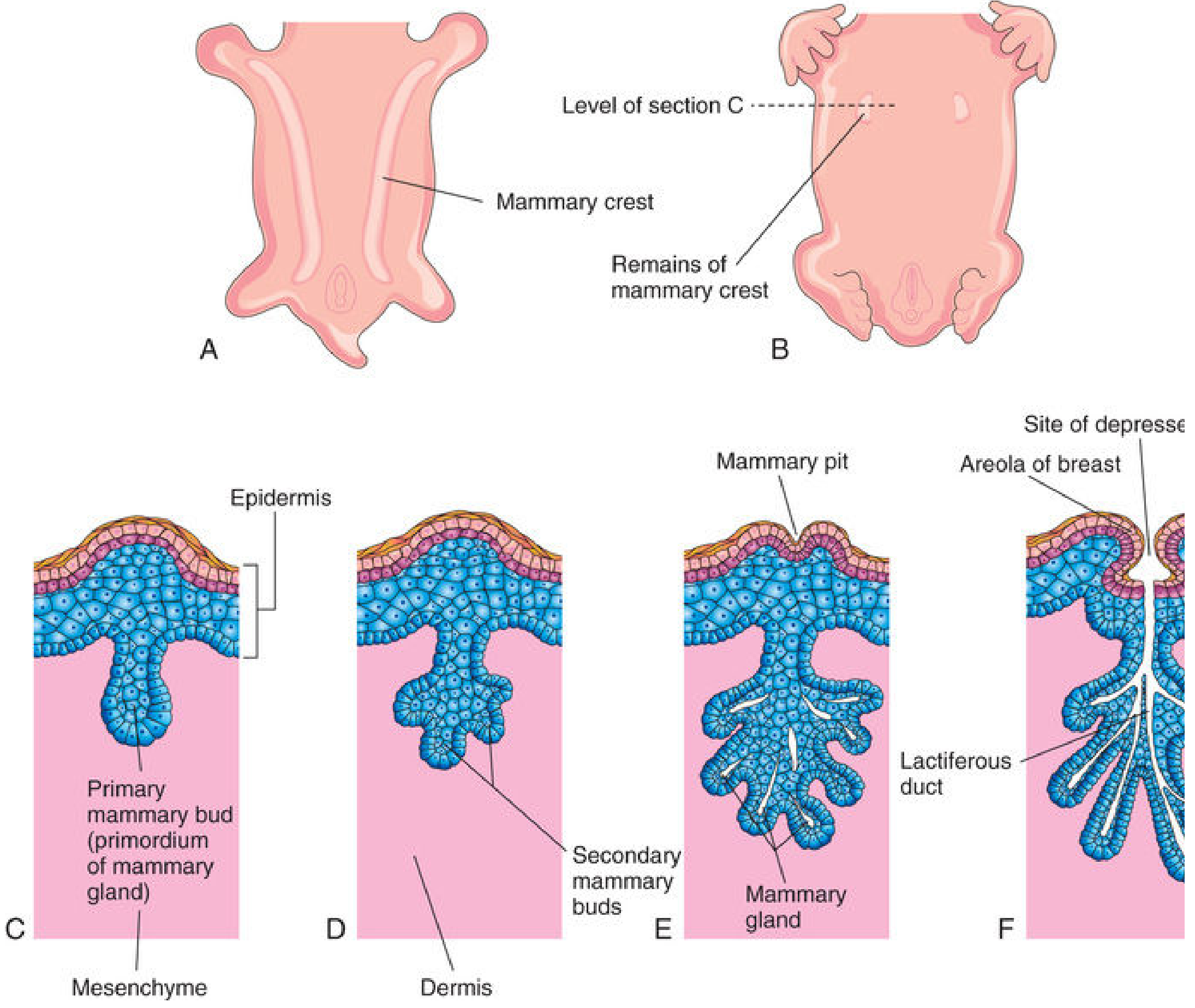
- 4th week: Mammary crests (ridges) appear as thickened ectoderm on each side of the ventral embryo surface, extending from axilla to inguinal region (milk lines)
- Most of the crest disappears; only the pectoral region persists on each side
- 5th week: Primary mammary buds form as downgrowths of epidermis into mesenchyme (signaled by PTHrP, TBX3, LEF1 transcription factors)
- Primary buds give rise to secondary mammary buds → develop into lactiferous ducts and their branches
- Canalization of the ducts is induced by placental sex hormones in late fetal life
- By term, 15-19 lactiferous ducts are formed; fibrous tissue and fat develop from surrounding mesenchyme
- Late fetal period: Epidermis depresses to form mammary pit (inverted nipple appearance at birth)
- After birth: Nipple rises due to proliferation of areolar connective tissue; smooth muscle differentiates from mesenchyme
- At birth (neonate): Glands in both sexes are identical and may be enlarged → may secrete a little milk ("witch's milk" / neonatal galactorrhea) due to maternal hormones crossing the placenta
- Puberty (girls): Estrogen causes fat accumulation and duct elongation; full development by ~19 years
- The Developing Human - Clinically Oriented Embryology, p. 1184-1185
APPLIED ANATOMY / CLINICAL CORRELATIONS
| Condition | Explanation |
|---|---|
| Supernumerary (accessory) nipples / breast (polymastia/polythelia) | Incomplete involution of the mammary crest; may appear anywhere along the milk line |
| Inverted nipple | Persistence of mammary pit; can also be caused by carcinoma due to tethering of lactiferous ducts |
| Peau d'orange skin | Lymphatic obstruction in breast carcinoma; skin edema between Cooper's ligament attachment points causes dimpling |
| Retraction of nipple/skin | Carcinoma invading and shortening Cooper's ligaments |
| Retromammary abscess | Pus collection in the retromammary space |
| Lymphedema of arm | Following mastectomy + axillary clearance; disrupts upper limb lymphatic drainage |
| Vertebral metastasis in breast cancer | Via intercostal veins → Batson's vertebral venous plexus (valveless) |
| Gynecomastia | Abnormal enlargement of male breast (due to estrogen excess or androgen deficiency) |
| Acute mastitis | Bacterial infection (usually S. aureus) entering through cracks in the nipple during lactation |
DIFFERENCES BETWEEN MALE AND FEMALE BREAST
| Feature | Female | Male |
|---|---|---|
| Glandular tissue | Well-developed | Rudimentary ducts only |
| Lobules | Present | Absent |
| Areola | Large, pigmented | Small |
| Size | Prominent | Rudimentary |
| Function | Secretory (lactation) | Non-functional |
SUMMARY TABLE: HORMONAL CONTROL
| Hormone | Action on Breast |
|---|---|
| Estrogen | Duct growth, fat deposition, pigmentation of areola |
| Progesterone | Lobulo-alveolar development |
| Prolactin | Milk production (synthesis) |
| Oxytocin | Milk ejection (myoepithelial contraction) |
| Placental lactogen | Augments prolactin action during pregnancy |