
Cyanotic child case presentation
Reading File
Finding Sources
Finding Sources
Searching PubMed
cyanotic congenital heart disease AND child
Searching the Web
cyanotic congenital heart disease clinical assessment child guideline
Reading File
Reading File
Reading File
Finding Sources
Searching Images
tetralogy of Fallot anatomy diagram cyanotic congenital heart disease child
Reading File
Reading File
Reading File
Finding Sources
Reading File
Cyanotic Child - Case Presentation
Identification
A ___-year/month-old child, ___ sex, resident of ___, was brought with bluish discoloration of lips and tongue for ___ duration, associated with ___.
Chief complaints
- Bluish discoloration of lips, tongue, and nail beds for ___
- Breathlessness / fast breathing for ___
- Feeding difficulty / poor weight gain / recurrent respiratory infection
- Episodes of sudden worsening cyanosis, crying, irritability, or loss of consciousness, if present
History of present illness
The child was apparently well until ___, when the parents first noticed bluish discoloration involving the tongue, oral mucosa, and lips, suggesting central cyanosis. State whether cyanosis:
- Began at birth, after the neonatal period, or recently
- Is continuous or intermittent
- Worsens on crying, feeding, defecation, fever, or exertion
- Is relieved by squatting or knee-chest position
- Is associated with respiratory distress, cough, wheeze, fever, choking, or aspiration
- Has episodic severe attacks with hyperpnea, irritability, limpness, syncope, or seizures
Ask specifically for hypercyanotic spells: sudden deepening of cyanosis with rapid breathing and agitation, classically triggered by crying, feeding, or defecation. They occur with acute reduction of pulmonary blood flow, often in tetralogy of Fallot. - The Harriet Lane Handbook, p. 47
Relevant negative history
- No fever, cough, or coryza, if excluding pneumonia/bronchiolitis
- No history of foreign-body aspiration
- No drug, nitrate, local anesthetic, or chemical exposure, if considering methemoglobinemia
- No breath-holding episode precipitated by anger or pain
- No altered sensorium, seizures, or focal neurologic deficit
Past history
- Previous similar episodes, hospitalization, ICU care, or oxygen requirement
- Known congenital heart disease, prior echocardiography, catheter intervention, cardiac surgery, or shunt
- Recurrent lower respiratory tract infections
- Growth and developmental delay
- History of polycythemia symptoms: headache, dizziness, irritability, or thrombosis
Birth and neonatal history
- Antenatal scans and fetal echocardiography
- Term/preterm, birth weight, mode of delivery
- Cyanosis from birth or after discharge
- NICU admission, ventilation, sepsis, prostaglandin infusion, or neonatal cardiac evaluation
Feeding and developmental history
- Breastfeeding difficulty, prolonged feeds, sweating while feeding, pauses during feeds
- Failure to thrive or poor weight gain
- Milestone attainment and school/exercise tolerance in an older child
Family history
- Congenital heart disease, sudden cardiac death, sibling death in infancy
- Consanguinity
- Maternal diabetes, infections, alcohol/drug exposure, or teratogenic medication during pregnancy
Examination
General examination
- Sick or comfortable appearing?
- Central cyanosis: lips, tongue, oral mucosa
- Peripheral cyanosis versus acrocyanosis
- Clubbing, especially in older children
- Growth parameters: weight, height/length, head circumference
- Pallor, edema, lymphadenopathy
- Respiratory rate, pulse, blood pressure in all limbs, temperature
- Preductal and postductal SpO₂: right hand and either foot
Central cyanosis with relatively little respiratory distress, a “comfortably blue” child, points toward a cardiac cause. Cardiac cyanosis often worsens with crying and has little improvement with oxygen, unlike most pulmonary causes. - Rosen’s Emergency Medicine, p. 3202
Respiratory system
- Tachypnea, retractions, nasal flaring, grunting
- Air entry and added sounds
- Signs of pneumonia, bronchiolitis, heart failure, or pulmonary edema
Cardiovascular system
- Precordial bulge, visible pulsations
- Apex beat location and character
- Parasternal heave
- Thrill
- Heart sounds: single S₂, loud P₂, fixed split S₂
- Murmur: location, timing, radiation, intensity
- Peripheral pulses, capillary refill, hepatomegaly, signs of heart failure
Other systemic examination
- CNS examination for hypoxic spells or stroke
- Abdomen: hepatomegaly, splenomegaly
- Signs of infective endocarditis or brain abscess where clinically suspected
Provisional diagnosis
Cyanotic congenital heart disease, likely tetralogy of Fallot (TOF), with/without hypercyanotic spells.
Supportive findings for TOF:
- Cyanosis increasing in infancy
- Squatting after exertion in an older child
- Ejection systolic murmur at the left upper sternal border
- Single loud S₂
- Right ventricular heave
- Clubbing and polycythemia in longstanding disease
- Chest radiograph may show a boot-shaped heart with reduced pulmonary vascular markings
- ECG may show right-axis deviation and right ventricular hypertrophy
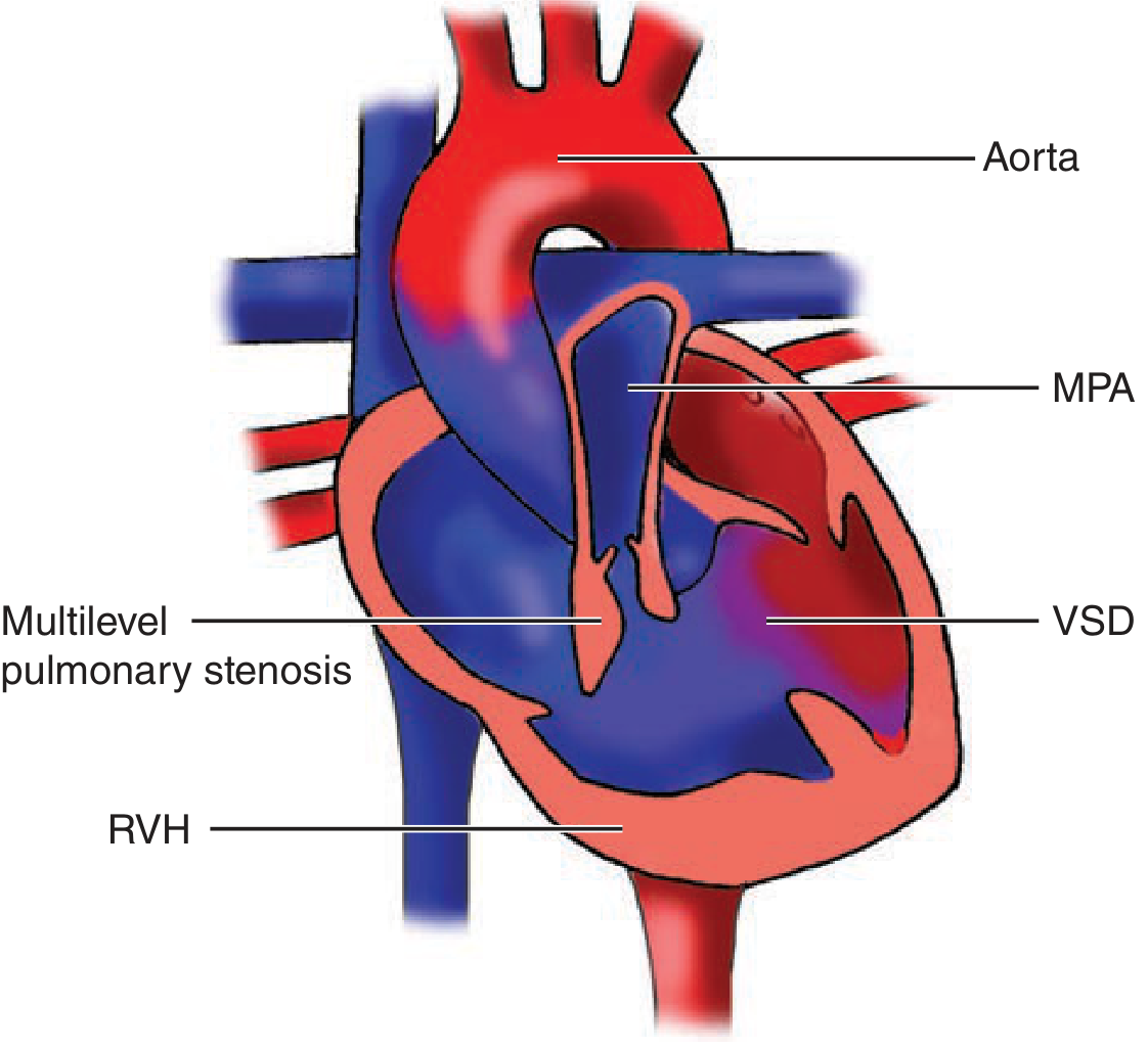
TOF consists of a large VSD, overriding aorta, right ventricular outflow tract obstruction, and right ventricular hypertrophy. The severity of RVOT obstruction determines cyanosis. - Schwartz’s Principles of Surgery, p. 812
Differential diagnosis
Cardiac
- Tetralogy of Fallot
- Transposition of great arteries
- Tricuspid atresia
- Total anomalous pulmonary venous return
- Pulmonary atresia with VSD
- Double outlet right ventricle
- Eisenmenger syndrome in an unrepaired large shunt lesion
Respiratory
- Severe pneumonia or bronchiolitis
- Pulmonary edema
- Asthma with severe respiratory failure
- Foreign-body aspiration
Hematologic/toxic/metabolic
- Methemoglobinemia
- Abnormal hemoglobin disorders
- Severe sepsis/shock
Investigations
Bedside and baseline
- Continuous pulse oximetry, including right hand and foot readings
- ABG with lactate and acid-base status
- CBC with hematocrit: secondary polycythemia may occur
- Blood glucose, electrolytes, renal function
- Chest radiograph
- ECG
Definitive cardiac assessment
- Urgent transthoracic echocardiography with Doppler: defines anatomy, shunt direction, RVOT obstruction, ventricular function, and pulmonary artery anatomy.
- Cardiology review and cardiac catheterization/CT angiography only when required for anatomy or intervention planning.
In a neonate, a hyperoxia challenge may help distinguish cardiac from pulmonary cyanosis, but it is now less commonly used because of hyperoxemia and alkalosis concerns. Marked failure of PaO₂ to rise supports a cardiac right-to-left shunt. - The Harriet Lane Handbook, p. 240
Management
If the child is acutely cyanosed or unstable
This is an emergency. Arrange ABC stabilization, continuous monitoring, IV/IO access, urgent pediatric cardiology input, and transfer to a pediatric cardiac center.
- Keep the child warm, calm, and minimally handled.
- Give oxygen while assessment proceeds, but recognize that a right-to-left shunt may respond poorly.
- Treat hypoglycemia, dehydration, acidosis, sepsis, or respiratory illness if present.
- Obtain urgent echocardiography where available.
If a hypercyanotic spell is suspected
- Calm the child and allow parental comforting.
- Knee-chest position in infants, or encourage squatting in an older child, to increase systemic vascular resistance and reduce right-to-left shunting.
- Administer oxygen if tolerated.
- Give carefully supervised fluid resuscitation if dehydration/hypovolemia is suspected.
- Escalate urgently to pediatric emergency/cardiology teams for appropriate sedation, vasoconstrictor therapy, beta-blockade, airway support, and definitive intervention if the spell persists.
A tet spell presents with extreme cyanosis, hyperpnea, tachypnea, and agitation. Knee-chest positioning or squatting helps by increasing systemic vascular resistance; persistent spells may need urgent surgery or palliation. - The Harriet Lane Handbook, p. 47
For a child with blue/grey lips or tongue, breathing difficulty, drowsiness, limpness, or altered responsiveness, seek emergency care immediately, as reflected in NHS emergency warning signs.
Definitive treatment
- Surgical repair or catheter-based intervention depends on the lesion.
- For TOF, definitive intracardiac repair is typically required, with timing individualized by anatomy, symptoms, pulmonary artery size, and spell frequency.
- Ongoing follow-up addresses growth, iron status, neurodevelopment, infective endocarditis risk counseling, and residual cardiac lesions.
Short Summary for Viva
“This is a ___-year-old child with central cyanosis since ___, worsened by crying/feeding/exertion and associated with ___, with clinical findings of clubbing, right ventricular heave, single loud S₂, and an ejection systolic murmur at the left upper sternal border. The likely diagnosis is cyanotic congenital heart disease, most likely tetralogy of Fallot. The child requires pulse oximetry, ABG, ECG, chest radiograph, urgent echocardiography, pediatric cardiology evaluation, and emergency management if a hypercyanotic spell is present.”
This is a shared conversation. Sign in to Orris to start your own chat.