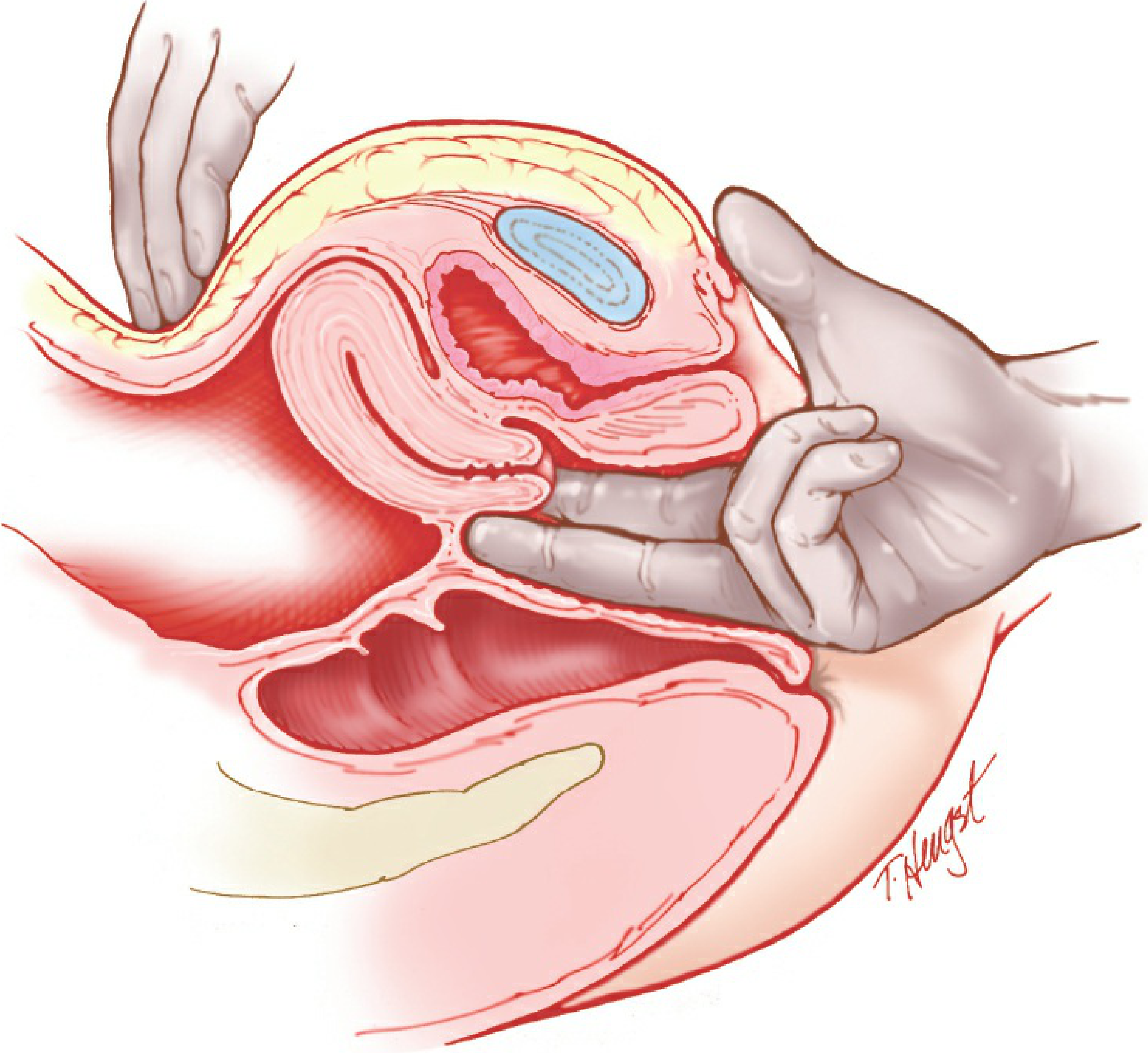
I now have the bimanual examination diagram and comprehensive textbook content. Let me compose the complete, detailed answer.# Mass Per Vaginum - Complete Detailed Explanation
1. Definition
"Mass per vaginum" (MPV) refers to the detection and systematic characterization of a pelvic or lower abdominal mass through the vaginal route, primarily using the bimanual pelvic examination. The term is both a clinical method and a clinical finding - it describes the information obtained when an examiner assesses a pelvic mass with fingers inside the vagina combined with a hand on the abdomen.
It is one of the most informative physical examination techniques in gynecology. It allows assessment of the uterus, adnexa, cervix, vaginal fornices, pelvic floor, and cul-de-sac, all without any imaging.
2. Anatomy Relevant to Per Vaginum Examination
Understanding what can be felt vaginally requires knowing the pelvic anatomy:
| Structure | Position | What is felt normally |
|---|
| Cervix | At vaginal vault | Smooth, firm, cylindrical; moves freely |
| Uterus | Anteverted/flexed above bladder | Pear-shaped, smooth, mobile, non-tender; ~7-8 cm |
| Anterior fornix | Between cervix and bladder | Empty space; reflects bladder |
| Posterior fornix | Behind cervix, deepest | Pouch of Douglas; normally empty |
| Lateral fornices (×2) | On either side of cervix | Ovaries palpable in thin women; tubes not normally felt |
| Broad ligament | Lateral | Not felt normally |
| Uterosacral ligaments | Posterolateral | Smooth cords; nodularity = endometriosis |
3. Technique of Per Vaginum (Bimanual) Examination
(Berek & Novak's Gynecology, Figure 1-3: The bimanual examination)
Patient Position
- Dorsal lithotomy position
- Legs in stirrups, hips at edge of table
- Bladder emptied beforehand (full bladder distorts findings)
Examiner Position
- Standing between the patient's legs
- Non-dominant hand on lower abdomen (suprapubic area)
Step-by-Step Technique
Step 1 - Lubrication and insertion
Apply lubricant to the gloved index and middle fingers. Insert gently into the vagina with palm upward. Warn the patient at each step.
Step 2 - Vaginal walls and fornices
Palpate the vaginal walls systematically - anterior, lateral, posterior. Feel each fornix (anterior, posterior, both lateral) for masses, fullness, tenderness, or nodularity.
Step 3 - Cervix
Feel the cervix - its position, consistency, length, surface, os. Then test for cervical motion tenderness (CMT) by gently moving the cervix side to side. Pain = positive CMT (excitation sign).
Step 4 - Uterus (bimanual)
Place vaginal fingers in the anterior fornix or behind the cervix. Press the abdominal hand firmly downward just above the pubic symphysis. The uterus is "caught" between both hands. Assess:
- Size (in weeks equivalent)
- Shape (pear-shaped vs irregular)
- Surface (smooth vs nodular)
- Consistency (firm vs soft vs hard)
- Position (anteverted / retroverted)
- Mobility (moves freely or fixed)
- Tenderness (any pain on palpation)
Step 5 - Adnexa (bimanual)
Move vaginal fingers into each lateral fornix in turn. The abdominal hand sweeps laterally corresponding to the same side. Structures are swept between both hands. In a normal woman, only the ovary may be palpable (almond-shaped, slightly tender). Assess:
- Presence of any mass
- Size, shape, surface
- Consistency (cystic vs solid)
- Mobility or fixity
- Tenderness
Step 6 - Posterior cul-de-sac (Pouch of Douglas)
Place vaginal fingers in the posterior fornix. Feel for any fluid, masses, or nodularity in the Pouch of Douglas. The abdominal hand palpates the suprapubic area simultaneously.
Step 7 - Rectovaginal examination (when indicated)
One finger in vagina, one in rectum. Assesses:
- Rectovaginal septum (endometriosis, fistula)
- Posterior uterine wall
- Uterosacral ligaments (nodularity = endometriosis)
- Posterior cul-de-sac deposits (ovarian cancer)
- Rectal wall
4. Parameters Assessed for a Mass Per Vaginum
When a mass is detected on PV examination, it is described using eight key parameters. Each gives specific diagnostic information.
4.1 Site / Origin of the Mass
This is the most important first determination - is the mass uterine or extra-uterine (adnexal)?
How to determine site:
- If the mass moves with the cervix when it is manipulated → uterine origin
- If the mass is felt separate from the uterus with a groove between them → adnexal origin
- If the uterus can be palpated separately from the mass → adnexal / extra-uterine
- If the mass fills the entire pelvis and the uterus cannot be delineated → clinical judgment + imaging needed
| Location | Likely Origin |
|---|
| Central pelvic mass, moves with uterus | Uterus itself (fibroid, adenomyosis, malignancy, pregnancy) |
| Lateral to uterus, separate groove | Ovarian / adnexal mass |
| Behind uterus, in posterior fornix | Pouch of Douglas collection, ovarian cyst, endometrioma |
| Anterior to uterus | Bladder, anterior fibroid |
| Fixed, bilateral, in fornices | Broad ligament disease, malignancy, TOA |
4.2 Size
Size is estimated in clinical terms and reported as:
- Centimeters (e.g., 8 × 6 cm mass in right fornix)
- Weeks of pregnancy equivalent for uterine enlargement:
| Gestational Equivalent | Anatomical Landmark |
|---|
| 6-8 weeks | Uterus just palpable, slightly enlarged, at pubic symphysis level |
| 10-12 weeks | Just above pubic symphysis, reaches the brim |
| 16 weeks | Midway between symphysis pubis and umbilicus |
| 20 weeks | At the umbilicus |
| 24 weeks | 4 fingerbreadths above umbilicus |
| 28 weeks | Halfway between umbilicus and xiphisternum |
This system is used because clinicians are intimately familiar with the landmark positions of the gravid uterus. In this patient's case - "16-week uterine size" means the uterus reaches halfway between the symphysis and umbilicus, indicating it is approximately 14-16 cm in its long axis.
4.3 Surface
This is felt through the vaginal wall as the mass is moved between both hands.
| Surface Character | Description | Interpretation |
|---|
| Smooth | Uniform, no irregularities | Simple ovarian cyst, pregnant uterus, adenomyosis, serous cystadenoma |
| Nodular / Irregular | Bumpy, lobulated contour | Multiple fibroids (each lump = one fibroid), malignancy, endometrioma |
| Boss-like projection | Single rounded protrusion | Pedunculated fibroid, subserosal fibroid |
| Papillary / rough | Rough surface with projections | Ovarian malignancy (papillary serous carcinoma) |
In this case: Irregular surface → multiple fibroids distorting the uterine contour, as confirmed by USG.
4.4 Consistency
What the mass feels like under pressure:
| Consistency | Feel | Interpretation |
|---|
| Cystic | Fluctuant, fluid-filled; "gives way" under pressure | Simple or complex ovarian cyst, hydrosalpinx, chocolate cyst |
| Soft / doughy | Uniform softness | Pregnant uterus, lipoma, occasionally adenomyosis |
| Firm / rubbery | Solid feel like an eraser or rubber ball | Fibroid uterus (most characteristic), leiomyoma |
| Hard | Stony hard, like bone | Calcified fibroid, dermoid (teratoma with calcification), bone-containing teratoma |
| Mixed / heterogeneous | Variable feel in different areas | Degenerated fibroid, malignant tumor, dermoid cyst |
| Tense-cystic | Tense fluid but less fluctuant | Large functional cyst, mucinous cystadenoma, full bladder |
In this case: Firm consistency → fibroid uterus. The whorled bundles of smooth muscle in leiomyomas give them their characteristic firm, rubbery feel.
4.5 Mobility
Assessed by attempting to move the mass with vaginal fingers while the abdominal hand restrains:
| Mobility | Description | Interpretation |
|---|
| Freely mobile | Moves easily in all directions | Benign pedunculated fibroid, simple ovarian cyst (benign) |
| Limited mobility | Moves partially; some restriction | Large intramural fibroid (limited by its own bulk), Broad ligament fibroid |
| Fixed / immobile | Cannot be moved | Malignancy (pelvic wall invasion), PID (adhesions), endometriosis (frozen pelvis), TOA |
Remember the rule:
- Benign masses tend to be mobile
- Malignant or inflammatory masses tend to be fixed
In this case: "Limited mobility" - this is not a red flag for malignancy here. A very large multi-fibroid uterus (16-week size) has mechanical restriction of movement due to its sheer bulk filling the pelvis, not due to infiltration of pelvic sidewalls. This must be distinguished from true fixity due to malignancy.
4.6 Tenderness
The examiner notes whether manipulation of the mass causes pain.
| Tenderness | Interpretation |
|---|
| Non-tender | Fibroid, ovarian cyst (functional or benign), malignancy (early), pregnancy |
| Tender | PID, TOA, twisted ovarian cyst (torsion), ectopic pregnancy, red degeneration of fibroid, endometrioma rupture |
| Cervical motion tenderness (CMT) | Peritoneal irritation: ectopic pregnancy, PID, TOA, appendicitis |
| Exquisitely tender | Torsion of ovary/fibroid, ruptured ectopic, ruptured TOA |
In this case: Non-tender mass + no CMT → strongly supports a benign, non-inflammatory etiology.
4.7 Relationship to the Uterus
| Relationship | Description | Interpretation |
|---|
| Continuous with uterus | Mass and uterus move together, no groove | Uterine fibroid, adenomyosis, uterine malignancy |
| Separate from uterus | Groove palpable between mass and uterus | Ovarian mass, adnexal mass, broad ligament mass |
| Bilateral | Both fornices feel full | Bilateral ovarian disease, TOA, malignancy with omental cake |
| Posterior | Mass in Pouch of Douglas | Ovarian cyst, endometrioma, pelvic abscess, sigmoid mass |
4.8 Condition of the Fornices
The fornices are the recesses in the vaginal vault around the cervix.
| Fornix Finding | Description | Interpretation |
|---|
| Free | Empty, non-tender, no masses | Ovaries/adnexa normal; uterine origin of mass |
| Full / obliterated | Fornix feels "doughy" or full | Parametrial involvement, broad ligament mass, pelvic spread |
| Tender fornix | Pain on palpating a fornix | PID, ovarian torsion, endometrioma, ectopic pregnancy |
| Nodular posterior fornix | Nodules felt in posterior fornix | Endometriosis (deposits in Pouch of Douglas), ovarian cancer peritoneal seedlings |
| Cystic lateral fornix | Fluid-filled mass in fornix | Ovarian cyst, paraovarian cyst, hydrosalpinx |
In this case: "Bilateral fornices free" → the ovaries and adnexa are not involved. This critically localizes the mass to the uterus itself.
5. Systematic Framework for Characterizing a Pelvic Mass on PV
Use this framework to describe any mass per vaginum systematically - the SCOTMAF mnemonic:
| Letter | Parameter | Example (this patient) |
|---|
| S | Site / Origin | Central, continuous with uterus (uterine origin) |
| C | Consistency | Firm / rubbery |
| O | cOntour / Surface | Irregular, nodular |
| T | Tenderness | Non-tender; no CMT |
| M | Mobility | Limited (mechanical) |
| A | Adnexa / Fornices | Bilateral fornices free |
| F | (si)ze in weeks/cm | 16-week equivalent |
6. What the Individual Findings Mean Diagnostically
"Uterus enlarged to 16 weeks"
- Indicates massive uterine enlargement
- Normally the non-pregnant uterus is 7-8 cm and non-palpable abdominally
- A uterus reaching 16-week size is ~14-16 cm long
- Causes of uterine enlargement on PV:
| Cause | Features |
|---|
| Multiple fibroids | Firm, irregular, lobulated |
| Adenomyosis | Smooth, globular, uniformly enlarged, often tender (especially premenstrual) |
| Intrauterine pregnancy | Soft, smooth; positive UCG |
| Endometrial carcinoma | May be enlarged; postmenopausal bleeding; irregular cervix sometimes |
| Uterine sarcoma | Rapidly enlarging; softer; sometimes tender |
"Firm consistency"
- Due to the whorled fibromuscular architecture of leiomyomas - dense smooth muscle bundles embedded in fibrous stroma
- Robbins describes leiomyomas as "sharply circumscribed, discrete, round, firm, gray-white tumors" with a "characteristic whorled pattern"
- Firm feel differentiates fibroids from:
- Cysts (fluctuant)
- Adenomyosis (doughy/boggy)
- Pregnancy (soft - Hegar's sign)
"Irregular surface"
- Each fibroid nodule protrudes beyond the uterine outline, creating a lobulated, bumpy surface
- The number and size of lobulations correlates roughly with the number of fibroids
- Smooth uterine surface despite enlargement would suggest adenomyosis or pregnancy
"Limited mobility"
- This is mechanical restriction, not true fixation
- A 16-week sized uterus physically occupies most of the pelvic cavity and cannot move freely
- True fixation (from pelvic wall infiltration) = malignancy / endometriosis / post-inflammatory adhesions
"Bilateral fornices free"
- Confirms the adnexa (ovaries, tubes) are uninvolved
- The mass is entirely of uterine origin
- Rules out: ovarian cyst, TOA, ectopic pregnancy, ovarian malignancy
"No cervical motion tenderness"
- Absence of CMT = no peritoneal irritation
- Rules out the entire PID spectrum (salpingitis, TOA)
- Rules out ectopic pregnancy
- Combined with negative UCG = ectopic definitively excluded
7. Differential Diagnosis of Pelvic Mass on PV Examination
| Condition | Site | Size | Surface | Consistency | Mobility | Tenderness | Fornices | Other clues |
|---|
| Fibroid uterus | Uterine | Variable (often large) | Irregular, nodular | Firm/rubbery | Variable (limited if large) | Non-tender | Free | Menorrhagia, parity |
| Adenomyosis | Uterine | Moderate (12-14 wks) | Smooth, globular | Doughy/boggy | Mobile | Tender (esp. premenstrual) | Free | Dysmenorrhea, multipara |
| Pregnancy | Uterine | Corresponds to dates | Smooth | Soft (Hegar's sign) | Mobile | Non-tender | Free | UCG positive, amenorrhea |
| Endometrial carcinoma | Uterine | Slightly enlarged | Smooth-irregular | Firm-soft | Mobile to fixed | Usually non-tender | Free or involved | Post-menopausal bleeding |
| Ovarian cyst (simple) | Adnexal | Variable | Smooth | Cystic/tense | Freely mobile | Non-tender | Filled (unilateral) | Separate from uterus |
| Endometrioma | Adnexal | 5-10 cm typically | Smooth-nodular | Tense-cystic | Restricted mobility | Tender | Tender fornix | Dysmenorrhea, chocolate cyst |
| Ovarian malignancy | Adnexal | Large | Irregular, nodular | Solid-mixed | Fixed | Non-tender to tender | Nodular / obliterated | Older age, ascites, weight loss |
| Tubo-ovarian abscess | Adnexal | Variable | Ill-defined | Doughy-cystic | Fixed | Exquisitely tender | Bilateral, tender, full | Fever, positive CMT, discharge |
| Ectopic pregnancy | Adnexal | Small-moderate | Ill-defined | Soft-cystic | Variable | Very tender | Tender | UCG positive, CMT positive |
| Dermoid cyst (Teratoma) | Adnexal | Variable | Smooth | Mixed (may feel hard) | Freely mobile (tends to be anterior) | Non-tender | Filled | Younger women |
| Pedunculated fibroid | Uterine/free | Variable | Smooth to irregular | Firm | Mobile | Non-tender | May appear separate | Stalk connecting to uterus |
| Broad ligament fibroid | Lateral | Large | Smooth | Firm | Very limited | Non-tender | Unilateral fullness | Displaced uterus |
8. Special Variant: Rectovaginal (RV) Examination
When indicated (suspected endometriosis, malignancy, RV fistula):
- One gloved finger in vagina, one in rectum simultaneously
- Assesses the rectovaginal septum, posterior uterine wall, uterosacral ligaments
- Nodularity or thickening of uterosacral ligaments = endometriosis (very specific finding)
- Fixed retroflexed uterus = pelvic adhesions or endometriosis
- Shelf-like mass felt anteriorly in rectum = Blumer's shelf sign = peritoneal metastases from ovarian/GI cancer
- Posterior cul-de-sac nodularity = Pouch of Douglas implants (ovarian malignancy, endometriosis)
9. Common Pitfalls in Per Vaginum Examination
| Pitfall | How to Avoid |
|---|
| Full bladder masking findings / creating a false mass | Always ask patient to empty bladder before examination |
| Missing a retroverted uterus | Fingers in posterior fornix; mass may be felt posteriorly |
| Confusing a pedunculated fibroid for an adnexal mass | Trace the stalk; it moves with the uterus |
| Missing a fixed retroflexed uterus as "adnexal mass" | Systematic bimanual on both sides; RV examination |
| Inadequate relaxation of patient causing false tenderness | Proper patient positioning, communication, and rapport |
| Obesity limiting examination quality | Deeper abdominal pressure; may be unreliable - proceed to imaging |
| Missing posterior cul-de-sac disease | Always perform posterior fornix palpation and RV if indicated |
10. Summary Table: PV Findings in This Patient
| Parameter | Finding | Interpretation |
|---|
| Site | Central, moves with cervix | Uterine origin confirmed |
| Size | 16-week equivalent | Massively enlarged uterus |
| Surface | Irregular, nodular | Multiple fibroids creating lobulations |
| Consistency | Firm/rubbery | Fibromuscular leiomyoma tissue |
| Mobility | Limited | Mechanical, due to large bulk - not malignant fixation |
| Tenderness | Non-tender | Benign process; no inflammation/torsion |
| CMT | Absent | No peritoneal irritation; excludes ectopic, PID |
| Fornices | Bilateral free | Adnexa uninvolved; purely uterine mass |
| Diagnosis | Multiple fibroid uterus | Confirmed by USG |
(Sources: Berek & Novak's Gynecology; Robbins, Cotran & Kumar Pathologic Basis of Disease; Creasy & Resnik's Maternal-Fetal Medicine)