
Aspect acetazolamide challenge
ASPECT acetazolamide challenge nuclear medicine SPECT cerebrovascular
acetazolamide challenge SPECT cerebrovascular reactivity
ASPECT score acetazolamide challenge nuclear medicine brain perfusion SPECT cerebrovascular reserve Moyamoya
https://pmc.ncbi.nlm.nih.gov/articles/PMC7051638
https://snmmi.org/common/Uploaded%20files/Web/Education%20an…
acetazolamide challenge cerebrovascular reserve SPECT Rogg criteria
acetazolamide challenge cerebrovascular reserve brain perfusion SPECT
acetazolamide challenge brain SPECT cerebrovascular reserve perfusion
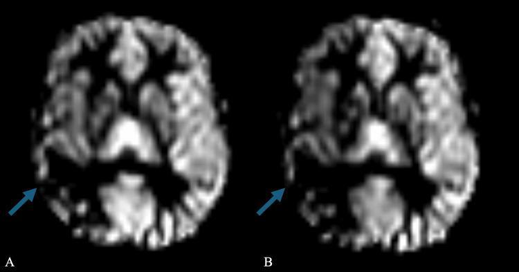
This diagnostic image displays two side-by-side axial brain MRI perfusion slices, labeled (A) Pre-Diamox and (B) Post-Diamox, used to evaluate cerebrovascular reserve. The images show a comparative analysis of blood flow within the brain parenchyma. Blue arrows point to the right middle cerebral artery (MCA) territory. In image A, the baseline signal intensity suggests existing perfusion deficits. In image B, following the administration of Diamox (acetazolamide), there is a noticeable reduction in signal intensity (darkening) in the right MCA territory compared to the baseline. This paradoxical response to a vasodilator indicates a 'steal phenomenon' or maximal baseline vasodilation with poor vascular reserve, often seen in conditions like radiation-induced cerebral vasculopathy or moyamoya-like syndromes. The educational focus is on the clinical application of Diamox-challenge MRI to assess hemodynamic compromise and the risk of ischemic events.
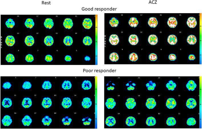
Comparison of quantitative Single-Photon Emission Computed Tomography (SPECT) axial brain scans evaluating regional cerebral blood flow (rCBF) and cerebrovascular reactivity (CVR). The image is divided into four sections: 'Rest' (baseline) and 'ACZ' (after Acetazolamide injection), comparing a 'Good responder' (upper panels) and a 'Poor responder' (lower panels). The upper panel shows axial slices of a patient with preserved CVR, demonstrating a diffuse, robust increase in signal intensity from baseline (green/blue) to post-ACZ challenge (yellow/red/white), indicating normal vasoreactivity. In contrast, the lower panel depicts a patient with a prior stroke and impaired CVR, showing minimal change in signal intensity and distribution between the rest and ACZ states, primarily maintaining low-intensity blue/green hues. A color scale on the right of each quadrant correlates warmer colors (yellow/red) with higher quantitative blood flow. This diagnostic image illustrates the assessment of hemodynamic reserve in cerebrovascular disease using pharmacological challenge.
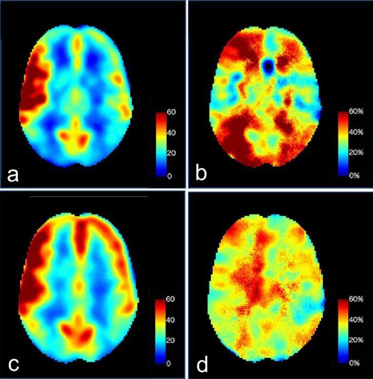
This diagnostic visual contains four axial brain slices (a-d) using Arterial Spin Labeling (ASL) perfusion MRI to assess cerebral blood flow (CBF) and cerebrovascular reactivity (CVR). The images utilize a color-coded pseudocolor scale where red/yellow indicates high perfusion and blue indicates low perfusion. Panels (a) and (c) represent baseline pre-acetazolamide states, both showing focal hyperperfusion in the right frontotemporal region. Panels (b) and (d) demonstrate the response to an acetazolamide challenge. Image (b) shows impaired CVR in the previously hyperperfused right frontotemporal zone (appearing blue/green) relative to the robust augmentation in the surrounding right hemisphere and normal response in the left hemisphere. Follow-up image (d) demonstrates a normalized bilateral augmentation response, indicating a recovery of cerebrovascular reserve. This series is used in neuroradiology to evaluate hemodynamics in cases of vasculitis or severe carotid stenosis, illustrating the concept of 'luxury perfusion' and subsequent recovery of normal vascular reactivity following clinical management.
ASPECT Acetazolamide Challenge (Diamox SPECT)
What Is It?
Mechanism of Action
- CO2 accumulation at the tissue level (by blocking CO2 conversion to HCO3-)
- CO2 is a potent cerebral vasodilator
- In normal vessels, arterioles dilate - CBF increases 20-30%
- In compromised vessels (already maximally dilated due to chronic hypoperfusion), no further vasodilation is possible
- A paradoxical decrease in perfusion in affected regions = the steal phenomenon (blood diverted to normally reactive territories)
Indications
| Condition | Rationale |
|---|---|
| Moyamoya disease/vasculopathy | Pre- and post-surgical planning (bypass surgery) |
| Symptomatic carotid stenosis / occlusion | Stratify stroke risk; guide revascularization |
| TIA / completed ischemic stroke | Differentiate infarct from ischemic penumbra |
| Atherosclerotic cerebrovascular disease | Hemodynamic vs. embolic etiology |
| Arteriovenous malformations | Assess surrounding steal |
| Vascular vs. neuronal dementia | Distinguish causes of perfusion defects |
Radiopharmaceuticals Used
- Tc-99m HMPAO (hexamethylpropylene amine oxime / Ceretec)
- Tc-99m ECD (ethyl cysteinate dimer / Neurolite)
- ¹⁵O-water PET - considered more sensitive; useful when SPECT is negative but clinical suspicion remains high (some studies show SPECT may be false-negative in up to 44% of cases where PET detects impaired CVRC)
Protocol
2-Day Protocol (Preferred)
| Day | Step |
|---|---|
| Day 1 (Stress/Challenge) | IV ACZ 1000 mg over 10 minutes → wait 15-20 min → inject radiotracer (555-1110 MBq / 15-30 mCi) → 30 min uptake in quiet, dim room → SPECT acquisition (~15-20 min) |
| Day 2 (Baseline) | Only needed if Day 1 is abnormal; same quiet room protocol, no ACZ |
If Day 1 (challenge) is completely normal, baseline may be omitted.
1-Day Protocol
- Baseline scan performed first (lower dose)
- Stress scan done same day at at least twice the baseline dose
- Less preferred due to tracer residual activity interfering
Pre-scan Preparation
- Avoid caffeine, alcohol, drugs affecting intracranial perfusion for ≥24 hrs
- No smoking on the day of the exam
- May eat and take regular medications
- Patient void before starting
- Monitor HR and BP throughout ACZ administration
Contraindications to Acetazolamide
- Stroke within the past 3 days
- Sulfonamide allergy (ACZ is a sulfonamide derivative)
- Electrolyte disturbances
- Significant renal or hepatic disease
- Adrenocortical insufficiency
- Long-term use in chronic noncongestive angle-closure glaucoma
Interpretation: Rogg Criteria (Type I / II / III)
| Type | Perfusion at Rest | Post-ACZ Response | Interpretation |
|---|---|---|---|
| Type I | Normal | Normal increase (≥20%) | Preserved CVR - low ischemic risk |
| Type II | Reduced / normal | No change or blunted increase | Impaired CVR - moderate risk |
| Type III | Reduced | Further decrease (steal) | Exhausted CVR - highest ischemic risk |
- Type III response = "cerebrovascular steal phenomenon" - indicates vessels are maximally dilated at baseline; ACZ diverts blood to reactive territories. This predicts the highest risk of perioperative or spontaneous ischemia.
- In Moyamoya series: ~32.5% of assessed brain regions show Type III responses, predominantly in frontal and parietal lobes.
Quantitative Assessment
- Calculated per vascular territory (MCA, ACA, basal ganglia, border zones)
- RI most significantly correlates with clinical symptoms in carotid stenosis
- A significantly negative RI in the anterior border zone distinguishes symptomatic from asymptomatic carotid stenosis
Clinical Decision-Making Utility
- Pre-surgical planning in Moyamoya - Type III regions guide which hemispheres to revascularize first and predict postoperative ischemic risk
- Post-surgical follow-up - improvement from Type III → Type I confirms successful revascularization
- Carotid stenting - ACZ-CTP confirms improvement of CVR after stenting
- Risk stratification - Normal CVR on ACZ challenge suggests lower short-term stroke risk in chronic steno-occlusive disease
Limitations and Pitfalls
- SPECT resolution is lower than CT or MRI - spatial discrimination is limited
- 2-day protocol required due to tracer kinetics (Day 1 residual activity interferes)
- False negatives - up to 44% of SPECT-negative patients may have impaired CVRC on ¹⁵O-water PET (as shown by Seifert et al., JNM 2018)
- Cerebellum can show artefactual changes due to crossed cerebellar diaschisis
- Cannot provide quantitative CBF values as reliably as PET
- Side effects of ACZ: mild vertigo, tinnitus, paresthesias, nausea (self-limited); postural hypotension on rising
Summary
- Rogg et al., AJR 1989;153:605-612 - original Rogg criteria
- SNMMI Diamox SPECT Fact Sheet - protocol standards
- Goyal et al., Asia Ocean J Nucl Med Biol 2026 - recent Moyamoya series (PMID 41626117)
Slit ventricle syndrome
slit ventricle syndrome pathophysiology diagnosis management neurosurgery
https://pmc.ncbi.nlm.nih.gov/articles/PMC8459697
slit ventricle syndrome CT brain small ventricles hydrocephalus shunt
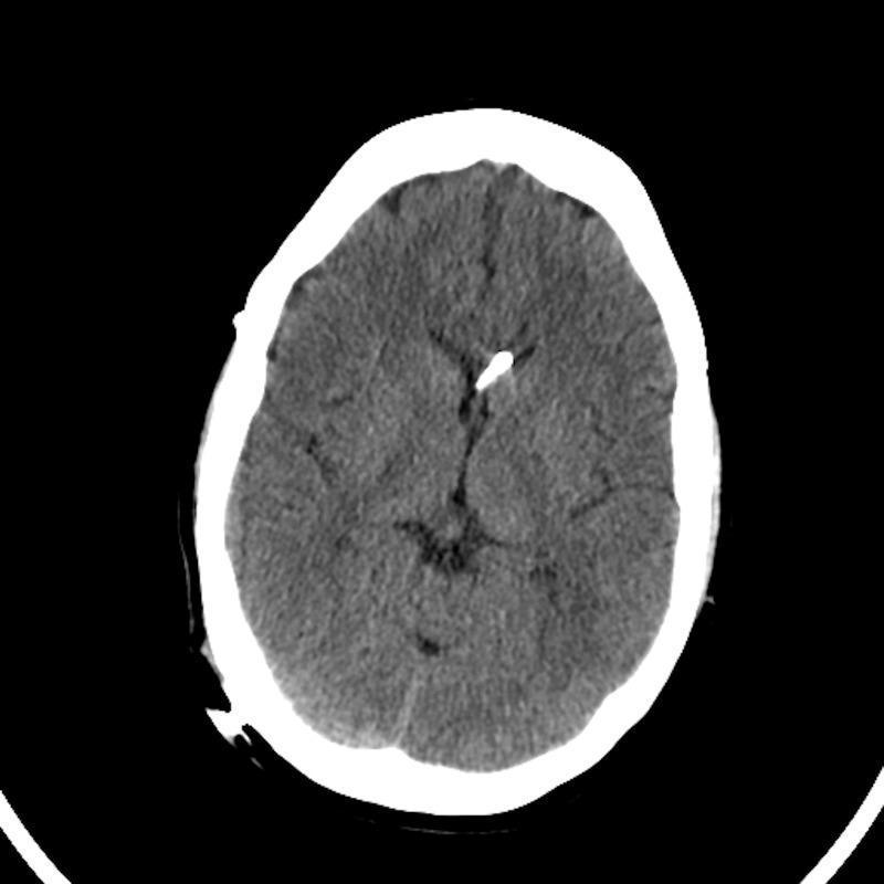
This diagnostic image is an axial non-contrast computed tomography (CT) scan of the brain, demonstrating features consistent with slit ventricle syndrome following surgical intervention. The lateral ventricles are markedly small and slit-like, showing severe compression or decompression from chronic shunting. A hyperdense, linear structure is visible within the right lateral ventricle, consistent with a ventricular shunt catheter. The brain parenchyma appears tightly compacted against the cranial vault, with evidence of effacement of the normal cortical sulci, suggesting restricted intracranial volume relative to the brain mass. The skull is intact and relatively smooth, following a cranial vault expansion procedure as indicated by the clinical context. This image illustrates the neuroimaging appearance of slit ventricles and the associated lack of cerebrospinal fluid space in patients with complex hydrocephalus or craniosynostosis history.
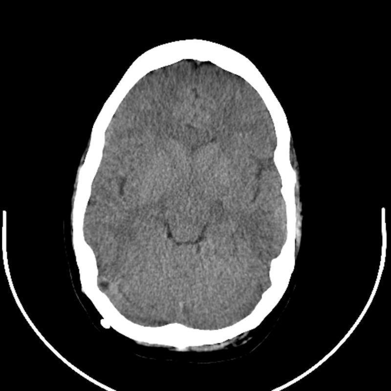
An axial non-contrast computed tomography (CT) scan of the head demonstrating features consistent with slit-ventricle syndrome in the context of chronic ventriculoperitoneal shunting. The primary finding is the presence of slit-like lateral ventricles, which appear remarkably small and narrow centrally within the brain parenchyma. The brain tissue shows a mottled gray attenuation and appears tightly compacted against the inner table of the cranial vault, with significantly effaced cortical sulci and minimal visible extracerebral cerebrospinal fluid (CSF) spaces. The cranial vault is represented by a thick, hyperdense (bright white) bony circumference. A small, focal hyperdensity is noted along the right inner table of the skull, potentially representing a shunt component or calcification. This diagnostic image illustrates the loss of intracranial compliance and ventricular collapse often seen in patients with long-standing CSF diversion, providing educational value for neurosurgery and neuroradiology training.
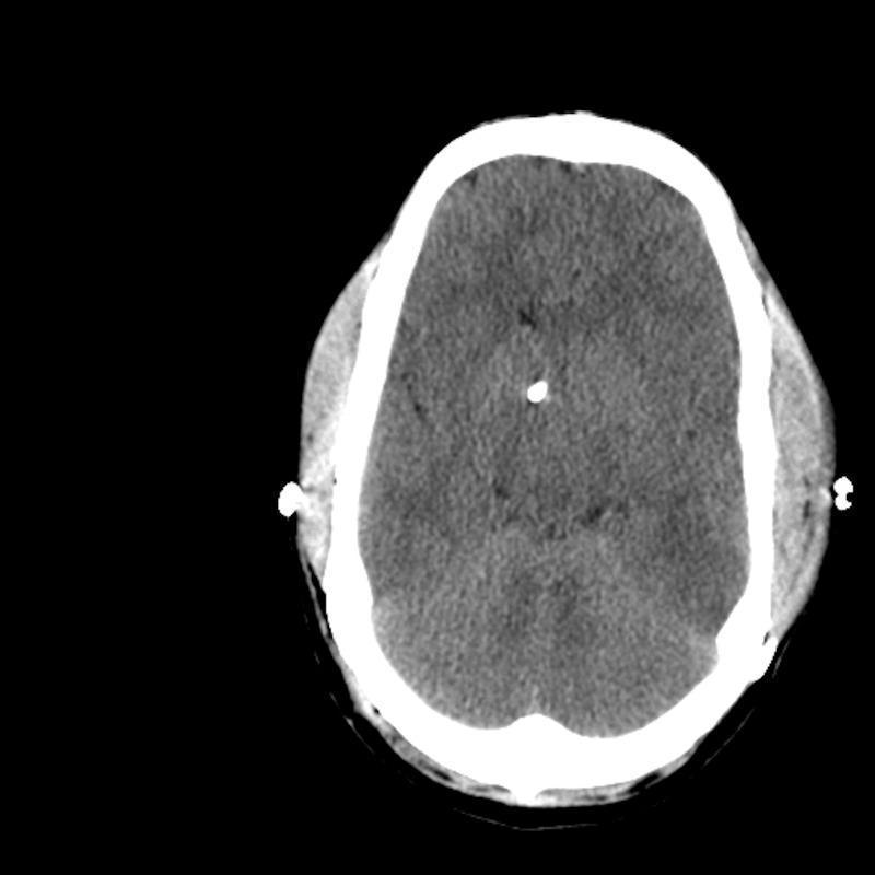
This diagnostic image is an axial non-contrast Computed Tomography (CT) scan of the brain, demonstrating features consistent with slit ventricle syndrome. The hallmark finding is the presence of extremely small, 'slit-like' lateral ventricles, indicating a significant reduction in cerebrospinal fluid (CSF) volume within the ventricular system. The brain parenchyma appears tightly compacted against the inner table of the skull, with a notable effacement of the cortical sulci and basal cisterns, suggesting increased intracranial pressure or a disproportionate brain-to-cranial vault ratio. A hyperdense punctate focus is visible near the midline, consistent with the distal tip of a ventricular catheter or shunt hardware. The surrounding skull shows intact bone structure, though the overall cranial volume appears relatively small for the parenchymal mass. This imaging is clinically significant for evaluating patients with chronic shunt dependence who present with intracranial hypertension despite appearing to have collapsed or 'normal' small ventricles on imaging.
slit ventricle syndrome shunt overdrainage treatment
Slit Ventricle Syndrome (SVS)
Definition
- Seen in approximately 10% of shunted patients (Tintinalli's)
- Symptoms typically appear 2-5 years after shunt placement
- Peak incidence in children aged 4-6 years
- More common in young children; also seen in adults
Pathophysiology
Overdrainage → Ventricular collapse
       ↓
Collapsed ventricular wall occludes proximal catheter orifice
       ↓
ICP rises (no drainage)
       ↓
Rising ICP disengages occluding tissue → drainage resumes
       ↓
Overdrainage again → cycle repeats
- Reduced brain compliance - the brain parenchyma loses its viscoelastic buffer capacity; even small volume changes cause large ICP spikes
- Underdeveloped subarachnoid spaces - in children shunted early, the subarachnoid CSF spaces never properly expand; normal ICP fluctuations (coughing, Valsalva) become symptomatic
- Altered cerebrovascular distensibility - chronic shunt therapy changes the biomechanical properties of brain vasculature
- Intermittent ventricular isolation - one overdrained ventricle collapses and occludes the catheter, causing contralateral ventricular enlargement and intracranial hypertension (some equate SVS with ventricular isolation - justifies neuroendoscopic treatment)
- Stiff, non-compliant ventricles - raised intraventricular pressure with no ventricular dilatation visible on imaging
- Headaches unrelated to the shunt - important differential to consider
Clinical Features
Cardinal Presentation
- Episodic (waxing and waning) headaches - often severe, with nausea and vomiting
- Postural component - symptoms are worse on standing/exercise (gravity accelerates CSF drainage via siphon effect) and relieved by lying down or Trendelenburg position
Additional Features in Children
- Irritability, ataxia, obtundation
- Repeated vomiting
- In infants: failure of skull growth despite normal brain size (cranium can fail to expand)
- ICP in the upright position drops to ~30 mmH2O
Signs of Acute Deterioration
- Decreased level of consciousness (highest correlation with shunt malfunction)
- Paralysis of upward gaze ("sunsetting") - third ventricle impingement on brainstem
- Dilated pupils, papilledema
Key distinguishing feature: contrast with shunt obstruction, where symptoms are not typically postural. In SVS, lying flat relieves symptoms; in outright shunt blockage, positional relief is less prominent.
CT Imaging
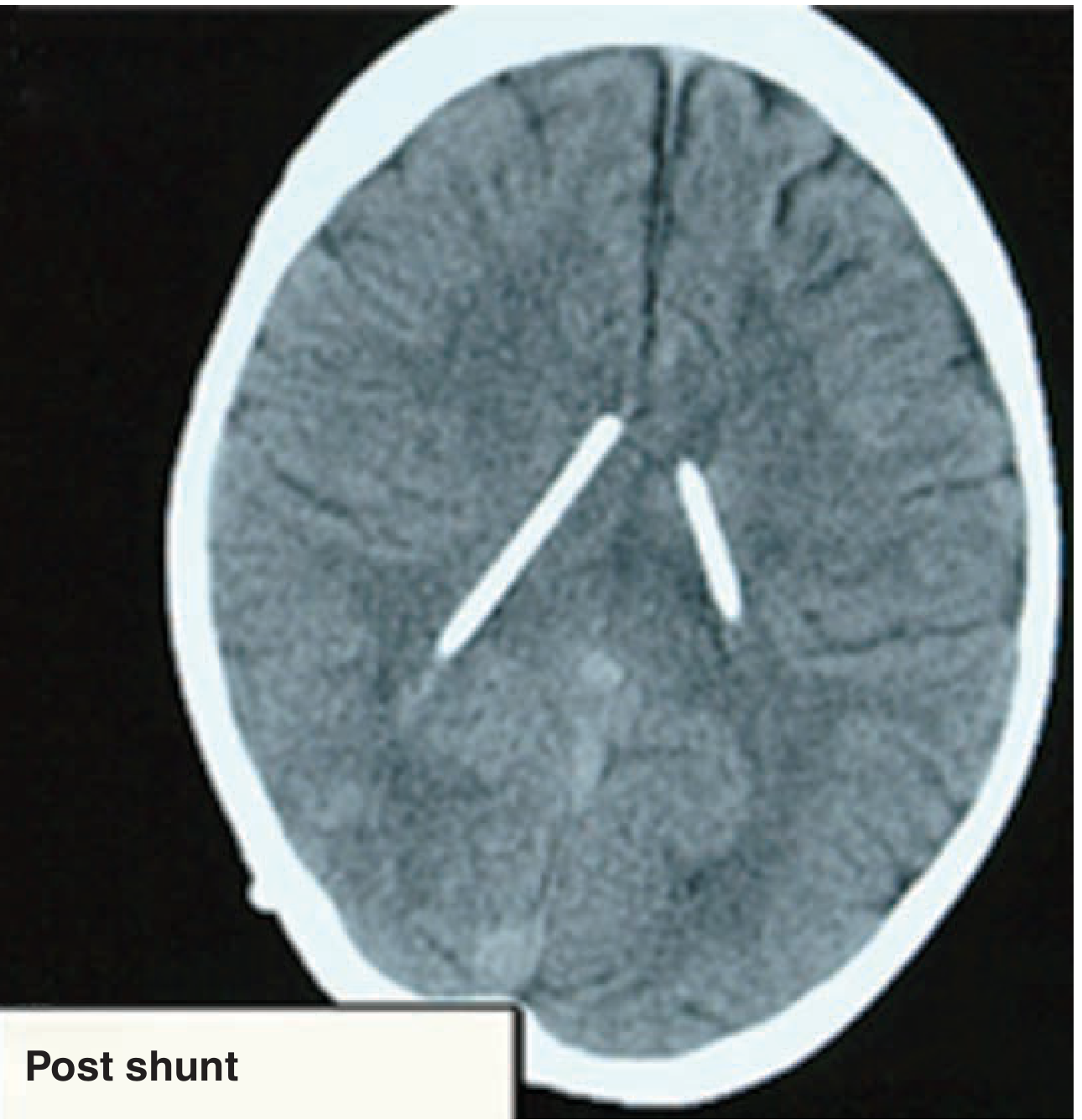
Shunt Valve Assessment
- In SVS, the shunt valve reservoir remains compressed on palpation (cannot refill) because the collapsed ventricle prevents CSF entry into the proximal catheter
- This distinguishes it from distal obstruction (difficulty compressing) and proximal obstruction with true blockage (slow refill >3 seconds)
- However: palpation has low PPV (~12%) - imaging is always required
Diagnosis
| Investigation | Findings |
|---|---|
| CT brain | Slit-like ventricles; absent subarachnoid spaces; shunt in place |
| Shunt series X-ray | Evaluate for kinking, disconnection, migration |
| Lumbar puncture | Assess opening pressure (ICP may be very low or transiently elevated) |
| Functional shunt study (Tc-99m scan) | Assess CSF flow through the shunt system |
| Non-invasive ICP monitoring | Recent reports support its use in guiding management (PMID 41120740) |
Management
Principle: Reduce CSF overdrainage
| Approach | Mechanism |
|---|---|
| Raise opening pressure of adjustable/programmable valve | Reduces flow, allows ventricles to re-expand |
| Antisiphon device (ASD) | Prevents valve flow when patient is upright (counteracts gravitational siphon) - most effective once established |
| Gravitational/anti-gravitational valves | Pressure-adjusting based on body position |
| Flow control valves | Self-regulating CSF flow rate |
From Adams & Victor: "The most effective measure has been the placement of an antisiphon device, which prevents valve flow when the patient stands."
- Useful in select patients with non-compliant ventricles
- Bypasses the ventricular system entirely; allows intracranial pressure normalization via lumbar drainage
- Recent reviews suggest LP shunts show promise in reducing overdrainage complications
| Procedure | Indication |
|---|---|
| Subtemporal decompression | Increases intracranial volume/compliance in non-compliant ventricle syndrome |
| Neuroendoscopic fenestration | Ventricular isolation - establishes communication between isolated compartments |
| Third ventriculostomy (ETV) | If aqueduct stenosis is contributing; avoids shunt dependency |
| Cranial vault expansion | Syndromic craniosynostosis with SVS; increases intracranial compliance |
Risk Factors and Associated Conditions
- Young age at shunting - brain/skull less compliant, subarachnoid spaces underdeveloped
- Small head circumference (<25th percentile)
- Congenital hydrocephalus (aqueduct stenosis, post-IVH)
- Secondary craniosynostosis (skull growth derangements from early over-drainage)
- Syndromic craniosynostosis (jugular outflow stenosis)
Complications of Overdrainage in General
- Low-pressure orthostatic headaches (worse upright, relieved by lying down)
- Subdural hygroma - extra-axial CSF collections
- Subdural haematoma - bridging vein rupture as brain pulls from dura
- Acquired Chiari malformation - cerebellar tonsillar descent from chronic low ICP
- Slit ventricle syndrome - loss of compliance, episodic high ICP
Summary Table
| Feature | Detail |
|---|---|
| Incidence | ~10% of shunted patients |
| Onset | Typically 2-5 years post-shunt (mean 4.3 yrs) |
| Age peak | 4-6 years |
| Core symptom | Episodic headache, worse upright, relieved supine |
| CT finding | Slit-like ventricles; no ventricular enlargement even with raised ICP |
| Mechanism | Overdrainage → collapsed ventricle occludes proximal catheter → cyclical ICP rise |
| Key distinction | From shunt obstruction: posture-dependent symptoms; from normal shunt function: episodic/waxing-waning course |
| First-line treatment | Raise valve opening pressure + antisiphon device |
| Surgical fallback | Subtemporal decompression, neuroendoscopy, ETV, LP shunt |
Benzodiazipines
benzodiazepines pharmacology mechanism classification clinical uses 2024
benzodiazepine GABA-A receptor mechanism diagram chloride channel
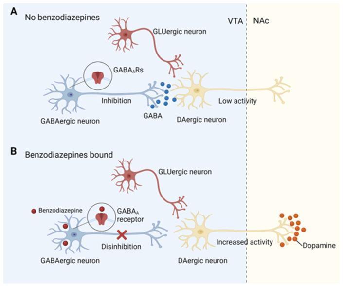
This pathophysiology diagram illustrates the neurobiological mechanism of benzodiazepine addiction through disinhibition of dopaminergic neurons in the mesolimbic reward system. The illustration is divided into two comparative panels. Panel A (No benzodiazepines) shows a baseline state in the Ventral Tegmental Area (VTA), where a blue GABAergic interneuron releases GABA molecules to inhibit a yellow dopaminergic (DAergic) neuron via GABAA receptors, resulting in 'Low activity' and minimal dopamine release in the Nucleus Accumbens (NAc). A GLUergic neuron is also shown nearby. Panel B (Benzodiazepines bound) depicts the introduction of red benzodiazepine molecules, which bind to GABAA receptors on the GABAergic interneuron. This binding induces 'Disinhibition' of the downstream DAergic neuron (indicated by an 'X' over the inhibitory synapse). Consequently, the DAergic neuron exhibits 'Increased activity,' leading to a significant rise in dopamine release (red molecules) within the NAc. The diagram highlights how benzodiazepines modulate neuronal signaling to trigger the brain's reward circuitry.
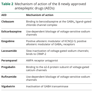
Table 2 Mechanism of action of the 8 newly approved antiepileptic drugs (AEDs) <table><thead><tr><th>AED</th><th>Mechanism of action</th></tr></thead><tbody><tr><td>Clobazam</td><td>Binding to benzodiazepine at the GABA<sub>A</sub> ligand-gated chloride channel complex</td></tr><tr><td>Eslicarbazepine</td><td>Use-dependent blockage of voltage-sensitive sodium channels</td></tr><tr><td>Ezogabine</td><td>Positive allosteric modulator of KCNQ2-5; positive allosteric modulator of GABA<sub>A</sub> receptors</td></tr><tr><td>Lacosamide</td><td>Slow inactivation of voltage-gated sodium channels; binds to CRMP-2</td></tr><tr><td>Perampanel</td><td>AMPA receptor antagonist</td></tr><tr><td>Pregabalin</td><td>Binding to the α2-δ protein subunit of voltage-gated calcium channels</td></tr><tr><td>Rufinamide</td><td>Use-dependent blockage of voltage-sensitive sodium channels</td></tr><tr><td>Vigabatrin</td><td>Inactivation of GABA transaminase</td></tr></tbody></table>
Benzodiazepines
Chemistry and Structure
Mechanism of Action
- Pentameric ligand-gated chloride channel
- Composed of 5 subunits: typically 2α + 2β + 1γ (most common isoform: 2α1 + 2β2 + 1γ2)
- GABA binding site: between α and β subunits (two sites)
- Benzodiazepine binding site (BZ site): between α and γ subunit - distinct from barbiturate binding site
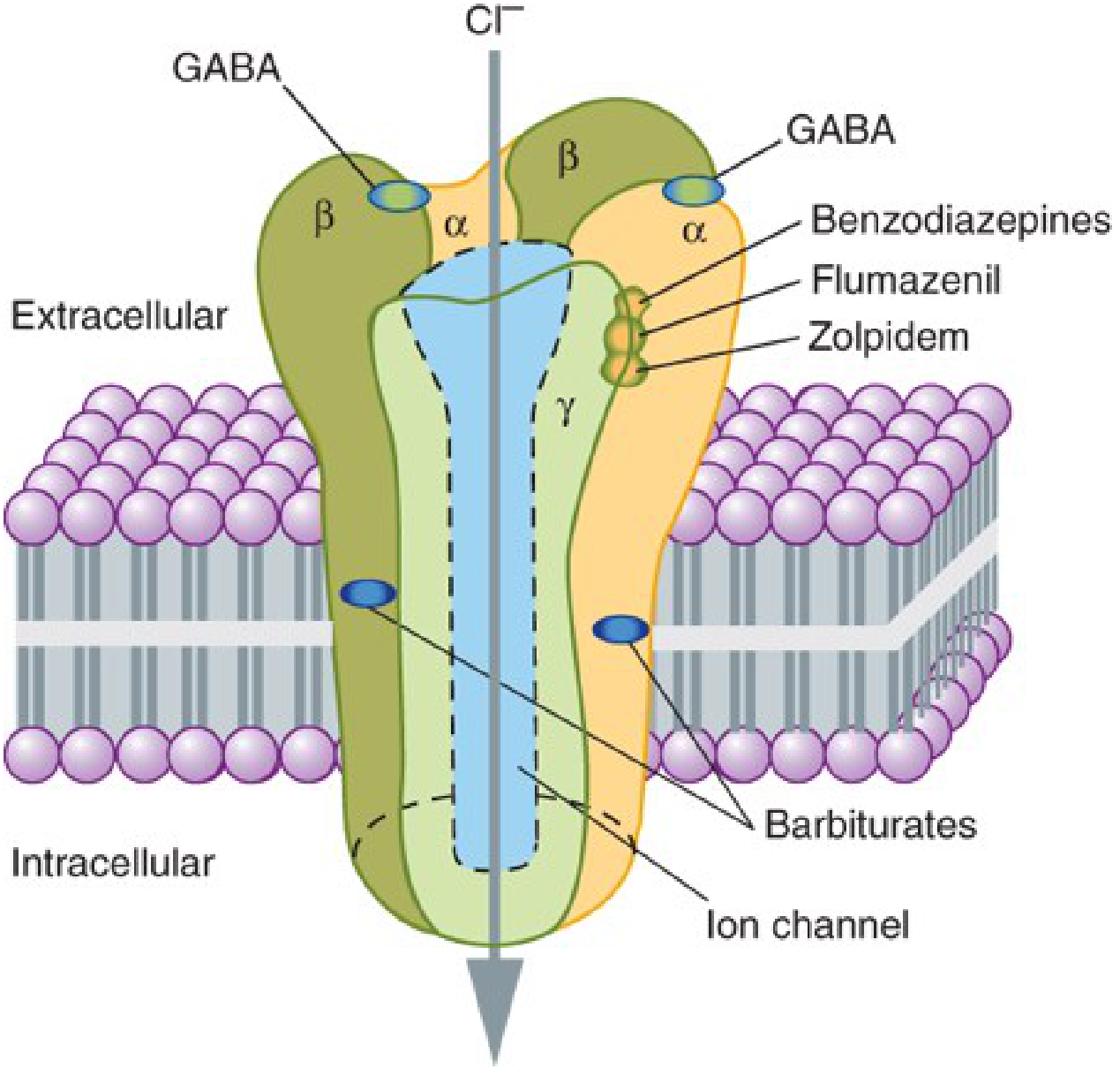
- BZ1 receptor (α1-containing) - mediates sedation, amnesia, anticonvulsant effect
- BZ2 receptor (α2, α3, α5-containing) - mediates anxiolytic, muscle relaxant, cognitive effects
- Clobazam: the only benzodiazepine with greater affinity for BZ2 than BZ1
| Ligand Type | Example | Effect |
|---|---|---|
| Nonselective full agonist | Diazepam, lorazepam | Maximum enhancement of GABA-A, all BZ subtypes |
| Selective full agonist | Zolpidem | Only BZ1 - less psychomotor impairment |
| Partial agonist | Bretazenil | Reduced efficacy, fewer adverse effects |
| Antagonist | Flumazenil | Occupies BZ site, no intrinsic activity - reverses BZD effects |
| Inverse agonist | Diazepam-binding inhibitor (DBI/ACBP) | Reduces GABA-A affinity - pro-convulsant, anxiogenic |
Pharmacological Effects
| Effect | Notes |
|---|---|
| Anxiolytic | Disinhibition of punishment-suppressed behavior; dose-dependent |
| Sedation / hypnosis | Decreases sleep latency; increases stage 2 NREM; decreases REM and slow-wave sleep |
| Anticonvulsant | Suppress seizure spread; first-line for status epilepticus |
| Muscle relaxant | Via spinal cord interneuron inhibition |
| Anterograde amnesia | Dose-dependent; useful peri-procedurally |
| Anxiolytic | Not antidepressant (contrast neuroactive steroids acting at benzodiazepine-insensitive GABA-A sites) |
Classification by Half-Life
| Duration | Drug | Half-life | Notes |
|---|---|---|---|
| Ultra-short | Triazolam | ~4 h | Rebound insomnia, daytime anxiety |
| Short | Midazolam, oxazepam, lorazepam, temazepam | 1-24 h | Good for elderly (LOT: Lorazepam, Oxazepam, Temazepam - no active metabolites, conjugation only) |
| Intermediate | Alprazolam, clonazepam | 12-40 h | Alprazolam: severe withdrawal syndrome |
| Long | Diazepam, chlordiazepoxide, flurazepam | 20-100 h + active metabolites | Self-tapering; preferred for alcohol withdrawal |
LOT rule (Katzung): Lorazepam, Oxazepam, Temazepam are metabolized solely by glucuronide conjugation - safe in liver disease, elderly, neonates (no CYP oxidation, no active metabolites)
Individual Drug Profiles (from Goodman & Gilman's)
| Drug | Therapeutic Uses | Key Features |
|---|---|---|
| Diazepam | Anxiety, alcohol withdrawal, status epilepticus, muscle relaxant, premedication | Prototype benzodiazepine; long-acting with active metabolites |
| Lorazepam | Anxiety, alcohol withdrawal, premedication, status epilepticus | Conjugation only; no active metabolites; IM reliable |
| Midazolam | Procedural sedation, preanesthesia, ICU | Rapid onset/offset; water-soluble at pH <4; IV/IM/IN |
| Remimazolam | Procedural sedation | Very rapidly inactivated (ester hydrolysis); reversible with flumazenil |
| Alprazolam | Anxiety disorders, panic disorder, agoraphobia | Severe withdrawal syndrome on discontinuation |
| Clonazepam | Seizure disorders, acute mania, certain movement disorders | Tolerance develops to anticonvulsant effect |
| Clobazam | Lennox-Gastaut syndrome, epilepsy | Active metabolite with long half-life; CYP2C19 metabolizer status affects dosing |
| Chlordiazepoxide | Anxiety, alcohol withdrawal | Long-acting; self-tapering via active metabolites |
| Oxazepam | Anxiety, alcohol withdrawal | Conjugation only; good in elderly/hepatic impairment |
| Flurazepam / Quazepam | Insomnia | Active metabolites accumulate; daytime sedation risk |
| Temazepam | Insomnia | Mainly conjugation |
| Triazolam | Insomnia | Very short t½; rebound anxiety/insomnia |
| Clorazepate | Anxiety, seizure disorders | Prodrug - converted to nordazepam during absorption |
Clinical Indications (FDA-Approved)
- Anxiety disorders (GAD, panic disorder, social anxiety, PTSD acute phase)
- Status epilepticus - IV lorazepam or diazepam; first-line
- Alcohol withdrawal (and other sedative-hypnotic withdrawal)
- Insomnia - short-term only
- Procedural sedation/amnesia (midazolam, remimazolam)
- Skeletal muscle spasm (diazepam)
- Acute mania (adjunctive - clonazepam)
- Spastic neurologic disorders (clonazepam)
Effects on Sleep Architecture
- Decrease sleep latency
- Increase stage 2 NREM duration
- Decrease REM sleep - REM rebound on abrupt withdrawal
- Decrease stage 3/4 slow-wave (deep) sleep
- Use >1-2 weeks → tolerance to hypnotic effects
Adverse Effects
| Adverse Effect | Comment |
|---|---|
| Sedation, psychomotor impairment | Impairs driving and occupational function |
| Anterograde amnesia | Useful for procedures; problematic otherwise |
| Respiratory depression | Especially with opioids/alcohol; less than barbiturates alone |
| Paradoxical reactions | Agitation, aggression, disinhibition (especially in children, elderly, brain-injured) |
| Falls and fractures | Particularly in elderly |
| Cognitive impairment | With chronic use |
| Dependence and withdrawal | Physiologic dependence with even therapeutic doses over weeks |
| Tolerance | Down-regulation of BZ receptors with chronic use |
| Rebound anxiety/insomnia | Especially short-acting agents |
Tolerance and Dependence
- Tolerance develops through down-regulation of brain benzodiazepine receptors (pharmacodynamic tolerance) and, for barbiturates, metabolic enzyme induction
- Partial cross-tolerance with alcohol and other sedative-hypnotics
- Physiologic dependence can develop with therapeutic doses taken chronically
- Severity of withdrawal correlates with:
- Higher doses used
- Shorter half-life of the drug (short t½ agents worst)
- Duration of use
Withdrawal Syndrome
- Anxiety, insomnia, tremor, sweating
- Nausea, tachycardia, hypertension
- Seizures (potentially life-threatening)
- Psychosis
Triazolam (t½ ~4 h) can produce inter-dose withdrawal symptoms between nightly doses. - Katzung, p. 608
- Convert to equivalent diazepam dose (long t½ = smoother taper)
- Gradual dose reduction - not abrupt cessation
- "Step-wise reduction proportionate to existing dose - decrements become smaller as dose lowers" (~⅛ of daily dose every 2 weeks)
- Taper over weeks to months (up to 6 months for some patients)
- CBT alongside dose reduction - evidence-based for successful deprescription
- No pharmacological add-on proven to facilitate withdrawal (2018 Cochrane review)
- Do NOT use another dependence-forming medicine, sodium valproate, or buspirone for withdrawal symptoms
Flumazenil (Benzodiazepine Antagonist)
- Mechanism: Competitive antagonist at the BZ binding site (α-γ interface); reverses sedation and amnesia but less reliably reverses respiratory depression
- Dose: 0.2 mg IV, repeated every 1 minute to maximum 3 mg
- Half-life: ~1 hour (brain t½ ~30 min) - re-sedation may occur with longer-acting benzodiazepines
- Also reverses zolpidem, zaleplon, eszopiclone (all share the same BZ site)
| Contraindication | Reason |
|---|---|
| Physical dependence on benzodiazepines | Precipitates acute withdrawal seizures |
| Co-ingestion of cyclic antidepressants | Seizures unmasked by BZD reversal |
| Unknown overdose agent | May be protecting against seizure from co-ingestant |
| Seizure disorder controlled by benzodiazepines | Removes anticonvulsant protection |
| Suspected raised ICP / severe head injury | Adverse cerebral hemodynamic effects |
If flumazenil-precipitated seizures occur, treat with phenobarbital or propofol (not another benzodiazepine - the BZ site is blocked). - Tintinalli's, p. 3021
- Isolated BZD overdose: rarely needs treatment beyond monitoring
- Activated charcoal: useful if ingested recently
- Respiratory support if mixed overdose
- Do NOT induce emesis (risk of aspiration with mental status depression)
Special Populations
| Population | Consideration |
|---|---|
| Elderly | Increased sensitivity; prolonged half-life; falls risk; prefer LOT agents |
| Hepatic impairment | Prefer lorazepam, oxazepam, temazepam (conjugation only) |
| Pregnancy | Neonatal withdrawal syndrome; cleft palate (first trimester, older data) |
| Children | Paradoxical reactions more common |
| COPD/OSA | Avoid or use with extreme caution - respiratory depression risk |