
2 Diagnostic tests 2.1. PHO Soln Amount Urine pregnancy test / card test 2.2 Estimation of hemoglobin using Sahli's hemoglobino meter * 2.3 Advice/assisting testing* HIV HBsAg VDRL Rh typing and Grouping 2.4 Preparation of peripheral smear for malaria 2.5 Urine testing for albumin and sugar 2.6 Preparation of mother for USG Administration of Td/TT 4 Prescription of iron & folic acid and calcium tablets 5 Administration of Iron sucrose .6 Identification and Assessment of Maternal physiological changes in pregnancy 10 Identification of minor disorders during pregnancy and their nursing management Answer me for all the questions for viva detaily in obg refer dutta book
OBG VIVA - COMPREHENSIVE ANSWERS
(PHO Practical Topics - Based on DC Dutta's Obstetrics)
2.1 - URINE PREGNANCY TEST / CARD TEST
Principle
- Detects hCG (human Chorionic Gonadotropin) in urine
- hCG is produced by the syncytiotrophoblast of the developing placenta
- It is an immunochromatographic assay (lateral flow immunoassay)
- Uses monoclonal antibodies against the beta subunit of hCG
When does hCG appear?
- hCG is detectable in blood from 8-10 days after fertilization (day 23-25 of cycle)
- In urine from 14 days after conception (i.e., at the time of missed period)
- Peaks at 8-12 weeks of gestation (60,000-100,000 mIU/mL), then falls
Procedure (Card Test)
- Collect first morning urine (most concentrated - highest hCG)
- Place the card on a flat surface
- Add 3 drops of urine into the sample well (S) using the dropper
- Read result at 5 minutes (not more than 10 minutes)
Reading the Result
- Positive: Two lines (C + T) - even a faint T line is POSITIVE
- Negative: One line (C only)
- Invalid: No line at C - repeat the test
Sensitivity
- Most card tests can detect hCG as low as 20-25 mIU/mL
- Some sensitive tests detect up to 10 mIU/mL
False Positive Causes
- Hydatidiform mole, Choriocarcinoma (very high hCG)
- Recent abortion or delivery (hCG still in blood)
- hCG injection given (fertility treatment)
- Ectopic pregnancy
- Some drugs (methadone, promethazine)
- Proteinuria or hematuria (interferes with test)
False Negative Causes
- Very early pregnancy (too little hCG)
- Very dilute urine
- Hook effect - very high hCG levels (e.g., molar pregnancy) can paradoxically give false negative with some kits
- Expired kit
- Ectopic pregnancy (low hCG production)
Clinical Importance
- Confirmatory test for pregnancy diagnosis
- Useful in ANC registration
2.2 - ESTIMATION OF HEMOGLOBIN USING SAHLI'S HEMOGLOBINOMETER
Principle
- Blood is mixed with N/10 HCl (dilute hydrochloric acid)
- Hemoglobin is converted to acid hematin (brown color)
- Diluted with water until color matches the standard brown comparator glass
- Reading is noted on the graduated tube
Apparatus
- Sahli's hemoglobinometer (graduated tube, standard glass, pipette)
- N/10 HCl
- Distilled water
- Stirring rod
- Sahli's pipette (20 cu.mm capacity)
Procedure
- Add N/10 HCl up to the 2 mark (20%) on the graduated tube
- Clean the fingertip (ring finger, left hand) with spirit swab, allow to dry
- Prick the fingertip with a sterile lancet
- Wipe away the first drop of blood
- Draw blood into the Sahli's pipette up to the 20 cu.mm mark
- Blow the blood into the HCl in the tube immediately
- Rinse the pipette 2-3 times with the mixture
- Wait for 10 minutes (acid hematin formation)
- Add distilled water drop by drop, stirring constantly
- Compare with the standard glass until the color matches
- Note the reading on the graduated tube
Reading
- Expressed as g/dL or % of normal (Sahli's scale goes up to 17.3 g/dL = 100%)
Normal Values in Pregnancy
- Normal non-pregnant female: 12-14 g/dL
- During pregnancy: Hb falls due to hemodilution
- Physiological anemia of pregnancy - Hb may fall to ~10-11 g/dL
- WHO defines anemia in pregnancy as Hb < 11 g/dL (1st and 3rd trimester) or < 10.5 g/dL (2nd trimester)
- Mild anemia: 10-10.9 g/dL
- Moderate anemia: 7-9.9 g/dL
- Severe anemia: < 7 g/dL (High risk)
Significance in ANC
- Routine test at first ANC visit, then at 28 and 36 weeks
- Guides supplementation and treatment
Limitations of Sahli's Method
- Subjective - depends on visual comparison (observer error)
- Less accurate than Cyanmethemoglobin (CNHb) method
- Not suitable for all types of Hb (e.g., fetal Hb)
- Cyanmethemoglobin method is the gold standard
2.3 - ADVICE/ASSISTING TESTING: HIV, HBsAg, VDRL, Rh TYPING AND BLOOD GROUPING
Why These Tests?
HIV Testing in Pregnancy
- Confidentiality assured
- Purpose of test
- Window period explanation (3 weeks to 3 months after exposure)
- Consequences of positive/negative result
- Prevention of MTCT if positive
- Treatment options (ART)
- Test 1 (E1) - Most sensitive ELISA/Rapid test
- If Reactive - Test 2 (E2) - Different antigen/format
- If Reactive - Test 3 (E3) - Third test (Western Blot / another ELISA)
- All three reactive = HIV Positive
- If Negative: Reinforce safe behavior, explain window period
- If Positive: Disclose carefully, link to ART center, counsel on MTCT prevention, infant feeding (exclusive formula or exclusive breastfeeding - not mixed)
- Prophylactic ART (Option B+) - start ALL HIV positive pregnant women on TDF + 3TC + EFV (Tenofovir + Lamivudine + Efavirenz) regardless of CD4 count - continue lifelong
- Institutional delivery
- Neonatal prophylaxis: Nevirapine syrup for 6 weeks to baby
- Avoid mixed feeding
HBsAg Testing (Hepatitis B Surface Antigen)
- Check HBeAg (marker of high infectivity)
- Baby at delivery: Give Hepatitis B vaccine (0.5 mL IM) + HBIG (Hepatitis B Immunoglobulin) 0.5 mL IM within 12 hours of birth - at different sites
- Complete the 3-dose vaccine series (0, 1, 6 months) - under universal immunization program (EPI)
- Breastfeeding is NOT contraindicated if baby receives immunoprophylaxis
VDRL (Venereal Disease Research Laboratory Test)
- Detects reagin antibody (IgG, IgM) against cardiolipin-lecithin antigen
- Patient's serum mixed with VDRL antigen on a slide
- Rotate at 180 rpm for 4 minutes
- Read under microscope
- Reactive = flocculation (clumping) seen - possible syphilis
- Non-reactive = No flocculation - likely no syphilis
- Untreated syphilis causes: Congenital syphilis, stillbirth, IUGR, hydrops fetalis
- Treatment: Benzathine Penicillin G 2.4 million units IM single dose (or 3 doses weekly for late syphilis)
- Partner must also be treated
- SLE, RA, viral infections (EBV, hepatitis), malaria, leprosy, old age, pregnancy itself
- Biological false positive (BFP) - that is why confirmatory test is always needed
Rh Typing and Blood Grouping
- Blood group determined by antigens on RBC surface (A, B, both, or none)
- Four groups: A, B, AB, O
- Universal donor: O negative
- Universal recipient: AB positive
- Rh antigen (D antigen) - present = Rh positive, absent = Rh negative
- ~85% Indians are Rh positive, 15% Rh negative
- Occurs when Rh negative mother carries Rh positive fetus
- During delivery, fetal RBCs enter maternal circulation → mother forms anti-D antibodies
- In subsequent Rh positive pregnancy, anti-D crosses placenta → destroys fetal RBCs → Hemolytic Disease of Newborn (HDN) / Erythroblastosis fetalis
- Give Anti-D immunoglobulin (Rho-D Ig, 300 micrograms IM):
- At 28 weeks prophylactically
- Within 72 hours of delivery of Rh positive baby
- After every sensitizing event: abortion, amniocentesis, CVS, ectopic pregnancy, trauma
- Indirect Coombs Test (ICT) - done at booking and at 28 weeks in Rh negative mothers to detect if already sensitized
- If ICT positive - monitor with serial MCA Doppler
2.4 - PREPARATION OF PERIPHERAL SMEAR FOR MALARIA
Purpose
- To detect malaria parasites (Plasmodium falciparum, P. vivax, P. malariae, P. ovale)
- Two smears prepared: Thick smear (screening - more sensitive) and Thin smear (species identification)
Materials
- Clean, grease-free glass slides
- Lancet/needle (sterile)
- Spirit swab
- Leishman's stain (for thin smear) / Giemsa stain (for thick smear)
- Microscope
- Immersion oil
Procedure
Thick Smear:
- Clean fingertip, prick with lancet
- Wipe first drop
- Touch slide to 2nd drop - collect 2-3 drops, spread in a circle ~1-2 cm diameter
- Allow to dry in air - do NOT heat fix (haemolysation needed)
- Stain with 3% Giemsa for 30-45 minutes OR 10% Giemsa for 10 min
- Wash gently with buffer water pH 7.2
- Allow to dry, examine under oil immersion
Thin Smear:
- Collect one drop of blood on slide
- Use spreader slide at 30-45° angle to spread in a thin film (feather edge)
- Air dry, then heat fix briefly (pass through flame 3-4 times)
- Flood with Leishman's stain for 2 minutes
- Add equal volume of buffer water (pH 7.2), mix, wait 10 minutes
- Wash, dry, examine under oil immersion
Reading
- Thick smear: Look for ring forms, trophozoites, gametocytes
- Thin smear: Species identification based on morphology
Malaria in Pregnancy - Why Important?
- Malaria is particularly dangerous in pregnancy
- Causes: Severe maternal anemia, cerebral malaria, hypoglycemia, preterm labor, IUGR, stillbirth, maternal death
- P. falciparum sequesters in placenta (placental malaria)
- Treatment: Chloroquine (safe in pregnancy for P. vivax); for P. falciparum - Artesunate-based combination therapy (ACT) in 2nd/3rd trimester; Quinine + Clindamycin in 1st trimester
- Primaquine is CONTRAINDICATED in pregnancy (hemolytic)
- Chemoprophylaxis: SP (Sulfadoxine-Pyrimethamine) IPTp in endemic areas
2.5 - URINE TESTING FOR ALBUMIN AND SUGAR
Urine Testing for Albumin (Proteinuria)
- Proteinuria in pregnancy is a sign of Pre-eclampsia (HTN + proteinuria after 20 weeks)
- Also seen in: UTI, renal disease, orthostatic proteinuria
- Fill test tube 2/3 with urine
- Boil the upper 1/3 over flame until it boils
- A white turbidity may appear (due to phosphates or albumin)
- Add 2-3 drops of 1% acetic acid
- Boil again
- Phosphates dissolve in acid → turbidity disappears = albumin ABSENT
- If turbidity persists or increases after acid → albumin PRESENT
- Trace: Slight turbidity (seen through the tube)
- 1+: Definite turbidity without floccules
- 2+: Heavy turbidity with fine granules
- 3+: Flocculation
- 4+: Solid coagulum (clot)
- Reagent strip impregnated with tetrabromophenol blue
- Color changes from yellow to blue-green in presence of albumin
- Read at 60 seconds
- Grading: Negative, Trace, 1+, 2+, 3+
Urine Testing for Sugar (Glucose)
- Glycosuria in pregnancy may indicate Gestational Diabetes Mellitus (GDM)
- Note: Renal threshold for glucose may be lower in pregnancy (due to increased GFR), so glycosuria may occur without diabetes - requires further confirmation with OGTT
- Take 5 mL of Benedict's reagent in a test tube
- Add 8 drops of urine
- Mix and boil for 5 minutes (or place in boiling water bath)
- Allow to cool, observe color change
- Blue (no color change) = Negative (no reducing sugars)
- Green precipitate = Trace (+)
- Yellow precipitate = 1+ (0.5-1%)
- Orange precipitate = 2+ (1-1.5%)
- Brick red precipitate = 3+ or 4+ (>2%)
- Uses glucose oxidase-peroxidase reaction
- Specific for glucose only (Benedict's detects all reducing sugars)
- Reliable, rapid
2.6 - PREPARATION OF MOTHER FOR USG (ULTRASONOGRAPHY)
Types of USG in Pregnancy
| Trimester | Weeks | Purpose |
|---|---|---|
| 1st Trimester | 6-9 weeks | Confirm intrauterine pregnancy, viability, gestational age, dating |
| 1st Trimester | 11-14 weeks | NT scan (Nuchal Translucency) - Down syndrome screening |
| 2nd Trimester | 18-20 weeks | Anomaly scan (TIFFA) - fetal structure survey |
| 3rd Trimester | 28-32+ weeks | Growth scan, liquor, placenta, Doppler |
Preparation of Mother for USG
Transabdominal USG (most common):
- Full bladder required in early pregnancy (1st trimester) - bladder acts as acoustic window
- Ask patient to drink 4-6 glasses of water (1-1.5 L) 1 hour before the scan and NOT pass urine
- No special diet preparation needed
- Explain procedure to allay anxiety
- Ask her to lie in dorsal supine position (tilt to left if >20 weeks to avoid aortocaval compression)
- Expose abdomen from xiphisternum to symphysis pubis
- Warm coupling gel applied to abdomen
Transvaginal USG (TVS):
- Empty bladder required (opposite to transabdominal)
- Ask patient to empty bladder before procedure
- Explain the procedure - may be slightly uncomfortable
- Obtain verbal consent
- Done for: Very early pregnancy, low-lying placenta, cervical length measurement, threatened abortion
Role of Nurse:
- Counsel patient about the procedure
- Ensure proper bladder filling for TAU or emptying for TVS
- Position patient appropriately
- Assist doctor/sonographer
SECTION 3 (implied) - ADMINISTRATION OF Td/TT VACCINE
Why Given in Pregnancy?
- To prevent Neonatal Tetanus (Tetanus Neonatorum) and maternal tetanus
- Neonatal tetanus occurs when cord is cut with unsterile instruments
- Tetanus Toxoid generates maternal antibodies which cross placenta → protect neonate
Schedule (as per MoHFW India - National Immunization Schedule):
| Category | Schedule |
|---|---|
| Previously unimmunized / unknown | TT-1 as early as possible in pregnancy; TT-2 at least 4 weeks after TT-1 |
| Received 2 TT doses in previous pregnancy (within 3 years) | 1 TT dose (booster) |
| Fully immunized in childhood (5 doses complete) | 1 TT booster dose |
Dose and Route:
- 0.5 mL intramuscular (IM)
- Site: Deltoid muscle (anterolateral mid-thigh in infants)
- Left arm preferred (right arm for other injections - easy identification)
- Needle: 23G, 1-1.5 inch
Contraindications:
- Severe allergic reaction (anaphylaxis) to previous dose
- Acute febrile illness (defer vaccination)
- Not contraindicated in pregnancy - safe
Side Effects:
- Local: Pain, redness, swelling at injection site
- Systemic: Mild fever, malaise (uncommon)
- Rare: Anaphylaxis (keep adrenaline ready)
Nursing Steps:
- Verify patient identity, check vaccination card
- Explain procedure and get verbal consent
- Check expiry date of vial, VVM (Vaccine Vial Monitor) color, cold chain maintenance
- Load 0.5 mL in syringe (aspirate from vial correctly)
- Clean deltoid with spirit swab - allow to dry
- Inject deep IM (90° angle) - do NOT massage after injection
- Document in MCP (Mother and Child Protection) card
- Observe for 15 minutes for anaphylaxis
- Advise: pain is normal, apply cold compress if needed, return if severe reaction
SECTION 4 - PRESCRIPTION OF IRON & FOLIC ACID AND CALCIUM TABLETS
Iron and Folic Acid (IFA) Supplementation
Why?
- Pregnancy increases iron requirement to ~27 mg/day (vs 18 mg/day non-pregnant)
- Required for: Increased maternal RBC mass, fetal RBC production, placenta
- Folic acid: Required for neural tube closure (day 18-28 of embryonic life), DNA synthesis, cell division
Government of India (National Health Mission) IFA Tablets:
- Large IFA tablet (given to pregnant women): Contains 100 mg elemental iron + 500 mcg (0.5 mg) folic acid
- IFA Small tablet (given to adolescents): Contains 45 mg elemental iron + 400 mcg folic acid
Dose and Schedule:
- Start at 12 weeks (after first trimester - to reduce nausea)
- In practice, start as soon as ANC registered
- One tablet daily for 180 days (6 months) during pregnancy and continue for 45 days postpartum
- If Hb < 10 g/dL: 2 tablets daily
Dutta's Advice on IFA:
- Advise to take tablet at night or after meals to reduce GI side effects
- Avoid with tea, coffee, antacids, milk (reduce absorption)
- Take with Vitamin C (citrus juice/lemon) to enhance absorption
- Side effects: Nausea, constipation, dark stools (warn patient)
Folic Acid Separately (Periconceptional):
- 5 mg folic acid daily from 3 months before conception and up to 12 weeks gestation
- Standard dose for low-risk: 0.4-0.5 mg
- High-risk (previous NTD child, epilepsy on valproate, diabetes): 5 mg/day
Calcium Supplementation
Why?
- Fetal calcium requirement is highest in 3rd trimester (200-250 mg/day for fetal bone mineralization)
- Pregnancy requires ~1200 mg calcium/day
- Calcium supplementation prevents / reduces risk of pre-eclampsia (mechanism: reduces vascular smooth muscle contraction, reduces PTH release)
WHO / India Recommendation:
- Calcium 1.5-2 g/day (in divided doses) for prevention of pre-eclampsia, especially in low calcium intake populations
- India: 500 mg elemental calcium TID (three times daily) - total 1500 mg/day
- Start from 20 weeks of pregnancy
Important Note:
- Do NOT take iron and calcium tablets at the same time - they compete for absorption
- Iron in the morning, Calcium at night (or separate by 2-4 hours)
SECTION 5 - ADMINISTRATION OF IRON SUCROSE
What is Iron Sucrose?
- Parenteral iron preparation - iron sucrose complex (Fe(III) hydroxide sucrose)
- Used when oral iron cannot be given or is insufficient
- Trade name: Venofer, Sucrofer
Indications in Pregnancy:
- Severe anemia (Hb 5-7 g/dL) with not enough time for oral therapy
- Intolerance to oral iron (severe GI side effects)
- Malabsorption syndromes
- Non-compliance with oral iron
- Gestational age > 32 weeks with moderate-severe anemia (not enough time for oral response)
Advantages over Iron Dextran:
- Lower risk of anaphylaxis
- Safer profile
- Can be given as IV infusion or slow IV push
Dose Calculation:
- Each session: 200 mg iron sucrose (10 mL of 20 mg/mL solution) in 100 mL normal saline, given over 30 minutes
- Maximum per session: 200 mg
- Given every alternate day or 2-3 times per week
- Maximum per week: 400-600 mg
Procedure (IV Iron Sucrose Administration):
- Check doctor's order, confirm patient identity, check allergies
- Prepare: Iron sucrose 200 mg (10 mL) + Normal saline 100 mL (total 110 mL)
- Ensure IV access (18-20G cannula)
- Perform test dose - in first infusion, observe for 15 minutes before giving full dose
- Infuse over 30 minutes (not faster than 100 mg/15 min)
- Keep patient under observation throughout
- Keep adrenaline (epinephrine) 1:1000, antihistamines, hydrocortisone ready (anaphylaxis kit)
- Monitor vital signs before, during, after infusion
Side Effects:
- Common: Nausea, vomiting, headache, dizziness, hypotension, metallic taste
- Serious: Anaphylaxis/anaphylactoid reaction (rare)
- Local: Phlebitis if extravasation (iron sucrose causes tissue staining unlike iron dextran)
- Iron overload if overdosed
Contraindications:
- Anemia NOT due to iron deficiency (hemolytic anemia, thalassemia)
- Evidence of iron overload
- Known hypersensitivity
Monitoring:
- Hb should rise by 0.8-1 g/dL per week with IV iron
- Re-check Hb after 2 weeks
- Once Hb > 10 g/dL, continue oral iron for maintenance
SECTION 6 - IDENTIFICATION AND ASSESSMENT OF MATERNAL PHYSIOLOGICAL CHANGES IN PREGNANCY
CARDIOVASCULAR CHANGES
- Blood volume increases 40-50% (plasma 50%, RBCs 30%) - peaks at 32-34 weeks
- Physiological hemodilution → Physiological anemia of pregnancy
- Cardiac output increases 30-50% (due to increased SV + HR)
- Heart rate increases by 10-20 bpm
- Blood pressure: Systolic unchanged; diastolic falls 10-15 mmHg in mid-pregnancy (due to reduced peripheral resistance from progesterone)
- Edema of legs common (increased venous pressure)
- Palpitations common (functional systolic murmur may be heard - innocent)
- Supine hypotensive syndrome (Aortocaval compression) - SBP falls >15 mmHg in supine position due to IVC compression by gravid uterus from 20 weeks onward
RESPIRATORY CHANGES
- Diaphragm displaced upward by 4 cm (by growing uterus)
- Tidal volume increases 40% (from 450 to 600 mL)
- Progesterone stimulates respiratory center → hyperventilation
- Respiratory rate slightly increases
- Functional Residual Capacity (FRC) decreases by 20%
- Dyspnea common (even at rest in late pregnancy)
- PaCO2 falls (chronic respiratory alkalosis) → compensated by renal HCO3 excretion
RENAL/URINARY CHANGES
- Kidneys enlarge slightly; collecting system dilates (due to progesterone and mechanical compression)
- GFR increases 50% → increased clearance of creatinine, urea, uric acid → serum creatinine/urea LOWER than normal in pregnancy
- Renal threshold for glucose decreases → glycosuria even without diabetes
- Frequency of micturition in 1st and 3rd trimester (pressure on bladder)
- Stress incontinence common
- Predisposition to UTI (due to ureteral dilation, vesicoureteral reflux)
GASTROINTESTINAL CHANGES
- Nausea and vomiting (morning sickness) - due to hCG and progesterone, peaks at 8-12 weeks
- Progesterone relaxes lower esophageal sphincter → heartburn/reflux (GERD)
- Gastric motility decreases → constipation
- Hemorrhoids common (constipation + IVC compression)
- Gums: Hyperemia, friable - pregnancy gingivitis
- Parotid: Excessive salivation (ptyalism)
- Pica - craving for unusual substances
MUSCULOSKELETAL CHANGES
- Relaxin and progesterone cause joint laxity and relaxation of pelvic ligaments
- Lumbar lordosis increases (waddling gait) - backache common
- Symphysis pubis widens (up to 8 mm) - diastasis symphysis pubis
- Center of gravity shifts forward
- Carpal tunnel syndrome - fluid retention compresses median nerve
SKIN AND HAIR CHANGES
- Linea nigra - dark pigmented line from umbilicus to symphysis (due to MSH)
- Chloasma/Melasma (mask of pregnancy) - brown patches on face
- Striae gravidarum (stretch marks) - abdomen, thighs, breasts
- Spider nevi, palmar erythema - due to elevated estrogen
- Hyperpigmentation of nipples, areola, perineum, axillae
- Hair: Increased hair in anagen phase during pregnancy (less shedding), then telogen effluvium (hair loss) postpartum
BREAST CHANGES
- Breast enlargement begins 8th week (under estrogen/progesterone)
- Colostrum secretion from 16th week onward
- Nipple enlargement and darkening of areola
- Montgomery's tubercles (sebaceous glands on areola) become prominent from 8th week
- Secondary areola (pale ring outside primary areola) appears around 20 weeks
UTERINE CHANGES
- Non-pregnant uterus weighs ~60 g; at term → 1000 g
- Uterus grows from pear-shaped to ovoid
- Braxton-Hicks contractions - painless, irregular uterine contractions from early pregnancy, more apparent from 16 weeks
- Uterine soufflé (soft blowing murmur synchronous with maternal pulse) audible by stethoscope
- Hegar's sign - softening of isthmus (8-10 weeks)
- Fundal height measurement: Fundus at umbilicus = 20 weeks; xiphisternum = 36 weeks; rises 4 cm/4 weeks
WEIGHT GAIN IN PREGNANCY (Dutta's)
- Total weight gain: 10-12 kg (normal BMI)
- Breakdown: Fetus (3.4 kg), placenta (0.6 kg), amniotic fluid (0.8 kg), uterus (0.9 kg), blood (1.5 kg), breast (0.4 kg), fat/fluid (2-4 kg)
SECTION 10 - MINOR DISORDERS OF PREGNANCY AND NURSING MANAGEMENT
1. NAUSEA AND VOMITING (Morning Sickness)
- Reassure - usually settles by 14-16 weeks
- Small, frequent meals (6 meals/day) - avoid empty stomach
- Avoid spicy, fatty, odorous foods
- Ginger tea, crackers helpful
- Rest adequately
- Avoid triggers
- Drugs: Pyridoxine (Vitamin B6) 10-25 mg TID, Doxylamine, Promethazine (Phenergan), Metoclopramide
- Hyperemesis Gravidarum (severe): IV fluids, IV antiemetics, correct electrolytes, thiamine supplementation
2. HEARTBURN / PYROSIS
- Small frequent meals
- Avoid spicy/fatty/acidic food, caffeine
- Sit upright after meals for 1 hour
- Elevate head end of bed
- Avoid tight clothing
- Antacids: Magnesium trisilicate or aluminium hydroxide (safe in pregnancy) - avoid sodium bicarbonate (Na load, fluid retention)
- Omeprazole or ranitidine if severe
3. CONSTIPATION
- High fibre diet (fruits, vegetables, whole grains)
- Increase water intake (2-3 L/day)
- Regular exercise (walking)
- Avoid straining at stool (risk of piles, varicose veins)
- Laxatives: Ispaghula (psyllium husk) - bulk forming; Lactulose - osmotic (safe in pregnancy); avoid castor oil (stimulates uterine contractions)
4. BACKACHE (Lumbosacral Pain)
- Correct posture
- Avoid prolonged standing
- Use low-heeled shoes
- Sleep on firm mattress
- Apply local heat
- Pelvic tilting exercises
- Analgesics: Paracetamol (safe); NSAIDs avoided in 1st and 3rd trimester
- Physiotherapy if severe
5. VARICOSE VEINS
- Elevate legs when sitting
- Avoid prolonged standing
- Wear elastic compression stockings (support hose)
- Sleep with legs elevated
- Regular walking to promote venous return
- Avoid crossing legs
- Usually regress after delivery
6. HEMORRHOIDS (PILES)
- High fibre diet, adequate hydration
- Avoid constipation (see above)
- Sitz baths (warm water)
- Local application of soothing creams
- Avoid prolonged sitting on toilet
- Usually improves postpartum
7. ANKLE EDEMA (Dependent Edema)
- Rest with limbs elevated
- Avoid prolonged standing
- Support stockings
- Normal sodium diet (don't restrict excessively)
- Distinguish from pathological edema of pre-eclampsia (generalized, facial, non-pitting may become pitting, with HTN + proteinuria)
8. FREQUENCY OF MICTURITION
- 1st trimester: Growing uterus presses on bladder
- 3rd trimester: Engagement of head compresses bladder
- Reassure it is normal
- Avoid reducing fluid intake
- Rule out UTI (urine culture if symptoms of burning/pain)
- Pelvic floor exercises (Kegel's)
9. LEUCORRHOEA (Vaginal Discharge)
- Reassure: Physiological discharge is white/clear, non-offensive, non-irritating
- Personal hygiene - clean with water
- Cotton underwear
- Avoid douching
- If offensive, yellow, itchy → investigate for infection (BV, Candidiasis, Trichomoniasis)
10. LEG CRAMPS
- Calcium and magnesium supplementation
- Dorsiflexion of foot during cramp
- Massage
- Heat application
- Maintain hydration
11. INSOMNIA
- Use pillows for positional support (wedge pillow)
- Avoid caffeine after afternoon
- Relaxation techniques
- Sleep on left lateral position
- Mild sedatives only if essential (chlorpheniramine, diphenhydramine are safer)
12. DYSPNEA (Breathlessness)
- Reassure - mostly physiological
- Propped up position
- Avoid exertion
- Rule out pathological causes (anemia, pulmonary embolism, cardiac disease)
QUICK VIVA PEARLS TO REMEMBER
| Topic | Key Point |
|---|---|
| hCG peaks at | 8-12 weeks |
| Anemia in pregnancy definition | Hb < 11 g/dL (WHO) |
| Sahli's waits | 10 minutes (acid hematin) |
| Anti-D dose | 300 mcg IM within 72 hours |
| NT scan | 11-14 weeks |
| TIFFA scan | 18-20 weeks |
| Full bladder for USG | Transabdominal (1st trimester) |
| Empty bladder for USG | Transvaginal |
| TT dose | 0.5 mL IM, deltoid |
| Iron tablet | 100 mg elemental iron + 500 mcg folic acid |
| Take IFA with | Vitamin C, avoid tea/coffee |
| Calcium vs Iron | Give separately (2-4 hours apart) |
| Iron sucrose dose/session | 200 mg in 100 mL NS over 30 min |
| Iron sucrose contraindication | Non-iron deficiency anemia, iron overload |
| Physiological anemia peak | 32-34 weeks |
| Cardiac output increases | 30-50% in pregnancy |
| GFR increases | 50% in pregnancy |
| Hegar's sign | Softening of isthmus at 8-10 weeks |
| Braxton-Hicks | Painless irregular contractions |
| Morning sickness drug | Pyridoxine (B6) |
| Safe laxative in pregnancy | Lactulose / Ispaghula |
| Safe antacid | Magnesium trisilicate |
Find recent clinical guidelines on gestational diabetes management
"gestational diabetes"[MeSH Terms] AND "guideline"[pt]
gestational diabetes management clinical guidelines 2024 2025 ADA ACOG FIGO
"gestational diabetes mellitus"[MeSH Terms] AND management
https://pmc.ncbi.nlm.nih.gov/articles/PMC11296496
"gestational diabetes" AND (insulin OR metformin OR screening OR management)
PMID: 39084811
PMID: 37786390
PMID: 39761054
PMID: 38909619
Gestational Diabetes Mellitus (GDM): Recent Clinical Guidelines & Evidence (2023-2026)
1. SCREENING AND DIAGNOSIS
Who & When to Screen
One-Step vs. Two-Step Approach
| Approach | Recommended by | Test |
|---|---|---|
| One-step (preferred globally) | WHO, FIGO, EBCOG, ADA | 75g 2-hour OGTT |
| Two-step | ACOG, some North American centres | 50g GCT screen → if abnormal, 100g 3-hour OGTT |
IADPSG/WHO 2013 Diagnostic Cut-offs (One-step 75g OGTT):
| Timepoint | Glucose Threshold |
|---|---|
| Fasting | ≥ 5.1 mmol/L (92 mg/dL) |
| 1-hour | ≥ 10.0 mmol/L (180 mg/dL) |
| 2-hour | ≥ 8.5 mmol/L (153 mg/dL) |
2. GLYCEMIC TARGETS DURING PREGNANCY
| Measurement | Target |
|---|---|
| Fasting blood glucose | < 5.3 mmol/L (95 mg/dL) |
| 1-hour postprandial | < 7.8 mmol/L (140 mg/dL) |
| 2-hour postprandial | < 6.7 mmol/L (120 mg/dL) |
| HbA1c (if used) | < 6.0-6.5% |
3. LIFESTYLE MANAGEMENT (FIRST-LINE)
- Macronutrient distribution: ~40% carbohydrate, 35% fat, 25% protein
- Emphasis on low-glycaemic-index foods
- 3 moderate meals + 2-3 snacks per day
- Caloric restriction in obese women (25-30 kcal/kg ideal body weight)
- 30 minutes of moderate aerobic activity most days (walking, swimming)
- Reduces insulin resistance and postprandial glucose
4. PHARMACOTHERAPY - KEY 2023-2026 UPDATES
Insulin: Still the First-Line Agent
- Does not cross the placenta at therapeutic doses
- Most studied and most effective
- Analogs (aspart, lispro, detemir, glargine) are increasingly accepted as safe in pregnancy
Metformin: Key 2023-2025 RCT Evidence
- Dunne et al. (Ireland) - 510 participants, double-blind RCT
- Early metformin (vs. placebo) did not significantly reduce the composite primary outcome (insulin initiation or elevated fasting glucose at 32/38 weeks)
- However, metformin group had: less gestational weight gain, better capillary glycaemic control, smaller neonates (lower rates of macrosomia >4 kg)
- Conclusion: Early metformin showed no superiority on primary endpoint; secondary data support further investigation
- Rademaker et al. (25 Dutch centres) - 820 participants
- Sequential strategy of metformin → glyburide → insulin (if needed) vs. insulin alone
- 79% of oral-agent group maintained glycaemic control without needing insulin
- Primary outcome (large-for-gestational-age infants): 23.9% oral agents vs. 19.9% insulin - did NOT meet noninferiority criteria (margin 8%; CI exceeded margin)
- Maternal hypoglycaemia was higher with oral agents (20.9% vs. 10.9%)
- Conclusion: Oral agent strategy did not prove noninferior to insulin for preventing LGA births - reinforcing insulin's primacy
- Can be considered as adjunct to insulin (reduces insulin dose) or second-line in patients who refuse/cannot afford insulin
- Avoid when IUGR is suspected (metformin associated with more SGA infants)
- Long-term offspring effects unknown (metformin crosses placenta)
Glyburide (Glibenclamide): Falling Out of Favour
- Associated with higher rates of macrosomia and neonatal hypoglycaemia vs. insulin or metformin
- Crosses placenta - promotes placental glucose transporter expression
- No longer a favoured choice; used only if insulin is not affordable/feasible
Drugs NOT Recommended in Pregnancy:
- GLP-1 receptor agonists (semaglutide, liraglutide) - NOT safe
- SGLT-2 inhibitors - NOT safe
- DPP-4 inhibitors - NOT safe
- Thiazolidinediones - NOT safe
5. OBSTETRIC MANAGEMENT
| Issue | Recommendation |
|---|---|
| Aspirin | Low-dose aspirin 81 mg/day from 12 weeks if GDM + additional pre-eclampsia risk factors (obesity, hypertension, age ≥35, nulliparity) |
| Fetal surveillance | Growth scans every 4 weeks from 28-32 weeks; biophysical profile as indicated |
| Timing of delivery | Diet-controlled GDM: await spontaneous labor to 40-41 weeks; pharmacologically treated GDM: deliver by 39 weeks |
| Mode of delivery | Vaginal preferred; caesarean for obstetric indications (macrosomia >4.5 kg is a relative indication) |
| Intrapartum glucose | Maintain 4.0-7.0 mmol/L during labor; insulin drip if needed |
6. POSTPARTUM CARE
- All antidiabetic medications stopped at delivery
- Recheck 75g OGTT at 6-12 weeks postpartum (GDM resolves in most, but 50% develop T2DM within 10 years)
- Breastfeeding encouraged (reduces maternal T2DM risk and infant obesity risk)
- Long-term: Annual fasting glucose / HbA1c screening; lifestyle counselling for diabetes prevention
7. PATHOPHYSIOLOGY UPDATE (Lancet 2024)
- Early GDM (diagnosed before 24 weeks) and insulin-resistant GDM are distinct subtypes with higher complication risk
- Early placental dysfunction may be a cause rather than consequence of GDM
- Heterogeneity in GDM calls for subtype-specific management strategies
- This is shaping future guideline revisions toward earlier screening in high-risk women
8. KEY GUIDELINE SOURCES (2024-2026)
| Guideline | Key Recommendation |
|---|---|
| ADA Standards of Care 2026 | Insulin first-line; IADPSG screening criteria; universal 24-28 wk screen; metformin acceptable second-line |
| ACOG Practice Bulletin 190 (updated 2024) | Two-step screening still an option; insulin first-line; emphasises postpartum T2DM screening |
| NICE UK Guidelines | One-step screening; tight glucose targets; insulin or metformin both acceptable first-line |
| FIGO GDM Initiative | Universal one-step screening; particularly for LMIC; pragmatic care pathways |
| WHO 2013 (still current) | One-step 75g OGTT; IADPSG cut-offs |
Summary of Strongest Recent Evidence
| Study | Design | PMID | Key Finding |
|---|---|---|---|
| Rademaker et al., JAMA 2025 | RCT (n=820) | 39761054 | Oral agents (metformin/glyburide) not noninferior to insulin for LGA prevention |
| Dunne et al., JAMA 2023 | RCT (n=510) | 37786390 | Early metformin did not beat placebo on primary outcome; reduced macrosomia |
| Hivert et al., Lancet 2024 | Review | 38909619 | GDM is pathophysiologically heterogeneous; early subtypes carry higher risk |
| Mohan & Egan, Endocrinol Metab Clin 2024 | Review | 39084811 | Comprehensive synthesis of all current guidelines; insulin remains gold standard |
| Valent & Barbour, Obstet Gynecol 2024 | Review | 38870526 | Practical insulin management strategies for GDM and T2DM in pregnancy |
When will we check malarial smear test for pregnant women why? How?
malarial smear test peripheral blood smear procedure pregnancy when why thick thin smear
peripheral blood smear malaria thick thin Plasmodium falciparum vivax microscopy
MALARIAL SMEAR TEST IN PREGNANT WOMEN
When? Why? How? - Complete Viva Answer
WHEN IS THE MALARIAL SMEAR DONE IN PREGNANCY?
Indications / Timing:
- At any ANC visit with fever (temperature ≥ 37.5°C / 99.5°F) - fever in pregnancy must be investigated for malaria immediately
- At every ANC visit in malaria-endemic areas (parts of India like Odisha, Jharkhand, Chhattisgarh, North-East states, tribal belts) - routine screening
- When the pregnant woman presents with:
- Sudden high-grade fever with chills and rigors
- Headache, body ache, nausea, vomiting
- Anaemia not explained by nutritional causes
- Pallor with splenomegaly
- If fever does not respond to routine antibiotics within 48-72 hours
- During first ANC registration in high-burden states (Odisha, North-East India) - routine test along with Hb, blood group, HIV, HBsAg, VDRL
Best time to collect the smear - ideally at or just after a fever spike/paroxysm, as parasite density is highest at that time. However, do NOT delay smear collection waiting for a fever spike - collect immediately.
WHY IS MALARIAL SMEAR IMPORTANT IN PREGNANCY?
Why the mother is more vulnerable:
- Pregnancy reduces cell-mediated immunity (immune tolerance for the fetus)
- Gravid women lose partial immunity to malaria - especially primigravidae and secundigravidae
- Increased susceptibility to severe/complicated malaria
Dangers of Malaria in Pregnancy (Harrison's 2025 / DC Dutta):
| Complication | Mechanism |
|---|---|
| Severe anaemia | Destruction of parasitised RBCs + hemolysis; worsened by pre-existing nutritional anaemia |
| Hypoglycaemia | P. falciparum consumes glucose + increased insulin secretion; worsened by quinine treatment |
| Acute pulmonary oedema | Especially in areas of unstable transmission; life-threatening |
| Cerebral malaria | P. falciparum; seizures, coma |
| Maternal death | Malaria-induced anaemia → haemorrhage at childbirth → death |
| Complication | Notes |
|---|---|
| Low birth weight (most common) | Falciparum - average reduction of ~170 g; P. vivax - ~110 g reduction |
| Preterm labour | Fetal distress triggers early delivery |
| Stillbirth / Fetal death | Common in severe malaria |
| IUGR (intrauterine growth restriction) | Placental malaria impairs nutrient transfer |
| Congenital malaria | Occurs in <5% of newborns of infected mothers |
| Spontaneous abortion | Especially in early pregnancy/1st trimester |
Special Danger - Placental Malaria:
- P. falciparum has a unique ability to sequester (accumulate) in the placental microcirculation by binding to chondroitin sulphate A (CSA) on placental receptors
- This causes placental malaria even when peripheral blood smear may appear negative or low-density
- This is why peripheral smear may miss placental malaria in some cases
Why in Primigravidae?
- First pregnancy - no prior placenta-specific immunity (no anti-CSA antibodies)
- Primigravidae and secundigravidae are at highest risk of symptomatic placental malaria and LBW babies
HOW IS THE MALARIAL SMEAR PREPARED? - STEP BY STEP
- Thick smear - for DETECTION (sensitivity)
- Thin smear - for SPECIES IDENTIFICATION and parasite count
MATERIALS REQUIRED:
- Clean, grease-free glass slides (2 per patient)
- Sterile lancet (blood lancet / auto-lancet)
- Spirit/70% alcohol swab
- Sterile cotton
- Leishman's stain (for thin smear) or Giemsa stain (for thick smear) - Giemsa at pH 7.2 is preferred (Harrison's 2025)
- Buffer water (pH 7.2)
- Microscope with oil immersion lens (×100)
- Immersion oil
- Marking pencil / grease pencil
STEP-BY-STEP PROCEDURE:
STEP 1: Blood Collection
- Clean the ring finger or middle finger (left hand preferred) with spirit swab
- Allow to dry completely (alcohol kills RBCs)
- Prick with a sterile lancet - brisk single prick on the lateral side of fingertip
- Wipe away the first drop (contains tissue fluid)
- Allow a good drop to form naturally (do not squeeze)
STEP 2: THICK SMEAR PREPARATION
- Touch the slide to the second drop - allow 2-3 drops to fall onto the centre/right side of the slide
- Use the corner of another slide (or a sterile stick) to spread the blood in a circular motion - make a circle about 1.5-2 cm in diameter
- The thickness should allow you to just read newsprint through it (ideal thickness)
- Allow to air dry completely at room temperature (15-30 minutes)
- DO NOT heat fix - heat fixing would prevent haemolysis and staining would fail
- The dried thick smear is naturally haemolysed during the staining step (RBCs lyse, WBCs + parasites concentrate)
STEP 3: THIN SMEAR PREPARATION
- Place a small drop of blood at one end of the second slide (or other half of same slide)
- Place a spreader slide at a 30-45° angle in front of the drop
- Allow blood to spread along the width of the spreader by capillary action
- Push the spreader slide forward in one smooth, fast motion - this creates the thin film
- The ideal thin smear has a feather-like edge (cells in monolayer, not overlapping)
- Air dry the thin smear
- Heat fix (or methanol fix) - briefly pass through flame 2-3 times OR flood with absolute methanol for 30 seconds, then drain and air dry
- Heat fixing preserves RBC morphology for species identification
STEP 4: STAINING
- Flood thick smear with 3% Giemsa stain for 30-45 minutes (or 10% Giemsa for 10 minutes)
- Wash gently with buffer water pH 7.2 - do NOT use tap water (distorted morphology)
- Allow to dry in air (do not blot)
- Flood thin smear with Leishman's stain for 1-2 minutes (acts as fixative too)
- Add equal volume of buffer water (pH 7.2), mix gently
- Leave for 10-15 minutes (Giemsa effect)
- Wash gently with buffer water
- Allow to air dry - stand upright
STEP 5: MICROSCOPIC EXAMINATION
- Examine first under low power (×10) to find the best stained area
- Switch to ×40 to identify suspicious areas
- Apply immersion oil and examine under ×100 (oil immersion)
- Scan at least 100-200 fields before declaring negative
- Look for: ring forms, trophozoites, schizonts, gametocytes
- Count number of parasites per 200 WBCs (express as parasites/μL using total WBC count)
- WBC count of 8000/μL is assumed if unavailable
- Examine the feather edge (monolayer zone)
- Identify species by morphology (see table below)
- Express parasitaemia as % of RBCs infected (count parasitised RBCs per 1000 RBCs)
SPECIES IDENTIFICATION ON THIN SMEAR:
| Feature | P. falciparum | P. vivax |
|---|---|---|
| RBC size | Normal or smaller | Enlarged |
| Ring forms | Small, delicate, multiple/cell, appliqué (Maurer's clefts) | Larger rings, 1/cell |
| Trophozoite | Rarely seen in peripheral blood (sequesters) | Amoeboid, irregular |
| Schizont | Rare in peripheral blood | Present, 16-24 merozoites |
| Gametocyte | Banana/crescent shaped (pathognomonic) | Round/oval |
| Stippling | Maurer's clefts | Schüffner's dots |
| Multiple infection/cell | Common | Rare |
Important exam point: Banana-shaped (crescent) gametocytes = P. falciparum exclusively. This is pathognomonic!
NEGATIVE SMEAR - WHAT TO DO?
- A single negative smear does NOT exclude malaria
- Repeat smear every 8-12 hours for at least 2-3 days
- If 3 serial smears are negative and clinical suspicion remains - consider Rapid Diagnostic Test (RDT) for malaria antigens (HRP-2 for P. falciparum, pLDH for P. vivax)
- If peripheral smear negative but placental malaria suspected - RDT or PCR
TREATMENT OF MALARIA IN PREGNANCY (Brief - Park's / Dutta)
| Trimester | P. falciparum | P. vivax |
|---|---|---|
| 1st trimester | Quinine + Clindamycin (ACT not safe in 1st trimester) | Chloroquine |
| 2nd & 3rd trimester | ACT (Artesunate + SP) or Artemether-Lumefantrine | Chloroquine |
| All trimesters | Primaquine is CONTRAINDICATED (causes haemolysis in fetus) |
QUICK VIVA RECALL POINTS:
| Question | Answer |
|---|---|
| When to do smear in pregnancy? | Any fever, or at every ANC visit in endemic areas |
| Best time to collect blood? | During or just after fever spike |
| Two types of smears? | Thick (detection) + Thin (species ID) |
| Preferred stain? | Giemsa at pH 7.2 |
| Thin smear - fix or not? | YES - heat fix or methanol fix |
| Thick smear - fix or not? | NO - do NOT heat fix (lysis needed) |
| Magnification for reading? | ×100 oil immersion |
| Banana gametocyte = ? | P. falciparum |
| Schüffner's dots = ? | P. vivax |
| Negative smear - repeat? | Every 8-12 hours for 2-3 days |
| Why dangerous in pregnancy? | Severe anaemia, hypoglycaemia, placental malaria, LBW, preterm, stillbirth |
| Most at risk gravida? | Primigravidae and secundigravidae |
| Primaquine in pregnancy? | CONTRAINDICATED |
| P. falciparum drug in 1st trimester? | Quinine + Clindamycin |
| P. falciparum drug in 2nd/3rd trimester? | ACT (Artesunate-based) |
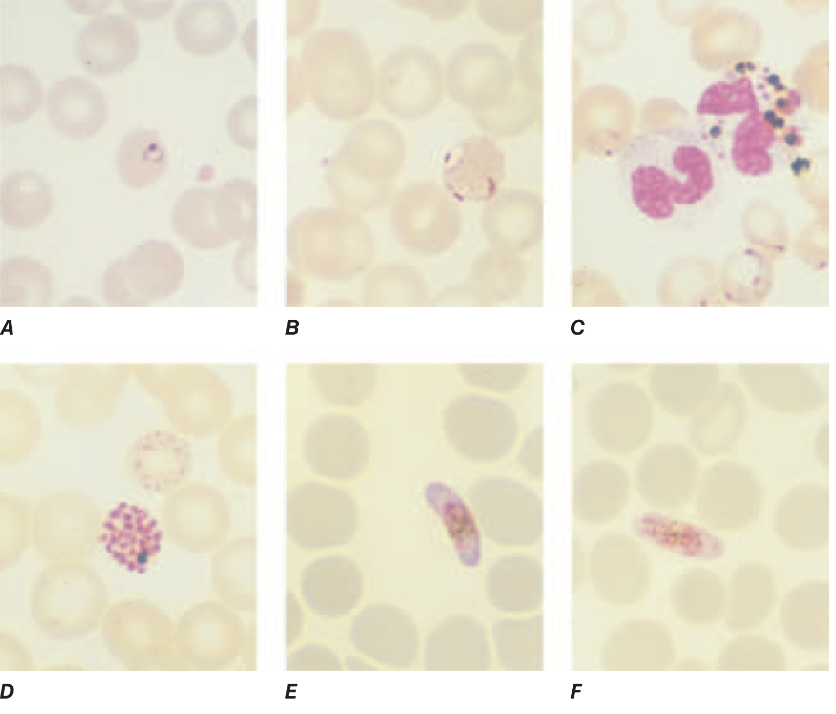
Reference Image - Thin Blood Film: P. falciparum (from Harrison's Principles of Internal Medicine 22nd Ed, 2025):
When will we take the test
WHEN TO TAKE MALARIAL SMEAR TEST IN PREGNANT WOMEN
1. WHENEVER FEVER IS PRESENT (Most Important)
- Any pregnant woman presenting with fever ≥ 37.5°C (99.5°F) must have a malarial smear done immediately
- Do NOT wait - malaria in pregnancy is an emergency
- Fever with chills and rigors = suspect malaria first
2. AT EVERY ANC VISIT IN ENDEMIC AREAS
- In malaria-endemic zones (Odisha, Jharkhand, Chhattisgarh, North-East India, tribal belts), the smear is taken routinely at every antenatal visit even without fever
- This is because many pregnant women in high-transmission areas are asymptomatically infected - they carry the parasite without obvious fever
3. AT THE FIRST ANC REGISTRATION
- At the first booking visit in endemic areas - malarial smear is included along with routine tests (Hb, blood group, HIV, HBsAg, VDRL)
4. SPECIFIC CLINICAL SITUATIONS - Take Smear Immediately:
| Situation | Why |
|---|---|
| Fever not responding to routine antibiotics after 48-72 hours | Malaria may be the cause |
| Unexplained severe anaemia in pregnancy | Malaria destroys RBCs |
| Fever + headache + body ache + nausea/vomiting | Classic malaria presentation |
| Fever + altered consciousness/seizures | Suspect cerebral malaria (emergency) |
| Fever + jaundice | Severe falciparum malaria |
| Pallor + splenomegaly + fever | Chronic or repeated malaria |
| Any fever in 1st trimester (high-risk period for fetal loss) | Malaria causes early abortion |
5. BEST TIMING WITHIN THE DAY:
- Ideally collect blood during or just after a fever spike/paroxysm - parasite density is highest at that time
- However - do NOT delay the test waiting for a fever spike
- Collect immediately whenever the patient presents
6. IF FIRST SMEAR IS NEGATIVE:
- Repeat every 8-12 hours for 2-3 days
- A single negative smear does NOT rule out malaria
- At least 3 serial negative smears are needed before ruling out malaria clinically
SIMPLE WAY TO REMEMBER:
"Any fever in pregnancy in an endemic area = Take malarial smear immediately" "No fever but in endemic area = Take at every ANC visit routinely" "Negative smear = Repeat every 8-12 hours × 3 times before ruling out"