
Everything about Ascitis
Reading File
Now let me search the medical library for comprehensive ascites coverage.
Finding Sources
Searching PubMed
ascites management cirrhosis
Excellent sources found. Let me now read the most comprehensive ones in parallel.
Reading File
Reading File
Reading File
Good data coming in. Now let me fetch the main Sleisenger dedicated ascites chapter and the ROSEN emergency block for clinical details.
Reading File
Reading File
Finding Sources
Excellent. Now let me get the Harrison's and Yamada's sections on SAAG, SBP, and the dedicated Sleisenger ascites chapter (block 23).
Reading File
Reading File
Let me read the Harrison's SAAG/diagnosis section and the Sleisenger SBP + management section for completeness.
Reading File
Reading File
Let me get the Harrison's SAAG/diagnosis content and the Sleisenger SBP section.
Reading File
Finding Sources
Now let me get the SBP and complications sections from Harrison's plus a SAAG diagram, and the refractory ascites/TIPS content.
Reading File
Reading File
I now have extensive material. Let me get the SAAG diagnostic flowchart image and the SBP treatment details from Harrison's.
Reading File
I now have comprehensive material from multiple authoritative textbooks. Let me compose the full answer.
Ascites - A Comprehensive Overview
Definition
Ascites (from Greek askos, "bag") is the pathological accumulation of free fluid within the peritoneal cavity. It is not a disease itself but a manifestation of an underlying disorder. The peritoneum normally secretes and absorbs small amounts of fluid continuously; ascites develops when this balance is disrupted by excess production or impaired absorption.
Epidemiology
- Cirrhosis accounts for approximately 75-80% of all ascites cases
- About 10% of ascites cases are malignant; of those, 80% stem from epithelial malignancies (colon, gastric, pancreatic, ovarian, breast, endometrial)
- Among cirrhotic patients, 50% will develop ascites within 10 years of diagnosis
- Prognosis: median survival 2 years for non-malignant ascites; 4 months for malignant ascites
- Sleisenger and Fordtran's Gastrointestinal and Liver Disease, p. 1800-1801
Pathophysiology
Cirrhotic Ascites (the dominant mechanism)
The development of cirrhotic ascites follows a well-defined cascade:
- Hepatic fibrosis distorts sinusoidal architecture and increases intrahepatic vascular resistance
- Stellate cell activation causes smooth-muscle contraction and further fibrosis
- Decreased eNOS production intrahepatically leads to increased intrahepatic vasoconstriction
- Paradoxically, systemic nitric oxide, VEGF, and TNF levels rise, causing splanchnic arterial vasodilation
- Splanchnic pooling reduces effective circulating volume - perceived by kidneys as hypovolemia
- Compensatory release of ADH, sympathetic activation, and RAAS stimulation follows
- Results in renal sodium and water retention, worsening portal hypertension and fluid accumulation
Non-cirrhotic Mechanisms
| Mechanism | Examples |
|---|---|
| Increased capillary pressure | Cardiac failure, constrictive pericarditis, Budd-Chiari syndrome, portal vein thrombosis |
| Reduced oncotic pressure | Nephrotic syndrome, protein malnutrition, malabsorption |
| Peritoneal inflammation/exudation | Peritoneal carcinomatosis, tuberculous peritonitis |
| Lymphatic obstruction | Trauma, tumor, lymphatic malformations |
| Pancreatic leakage | Pancreatitis, pancreatic duct disruption |
- Harrison's Principles of Internal Medicine 22E, p. 371
- Bailey and Love's Short Practice of Surgery 28th Edition
Causes (Differential Diagnosis)
By SAAG Classification
High SAAG (≥1.1 g/dL) - Portal Hypertensive Causes
- Cirrhosis (most common)
- Alcoholic hepatitis
- Cardiac ascites / congestive heart failure
- Budd-Chiari syndrome (hepatic vein thrombosis)
- Sinusoidal obstruction syndrome (veno-occlusive disease)
- Massive liver metastases
- Portal vein thrombosis
- Myxedema
Low SAAG (<1.1 g/dL) - Non-Portal Hypertensive Causes
- Peritoneal carcinomatosis
- Tuberculous peritonitis
- Pancreatic ascites
- Nephrotic syndrome
- Biliary ascites
- Bowel obstruction or infarction
- Serositis in connective tissue diseases
- Postoperative lymphatic leak
- Sleisenger and Fordtran's Gastrointestinal and Liver Disease, Box 93.2
Clinical Features
Symptoms
- Abdominal distension and discomfort
- Early satiety, nausea
- Dyspnea and orthopnea (from diaphragmatic elevation)
- Reduced exercise tolerance
- Weight gain
Physical Signs
| Sign | Description | Sensitivity |
|---|---|---|
| Flank dullness | Dullness on percussion of flanks | ~80% |
| Shifting dullness | Dullness shifts to dependent areas on repositioning | ~83% |
| Fluid wave / fluid thrill | Transmitted impulse across the abdomen | ~62%, more specific |
| Puddle sign | Dullness in the most dependent part in knee-elbow position | Small volumes |
| Bulging flanks | Visible protrusion of flanks | ~72% |
Typically, >1.5 L of fluid must accumulate before clinical detection; ultrasound can detect as little as 100-200 mL.
Associated findings suggesting the cause
- Caput medusae (portosystemic collaterals at umbilicus): cirrhosis / Budd-Chiari
- Spider nevi, palmar erythema, jaundice: cirrhosis
- Elevated JVP + peripheral edema: cardiac cause
- Hard, irregular, tender abdomen: peritoneal carcinomatosis
- Lymphadenopathy: malignancy or tuberculosis
Diagnosis
Initial Investigations
Serum tests: LFTs, albumin, prothrombin time/INR, CBC, BMP, BNP (to screen for cardiac cause), serum amylase/lipase
Diagnostic Paracentesis
This is the most important single test - should be performed in all patients with new-onset ascites and in all hospitalized patients with ascites within 12 hours of admission (paracentesis within 12 hours of hospitalization reduces mortality by enabling early detection of SBP).
Ascitic fluid appearance:
- Straw-colored, clear: typical cirrhotic/cardiac
- Turbid: infection or tumor
- Milky/white (triglycerides >200 mg/dL, often >1000 mg/dL): chylous ascites (lymphatic disruption)
- Dark brown: biliary ascites (bile duct perforation)
- Bloody: malignancy, trauma, hepatocellular carcinoma rupture
Ascitic fluid tests:
| Test | Purpose |
|---|---|
| Albumin (same day as serum) | Calculate SAAG |
| Total protein | Distinguish cardiac vs. cirrhotic high-SAAG |
| PMN cell count | Diagnose SBP (≥250 PMN/µL) |
| Culture (blood culture bottles at bedside) | Identify pathogen in SBP |
| Glucose, LDH | Distinguish SBP from secondary peritonitis |
| Amylase | Pancreatic ascites (typically >1000 U/L) |
| Cytology (≥50 mL) | Peritoneal carcinomatosis |
| AFB smear + culture | Tuberculosis |
| Triglycerides | Chylous ascites |
| Adenosine deaminase | TB in non-cirrhotic patients (>30-45 U/L has >90% sensitivity) |
The SAAG
The Serum-Ascites Albumin Gradient is the single most useful test for determining the etiology:
SAAG = Serum albumin - Ascitic fluid albumin (Both specimens drawn on the same day; expressed in g/dL)
- SAAG ≥ 1.1 g/dL = portal hypertension present (accuracy ~97%)
- SAAG < 1.1 g/dL = non-portal hypertensive cause
The SAAG reflects oncotic pressure that counterbalances portal pressure (per Starling's law). Importantly, SAAG does not change with diuresis.
Further stratification for high-SAAG ascites using ascitic protein level:
- Ascitic protein ≥ 2.5 g/dL: sinusoids are intact - cardiac ascites, early Budd-Chiari, sinusoidal obstruction syndrome
- Ascitic protein < 2.5 g/dL: damaged/scarred sinusoids - cirrhosis, late Budd-Chiari, massive liver metastases
Distinguishing SBP from Secondary Bacterial Peritonitis:
- SBP: glucose normal, LDH normal/low, single organism on culture
- Secondary peritonitis: glucose <50 mg/dL, LDH > serum LDH, multiple organisms on culture
- Harrison's Principles of Internal Medicine 22E, p. 372
- Sleisenger and Fordtran's Gastrointestinal and Liver Disease, p. 1810
Grading of Ascites (International Ascites Club)
| Grade | Description |
|---|---|
| Grade 1 | Mild - detectable only by ultrasound |
| Grade 2 | Moderate - moderate symmetric distension of abdomen |
| Grade 3 | Large/Tense - marked abdominal distension |
Management
1. Salt Restriction
- 2 g (88 mEq) sodium per day is the standard recommendation
- More restrictive diets are not recommended (compromise nutrition)
- Goal: negative sodium balance; urine sodium should exceed dietary intake
2. Diuretics (mainstay of treatment)
- Spironolactone (aldosterone antagonist) is more effective than loop diuretics as first-line
- Start: 100 mg/day (once daily in the morning)
- Titrate every 3-4 days to maximum 400 mg/day
- Furosemide: start 40 mg/day, escalate to 160 mg/day
- Add concurrently if ascites is tense, or if hyperkalemia develops with spironolactone alone
- Preferred ratio: spironolactone:furosemide = 100:40
- Weight loss targets:
- Without peripheral edema: no more than 500 g/day
- With peripheral edema: no more than 1 kg/day
- Goal: ~1 kg loss in first week, then ~2 kg/week
Diuretic side effects:
- Hypovolemic hyponatremia
- Hyperkalemia (spironolactone) or hypokalemia (furosemide)
- AKI from renal hypoperfusion
- Hepatic encephalopathy
- Painful gynecomastia (spironolactone) - can substitute amiloride 5-40 mg/day
- Muscle cramps (managed with albumin infusion or baclofen)
When to stop/withhold diuretics:
- Serum sodium < 125 mEq/L (severe hyponatremia)
- AKI
- Hepatic encephalopathy
- Severe hypokalemia <3 mEq/L (furosemide)
- Hyperkalemia >5.5 mEq/L (spironolactone)
Drugs to avoid: NSAIDs, ACE inhibitors, ARBs (all worsen renal function in cirrhotic ascites)
- Goldman-Cecil Medicine, p. 1621
- Sleisenger and Fordtran's Gastrointestinal and Liver Disease, p. 1803
3. Albumin Infusion
- Long-term albumin (40 g twice weekly for 2 weeks, then 40 g weekly) added to diuretics significantly reduces recurrent ascites and improves 18-month survival
- Albumin alone in the acute setting is of no benefit and may increase risk of pulmonary edema
4. Large-Volume Paracentesis (LVP)
- For tense/grade 3 ascites or when diuretics are insufficient
- Remove all or most of the fluid
- Mandatory albumin infusion: 6-8 g IV per liter of ascitic fluid removed (especially when >5 L removed) to prevent paracentesis-induced circulatory dysfunction (PICD)
- PICD: intravascular volume depletion, hemodynamic instability, electrolyte abnormalities, activation of RAAS
- After LVP, restart diuretics at lowest effective dose
- Rosen's Emergency Medicine, p. 2967-2974
5. Refractory Ascites
Definition: Ascites that cannot be mobilized or prevented from recurring despite:
- Maximum diuretic therapy (spironolactone 400 mg + furosemide 160 mg/day), OR
- Development of diuretic-related complications precluding full doses
Options for refractory ascites:
| Option | Notes |
|---|---|
| Repeated LVP | With mandatory albumin; frequency dictated by fluid reaccumulation rate |
| TIPS (Transjugular Intrahepatic Portosystemic Shunt) | More effective than LVP at preventing reaccumulation; increases hepatic encephalopathy risk; no survival benefit; unsuitable if bilirubin >3 mg/dL, severe coagulopathy, or overt HE |
| Midodrine (α1 agonist) | Counters splanchnic vasodilation, improves systemic hemodynamics |
| Alfapump system | Automated pump transferring ascites to bladder; high technical difficulty/renal dysfunction rates |
| Peritoneovenous shunt | Rarely used; reserved for malignant ascites refractory to all other treatment |
Note on beta-blockers in refractory ascites: While often prescribed for variceal prophylaxis, beta-blockers in patients with refractory ascites may be associated with decreased survival rates and should be used with caution.
Complications
1. Spontaneous Bacterial Peritonitis (SBP)
The most common and potentially lethal complication of cirrhotic ascites.
Definition: PMN count ≥250/µL in ascitic fluid (even if culture negative)
Pathogenesis: Enteric bacteria translocate across edematous bowel wall into ascitic fluid
Most common pathogens:
- Gram-negative rods: E. coli, Klebsiella
- Gram-positive: Streptococci, Enterococci
Presentation:
- Increase in abdominal girth
- Fever (not always present)
- Abdominal tenderness in only ~40% of patients
- Rebound tenderness uncommon
- New/worsening hepatic encephalopathy
- May be asymptomatic
Treatment:
- Cefotaxime 2 g IV every 12 hours x 5 days (standard)
- Repeat paracentesis at 48 hours to confirm PMN count decreased ≥25%
- IV albumin 1.5 g/kg on day 1, then 1 g/kg on day 3 to prevent hepatorenal syndrome
- Nosocomial SBP: consider carbapenem (multidrug-resistant organisms increasingly common)
Prophylaxis (when indicated):
- History of prior SBP
- Ascitic fluid total protein <1 g/dL
- Active GI bleeding
- Agents: norfloxacin or ciprofloxacin daily orally; IV ceftriaxone in hospitalized patients
- Harrison's Principles of Internal Medicine 22E, p. 372-373
2. Hepatorenal Syndrome (HRS)
- Functional renal failure complicating cirrhotic ascites
- Type 1 HRS: rapid, doubling of creatinine to >2.5 mg/dL in <2 weeks; median survival 2 weeks
- Type 2 HRS: moderate, steady renal failure; presents with refractory ascites; median survival 4-6 months
3. Hepatic Hydrothorax
- Ascitic fluid migrates via diaphragmatic fenestrae into pleural space (usually right-sided)
- Serum-to-pleural albumin gradient ≥1.1 g/dL is suggestive
- Treat: sodium restriction, diuretics; thoracentesis or TIPS if refractory
- Avoid chest tube placement (risk of high-output fistula and protein depletion)
4. Umbilical Hernia
- Tense ascites increases intraabdominal pressure - may cause umbilical herniation with risk of rupture
5. Abdominal wall complications
- Abdominal hernias, skin ischemia over tense ascites
Special Types of Ascites
Malignant Ascites
- Usually low-SAAG (peritoneal carcinomatosis) but can be high-SAAG if massive liver involvement
- Does not respond to sodium restriction or diuretics
- Managed with serial LVPs, drainage catheters, or peritoneovenous shunts
- Median survival ~4 months (longer if the underlying malignancy is chemosensitive)
Chylous Ascites
- Triglycerides >200 mg/dL (often >1000 mg/dL), milky appearance
- Causes: trauma, cirrhosis, lymphoma, retroperitoneal surgery, tuberculosis, congenital
- Managed with low-fat, medium-chain triglyceride diet; octreotide; or surgery
Pancreatic Ascites
- Amylase >1000 U/L in fluid
- Caused by pancreatic duct disruption (pancreatitis, trauma)
- Treated with somatostatin analogues, endoscopic ductal stenting, or surgical repair
Tuberculous Ascites
- Low-SAAG, lymphocyte-predominant, elevated adenosine deaminase (>30-45 U/L)
- AFB smear has sensitivity of only 0-3%; culture increases to 35-50%
- Laparoscopy with peritoneal biopsy is gold standard when cause unclear
- Treated with standard anti-tuberculous therapy
Cardiac Ascites
- High-SAAG (≥1.1), but ascitic protein ≥2.5 g/dL (intact sinusoids)
- Light yellow fluid with low specific gravity
- Elevated BNP levels help identify cardiac cause
- Treatment targets underlying cardiac condition
Prognosis
| Condition | Median Survival |
|---|---|
| Non-malignant ascites | ~2 years |
| Malignant ascites | ~4 months |
| Type 1 HRS | ~2 weeks |
| Type 2 HRS | ~4-6 months |
| Urine sodium <10 mEq/day | ~1.5 years |
| Urine sodium ≥10 mEq/day | ~4.5 years |
| HE cumulative 1-year survival | 20-40% |
MELD score (using bilirubin, INR, creatinine) is the best short-term predictor of mortality in cirrhosis with ascites. Ascites itself is an independent marker of poor prognosis.
- Sleisenger and Fordtran's Gastrointestinal and Liver Disease, p. 1800, 1812
Recent Evidence (2024-2025)
A 2025 systematic review (PMID 40429923) examined SGLT-2 inhibitors in liver cirrhosis, finding emerging evidence that these agents may have a role in ascites management and slowing disease progression, though safety data in decompensated cirrhosis remain limited. A 2025 systematic review on noninvasive markers (PMID 40131017) confirms elastography and laboratory indices can predict hepatic decompensation (including ascites development) before it occurs, supporting earlier intervention.
Summary Algorithm
NEW ASCITES
     |
Diagnostic Paracentesis + Serum Albumin (same day)
     |
Calculate SAAG
     |
     +-- SAAG ≥1.1 (Portal HTN) -----> Ascitic Protein?
     |                                    ≥2.5: Cardiac/Budd-Chiari
     |                                    <2.5: Cirrhosis/late Budd-Chiari
     |
     +-- SAAG <1.1 (Non-portal) ------> Malignancy / TB / Pancreatic / Nephrotic


PMN COUNT ≥250/µL → Diagnose SBP → Antibiotics + Albumin


MANAGEMENT (if cirrhotic):
  Grade 1-2: Sodium restriction (2 g/d) + Spironolactone ± Furosemide
  Grade 3 (tense): LVP + albumin 6-8 g/L removed → then maintenance diuretics
  Refractory: Serial LVP + albumin, or TIPS (if eligible), or Midodrine
  Await liver transplantation
Sources: Harrison's Principles of Internal Medicine 22E | Sleisenger and Fordtran's Gastrointestinal and Liver Disease | Goldman-Cecil Medicine | Rosen's Emergency Medicine | Bailey and Love's Short Practice of Surgery 28th Edition
Cellulitis
Reading File
Now let me search the library and PubMed in parallel.
Finding Sources
Searching PubMed
cellulitis skin infection management
Excellent sources. Let me read the most comprehensive ones in parallel - Andrews' Dermatology, Goldman-Cecil, and Sabiston Surgery.
Reading File
Reading File
Reading File
Good content. Let me now get the Rosen's EM and Goldman-Cecil block 44 sections, plus read the Sabiston further for necrotizing fasciitis differentiation.
Reading File
Reading File
Reading File
Excellent content. Now let me get the clinical images and the Goldman-Cecil block 59 continued section (antibiotics/treatment) plus the Family Medicine section for outpatient management details.
Reading File
Reading File
Loading Image
I now have comprehensive material from multiple authoritative textbooks plus clinical images. I have enough to compose a thorough, well-structured answer.
Cellulitis - A Comprehensive Overview
Definition
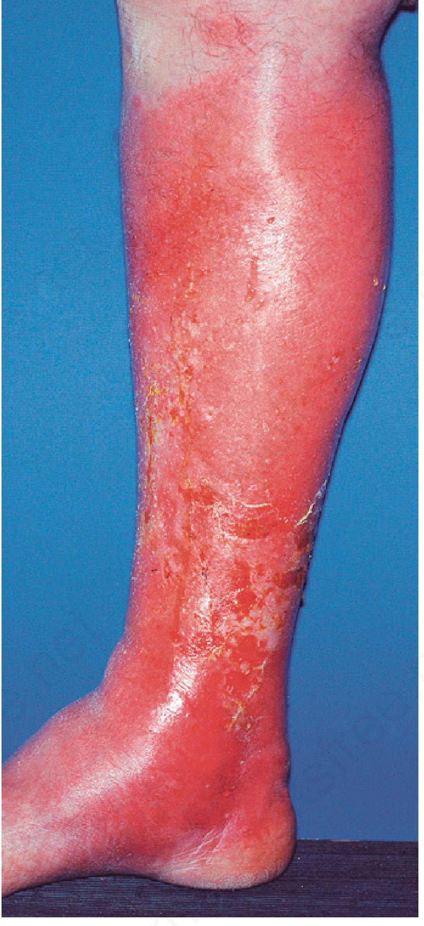
Cellulitis is an acute bacterial infection of the deeper dermis and subcutaneous fat, occurring when bacteria enter through a disrupted cutaneous barrier. It presents with the cardinal signs of inflammation - erythema, warmth, swelling, and tenderness - and sits in the middle of a spectrum of soft tissue infections: erysipelas (more superficial, sharply demarcated) at one end, and necrotizing fasciitis (deeper, necrotic, undermining) at the other.
Epidemiology
- One of the most common skin infections seen in primary care and emergency departments
- Most frequently involves the lower extremities (leg cellulitis accounts for the majority of cases)
- Annual incidence in the US: approximately 14.5 million cases
- A significant cause of hospitalization, particularly in the elderly and immunocompromised
Microbiology / Causative Organisms
| Organism | Context |
|---|---|
| Group A β-hemolytic Streptococcus (S. pyogenes) | Most common cause of non-purulent cellulitis (~75% of cases) |
| Staphylococcus aureus (MSSA) | Most common cause of purulent cellulitis; major remainder of non-purulent cases |
| MRSA (CA-MRSA) | Increasingly common; consider in risk groups |
| Gram-negative bacilli | Diabetics, immunocompromised patients |
| Vibrio vulnificus | Exposure to seawater/shellfish (particularly in liver disease) |
| Pseudomonas aeruginosa | Immunocompromised, water exposure |
| Aeromonas hydrophila | Freshwater exposure |
| Haemophilus influenzae | Periorbital cellulitis in children |
| Cryptococcus spp. | Immunocompromised |
| Atypical mycobacteria | Immunocompromised |
Key distinction: Non-purulent cellulitis is predominantly streptococcal. Purulent cellulitis (with abscess, pustules, or drainage) is predominantly staphylococcal.
- Andrews' Diseases of the Skin, p. 47
- Goldman-Cecil Medicine, p. 4321
Risk Factors / Predisposing Conditions
- Portal of entry: wounds, abrasions, insect bites, surgical incisions, IV drug use, skin ulcers
- Tinea pedis (very common portal of entry for lower extremity cellulitis)
- Lymphedema and chronic lymphatic obstruction
- Venous insufficiency and chronic lower extremity edema
- Obesity
- Diabetes mellitus
- Immunocompromised states (HIV, malignancy, steroid use, chemotherapy)
- Previous episodes of cellulitis (the single strongest predictor of recurrence)
- Saphenous vein harvest, lymph node dissection, or radiation therapy
- Prosthetic knee/hip surgery
Pathophysiology
Bacteria enter through a break in the skin barrier. In the dermis and subcutaneous tissue, they trigger an acute inflammatory response with neutrophil infiltration. The infection can spread rapidly along tissue planes. Lymphangitic spread produces the characteristic red streaks proximal to the lesion.
Why streptococcal cellulitis spreads so rapidly: Group A Streptococcus produces hyaluronidase, streptokinase, and DNAses that digest connective tissue and fibrin, facilitating rapid tissue plane invasion.
Clinical Features
Symptoms
- Localized pain and tenderness (hallmark)
- Warmth
- Swelling / edema
- Fever, malaise, chills (systemic features - not always present in mild cases)
- Red streaks (lymphangitis) spreading from the area
- Regional lymphadenopathy
Signs
| Feature | Description |
|---|---|
| Erythema | Diffuse, spreading redness - borders are NOT elevated or sharply demarcated (unlike erysipelas) |
| Edema | Soft tissue swelling; may pit on pressure |
| Warmth | Palpable heat over the affected area |
| Tenderness | Uniform or patchy |
| Lymphangitis | Streaks of erythema tracking proximally toward draining lymph nodes |
| Bullae/vesicles | Central part may develop vesicles - indicates severe infection |
| Skin necrosis | Late/severe; alarming sign requiring urgent evaluation for NSTI |
Stages of severity
- Mild: Local erythema and tenderness only; afebrile; no systemic toxicity
- Moderate: Fever, malaise, moderate spread, regional lymphadenopathy
- Severe: Systemic sepsis, rapid spread, bullae, skin necrosis, hemodynamic instability
Cellulitis vs. Erysipelas - Key Distinction
| Feature | Cellulitis | Erysipelas |
|---|---|---|
| Depth | Deeper dermis + subcutaneous fat | Upper dermis + superficial lymphatics |
| Border | Ill-defined, NOT raised | Well-defined, raised/palpable border |
| Skin color | Pink to red | Fiery red / salmon-red |
| Cause | Streptococcus, S. aureus | Exclusively S. pyogenes (Group A Strep) |
| Bullae | Possible in severe cases | Flaccid bullae day 2-3 (but rarely extends deep) |
| Face | Less common | Classic - nasolabial fold |
| Lymphatic involvement | May occur | Prominent |
Both can be managed with the same antibiotic spectrum, but erysipelas responds better to penicillin alone.
Diagnosis
Clinical Diagnosis
The diagnosis is primarily clinical - there is no single laboratory test that confirms cellulitis. Key features:
- History of skin breach or predisposing condition
- Classic local signs in a non-bilateral distribution
- Spreading erythema with advancing border
- Systemic signs (variable)
Laboratory Tests (selected cases)
| Test | Utility |
|---|---|
| CBC | Leukocytosis supports bacterial infection; eosinophilia suggests Wells syndrome mimic |
| CRP / ESR | Elevated in significant infection; useful for monitoring response |
| Blood cultures | Low yield (~2-4%) - only indicated if systemic sepsis, immunocompromised, or suspected bacteremia |
| Wound/surface swabs | Low yield unless open wound present |
| Aspiration culture | Consider in immunocompromised patients (saline injection followed by aspiration) |
| Skin biopsy | Rarely needed; shows neutrophilic infiltrate in dermis; useful to exclude mimics |
Imaging
- Ultrasound: Most useful investigation - distinguishes cellulitis from abscess
- Cellulitis: "cobblestone" appearance - hypoechoic stranding in a reticular pattern
- Abscess: discrete fluid-filled cavity (amenable to drainage)
- Also helps exclude deep vein thrombosis and assess for NSTI (gas tracking)
- CT/MRI: Reserved for suspected deep infection or necrotizing fasciitis; MRI is gold standard for NSTI
Differential Diagnosis - Mimics of Cellulitis
This is clinically important - a large proportion of "cellulitis" diagnoses are actually mimics:
| Condition | Distinguishing Features |
|---|---|
| Stasis dermatitis | Bilateral, no fever, no pain (itchy instead), centered over medial malleoli, hyperpigmentation |
| Lipodermatosclerosis | Chronic; "inverted wine bottle" appearance; skin never returns to normal even after treatment |
| DVT | Unilateral leg swelling; Doppler ultrasound confirms |
| Allergic/contact dermatitis | Pruritic (not painful), no fever, history of allergen exposure |
| Erythema migrans (Lyme disease) | Expanding red patch, central clearing, tick exposure, less painful |
| Eosinophilic cellulitis (Wells syndrome) | History of insect bite; eosinophilia (not neutrophilia); biopsy shows eosinophils |
| Gout / pseudogout | Joint centered, crystal analysis confirms |
| Inflammatory carcinoma of breast | Peau d'orange, no fever response to antibiotics |
| Toxic erythema of chemotherapy | Acral, recent chemotherapy history |
| Necrotizing fasciitis | "Pain out of proportion," wooden-hard skin, crepitus, sepsis - SURGICAL EMERGENCY |
Clinical pearl: Bilateral lower leg "cellulitis" is almost never true cellulitis. The most likely diagnosis is stasis dermatitis or chronic venous insufficiency.
Management
Step 1: Classify severity and purulence
The 2014 IDSA guidelines stratify management based on purulence (non-purulent vs. purulent) and severity (mild/moderate/severe):
Non-Purulent Cellulitis (no abscess, no pus)
Mild (outpatient):
- Cephalexin 250-500 mg PO 4x/day x 5-10 days
- Dicloxacillin 500 mg PO 4x/day x 5-10 days
- Amoxicillin-clavulanate (alternative)
- Clindamycin 300 mg PO 3x/day (penicillin allergy)
- Covers Streptococcus and MSSA
Moderate (requires IV, hospitalization):
- Cefazolin IV (first-line)
- Ceftriaxone IV
- Penicillin IV (if purely streptococcal)
- Clindamycin IV (β-lactam allergy)
Severe (sepsis/immunocompromised):
- Vancomycin IV + Piperacillin/tazobactam (empiric broad-spectrum)
- Or Vancomycin IV + Cefepime (2 g TID)
- Duration: until clinical improvement allows transition to oral
Purulent Cellulitis (with abscess, pus, or boils)
- Requires incision and drainage (I&D) as primary treatment
- MRSA coverage is standard:
- TMP-SMX 160/800 mg PO BID x 5-10 days
- Doxycycline 100 mg PO BID x 5-10 days
- Clindamycin 300-600 mg PO TID x 5-10 days
- Linezolid 600 mg PO BID (severe/resistant)
- Vancomycin IV (severe/inpatient MRSA)
MRSA Risk Groups Requiring CA-MRSA Coverage Even in Non-Purulent Cellulitis
- Children
- Athletes (skin-to-skin contact sports)
- Residents of long-term care facilities, military recruits, prisoners
- IV drug users
- Men who have sex with men
- Prior MRSA infection or colonization
Antibiotic Duration
- 5 days is sufficient for most mild-moderate cases (no evidence that >5 days improves outcomes in responsive infections)
- Extend to 10-14 days if inadequate response at 5 days
- Fluoroquinolones are no longer recommended for SSTIs due to tendinopathy, peripheral neuropathy, and CNS adverse effects
- Sabiston Textbook of Surgery, p. 638
- Goldman-Cecil Medicine, p. 4321-4322
- Andrews' Diseases of the Skin, p. 58
Adjunctive Measures
- Limb elevation - reduces edema and accelerates resolution
- Mark the border of erythema with a pen to monitor progression
- Compression stockings to reduce edema and prevent recurrence
- Treat tinea pedis aggressively to eliminate the portal of entry
- Moisturize skin to prevent fissuring
- Ice bags / cold compresses for local comfort
- Maintain good glycemic control in diabetics
Criteria for Hospitalization
- Systemic toxicity (fever >38.5°C, tachycardia, hypotension)
- Immunocompromised state (HIV, chemotherapy, diabetes with poor control)
- Rapidly spreading infection despite oral antibiotics
- Facial or periorbital cellulitis
- Failure of outpatient treatment (no improvement in 48-72 hours)
- Suspected deep infection or necrotizing fasciitis
- Extremes of age (infants, elderly)
- Comorbidities: cardiac failure, renal failure, hepatic failure
Recurrent Cellulitis
Defined as ≥2 episodes per year. Affects a significant minority of patients and leads to chronic lymphedema.
Risk factors for recurrence
- Chronic lymphedema (the biggest risk)
- Venous insufficiency
- Obesity
- Tinea pedis (untreated)
- Previous cellulitis episodes
Secondary Prevention
A 2026 systematic review (PMID 41275489) confirms that prevention strategies including compression therapy, treatment of predisposing skin conditions (tinea pedis), weight loss, and antibiotic prophylaxis in high-recurrence patients are effective.
Antibiotic prophylaxis (long-term):
- Penicillin V 250 mg PO twice daily - most evidence
- Erythromycin (penicillin allergy)
- Consider in patients with ≥2-3 episodes/year
- Trial period: minimum 6-12 months; reassess benefit
Chronic lymphedema consequence: Repeated bouts of bacterial lymphangitis eventually cause permanent obstruction of major lymphatic channels, resulting in a hypertrophic fibrosis called elephantiasis nostras - an irreversible end-stage complication.
Special Situations
Orbital / Periorbital Cellulitis
- Preseptal (periorbital): infection anterior to orbital septum; less severe; treat with IV antibiotics
- Orbital: infection posterior to septum; ophthalmic emergency - proptosis, ophthalmoplegia, visual loss; CT required; IV antibiotics + urgent ophthalmology/ENT consult; may need surgical drainage
Diabetic Foot Cellulitis
- Often polymicrobial (Streptococcus, Staphylococcus, gram-negatives, anaerobes)
- Higher risk of deep tissue involvement and osteomyelitis
- Broad-spectrum coverage required
- Probe-to-bone test and imaging to exclude osteomyelitis
Cellulitis After Lymph Node Dissection / Radiation
- May be caused by uncommon organisms
- Requires prolonged courses
- Associated with disrupted lymphatics - prone to recurrence
Post-Traumatic / Water-Exposure Cellulitis
- Vibrio vulnificus: seawater/shellfish exposure, especially in liver disease - can be rapidly fatal; treat with doxycycline + ceftriaxone
- Aeromonas hydrophila: freshwater exposure; treat with fluoroquinolone or TMP-SMX
- Pseudomonas: contaminated water or soil; ciprofloxacin or antipseudomonal beta-lactam
Complications
| Complication | Notes |
|---|---|
| Bacteremia / Sepsis | ~2-4% of cellulitis cases; higher in immunocompromised |
| Abscess formation | Secondary abscess may develop within cellulitic tissue |
| Necrotizing fasciitis | Most feared complication; requires urgent surgical debridement |
| Lymphangitis | Spread along lymphatics; may seed regional lymph nodes |
| Metastatic abscesses | Rare; septic emboli to distant sites |
| Osteomyelitis | Spread to underlying bone, especially in diabetics |
| Chronic lymphedema | Permanent consequence of recurrent attacks |
| Elephantiasis nostras | End-stage fibrosis from repeated lymphangitic insults |
| Post-streptococcal GN | Rare; if caused by nephritogenic strains of Strep |
| Death | Extremely rare in immunocompetent adults when treated promptly |
Necrotizing Fasciitis - When to Suspect
The critical distinction every clinician must make is cellulitis vs. necrotizing fasciitis (NSTI). Up to 71% of NSTIs are misdiagnosed initially.
Red flags suggesting NSTI (not simple cellulitis):
- Pain out of proportion to clinical appearance
- Skin that feels wooden-hard beneath the surface (fascial involvement)
- Crepitus (gas in tissues)
- Rapid progression despite antibiotics
- Skin color change: red → purple → blue → black (necrosis)
- Hemorrhagic bullae with dusky/necrotic skin
- Severe systemic sepsis disproportionate to local signs
- Anesthesia of overlying skin (nerve necrosis)
- Failure to respond to IV antibiotics within 24 hours
Any suspicion of NSTI = surgical emergency. Do not wait for imaging if the patient is deteriorating.
Recent Evidence (2024-2026)
A 2024 network meta-analysis (PMID 39240378) of RCTs confirmed that first-line antibiotics (penicillins, cephalosporins) remain highly effective for uncomplicated cellulitis/erysipelas, with no clear superiority of one agent over another for standard cases. MRSA-active agents (TMP-SMX, doxycycline, clindamycin) are preferred only when MRSA is suspected based on risk stratification.
Summary
CELLULITIS
     |
Assess: Purulent? ─── YES ──> I&D + MRSA antibiotics (TMP-SMX/doxycycline)
     |
     NO (non-purulent)
     |
Severity?
  Mild/outpatient ──> Cephalexin or Dicloxacillin PO x 5-10d
                      (+MRSA coverage if risk factors present)
  Moderate/hospital ─> Cefazolin or Ceftriaxone IV
  Severe/immunocomp ─> Vancomycin + Pip/Taz IV
     |
Adjunct: Elevate limb, mark borders, treat tinea pedis
     |
Monitor 48-72h:
  Improving? ─> Complete course
  Not improving? ─> Re-examine, consider mimic, image, escalate Abx, rule out NSTI
     |
Recurrent? ─> Compression, skin care, penicillin prophylaxis
Sources: Andrews' Diseases of the Skin | Goldman-Cecil Medicine | Sabiston Textbook of Surgery | Rosen's Emergency Medicine | Textbook of Family Medicine 9e | Medical Microbiology 9e
Pelvic inflammatory disease
Finding Sources
Searching PubMed
pelvic inflammatory disease treatment diagnosis
Excellent sources found. Let me now read the most comprehensive ones in parallel - Harrison's (most extensive at 11 paragraphs), Goldman-Cecil, and Berek & Novak's Gynecology.
Reading File
Reading File
Reading File
Excellent content. Now let me get the Harrison's continued PID section (treatment regimens, TOA, Fitz-Hugh-Curtis) and the Washington Manual regimens, and the radiology section.
Reading File
Reading File
Reading File
Good. I need the Harrison's full treatment regimens and the Fitz-Hugh-Curtis / TOA section specifically. Let me get that next section of Harrison's and the radiology PID imaging section.
Reading File
Reading File
Excellent - I now have the full treatment table from Harrison's. Let me get the continuation with TOA, Fitz-Hugh-Curtis, outcomes/sequelae, and the Red Book pediatric section for adolescent considerations.
Reading File
Reading File
I now have comprehensive material from Harrison's, Goldman-Cecil, Berek & Novak's, Washington Manual, and Red Book. I have everything needed to compile a thorough answer.
Pelvic Inflammatory Disease (PID)
Definition
Pelvic inflammatory disease (PID) is an ascending infection of the female upper genital tract - comprising any combination of endometritis, salpingitis, parametritis, oophoritis, tubo-ovarian abscess, and pelvic peritonitis. It begins in the endocervix and spreads upward to the uterus, fallopian tubes, ovaries, and peritoneum. PID is a clinical diagnosis implying upper genital tract infection and inflammation at any point along a continuum.
Epidemiology
- In the United States, more than 800,000 women develop PID annually
- Annual physician office visits for PID fell from ~400,000 in the 1980s to ~51,000 in 2014 (reflecting improved STI control and screening)
- Hospitalizations remain fairly constant at 70,000-100,000/year since 1995
- Most common in sexually active women of reproductive age, particularly those aged 15-25
- A significant proportion of cases are subclinical - going unrecognized with only mild or atypical symptoms, yet still causing tubal damage
- Harrison's Principles of Internal Medicine 22E, p. 1148
- Goldman-Cecil Medicine, p. 1493
Etiology / Microbiology
PID is polymicrobial in most cases:
| Organism | Role |
|---|---|
| Neisseria gonorrhoeae | Classic cause; more severe presentation; higher incidence in endemic regions |
| Chlamydia trachomatis | ~27-35% of cases; often subclinical/mild but causes significant tubal damage |
| Mycoplasma genitalium | Increasingly recognized; associated with endometritis and salpingitis; mild symptoms similar to chlamydial PID |
| Bacterial vaginosis organisms | Prevotella spp., Peptostreptococci, Gardnerella vaginalis - facilitate ascending spread by enzymatically altering cervical mucus |
| Other anaerobes | Bacteroides spp., peptostreptococci |
| Facultative organisms | E. coli, Haemophilus influenzae, group B streptococci |
| Respiratory pathogens | H. influenzae, Group A streptococci, pneumococci (less frequent) |
| Cytomegalovirus (CMV) | Implicated in some cases |
In recent studies, fewer than half of PID cases have laboratory evidence of gonorrhea or chlamydia, highlighting the polymicrobial nature.
- Harrison's Principles of Internal Medicine 22E, p. 1148
- Berek & Novak's Gynecology, p. 817
Pathophysiology
Microorganisms originating in the endocervix ascend upward through the uterine cavity into the fallopian tubes and peritoneum:
- Endocervicitis - initial infection at the cervix
- Endometritis - spread to the uterine lining; disrupts the normal cervical mucus barrier (especially with BV)
- Salpingitis - infection of the fallopian tubes; leads to inflammation, edema, and exudate
- Peritonitis - spread to the pelvic peritoneum; free fluid in pelvis
- Tubo-ovarian abscess (TOA) - complex abscess involving tube and ovary
- Perihepatitis (Fitz-Hugh-Curtis syndrome) - spread to liver capsule via peritoneal fluid
Why menstruation is a risk factor: The cervical mucus plug, which normally acts as a barrier, is lost during menstruation, allowing cervicovaginal organisms to ascend.
Immunopathology in chlamydial PID: Repeated C. trachomatis exposure triggers the greatest tissue inflammation and damage - immunologic memory amplifies the inflammatory response with each subsequent exposure, explaining why repeated infections cause progressively more tubal damage.
Risk Factors
| Risk Factor | Mechanism |
|---|---|
| Age 15-25 | Higher rates of STIs, more susceptible cervical ectopy |
| Multiple sexual partners / new partner | Increased STI exposure |
| Prior episode of PID | Damaged tubal defenses |
| Bacterial vaginosis | Disrupts cervical mucus barrier |
| IUD insertion (especially recent) | Breach of cervical barrier (risk highest in first 3 weeks) |
| Vaginal douching | Disrupts normal flora and barrier |
| No contraceptive use | Oral contraceptives thicken cervical mucus, providing protection |
| Dilation and curettage, hysterosalpingography, D&E | Iatrogenic ascent of organisms |
| Cesarean section | Disrupts uterine barrier |
Protective factors: Oral contraceptives (thicken cervical mucus), barrier contraceptives (condoms), and chlamydial screening programs
Clinical Features
Symptoms
- Lower abdominal/pelvic pain (most common) - bilateral, dull, aching; may be constant or intermittent
- Often begins during or soon after menstruation
- Abnormal vaginal discharge (mucopurulent)
- Fever (>38.3°C / >101°F)
- Abnormal uterine bleeding (menorrhagia, metrorrhagia)
- Dyspareunia (pain with intercourse)
- Vomiting, nausea
- Urinary symptoms (dysuria, frequency)
- Right upper quadrant pain - if Fitz-Hugh-Curtis syndrome develops
Important: Many women with PID have subtle or mild symptoms not readily recognized as PID. Subclinical PID (upper tract inflammation without overt symptoms) represents a large proportion of all cases and still causes tubal damage.
Physical Examination Findings
| Finding | Significance |
|---|---|
| Cervical motion tenderness (CMT) | Stretching of peritoneum when cervix is moved; suggests peritoneal involvement - the most specific physical sign |
| Uterine tenderness | Indicates endometritis |
| Adnexal tenderness (unilateral or bilateral) | Indicates salpingo-oophoritis |
| Adnexal fullness/mass | May indicate TOA or hydrosalpinx |
| Fever >38.3°C | Supports diagnosis |
| Mucopurulent cervical discharge | Indicates endocervicitis |
| Peritoneal signs (rebound, guarding) | Peritonitis; may indicate TOA rupture |
| Right upper quadrant tenderness | Fitz-Hugh-Curtis syndrome |
Diagnostic Criteria (CDC Minimum Criteria)
The CDC recommends empirical treatment for PID if a sexually active young woman or any woman at risk for STIs presents with pelvic or lower abdominal pain AND has at least one of the following on pelvic examination (in the absence of another identifiable cause):
- Cervical motion tenderness, OR
- Uterine tenderness, OR
- Adnexal tenderness
This low threshold is intentional - the goal is to avoid missing mild cases, as the consequences of untreated PID (infertility, ectopic pregnancy, chronic pelvic pain) are severe. Over-treatment is preferred over under-treatment.
Additional Criteria That Support the Diagnosis
| Finding | Value |
|---|---|
| Oral temperature >38.3°C | Supports diagnosis |
| Abnormal cervical or vaginal mucopurulent discharge | Suggests infection |
| Elevated WBCs on vaginal saline wet mount | Increases specificity |
| Elevated ESR or CRP | Systemic inflammation |
| Lab evidence of gonorrhea/chlamydia (NAAT positive) | Specific if positive, but absence does not exclude PID |
| Pyuria on urine microscopy | Supporting evidence |
| Adnexal mass on ultrasound | Suggests TOA |
Most Specific Additional Criteria (for uncertain cases)
- Histopathologic evidence of endometritis on endometrial biopsy (correlates well with salpingitis)
- Thickened, fluid-filled fallopian tubes with or without free pelvic fluid or tubo-ovarian complex on transvaginal ultrasound or MRI
- Laparoscopic evidence of PID (gold standard) - but invasive; reserved for complex cases or when diagnosis is uncertain
The clinical diagnosis of PID made by expert gynecologists is confirmed laparoscopically in only ~70% of cases, reflecting the imprecision of clinical diagnosis.
Laboratory Investigations
| Test | Purpose |
|---|---|
| NAAT for N. gonorrhoeae and C. trachomatis (endocervical/vaginal swab) | Identify causative organism; guides targeted therapy |
| Saline wet prep of vaginal discharge | PMNs (increased) support PID |
| Gram stain of cervical discharge | Gram-negative intracellular diplococci = gonorrhea |
| CBC | Leukocytosis common |
| ESR / CRP | Elevated in active infection; useful for monitoring |
| Urine pregnancy test (β-hCG) | MUST exclude ectopic pregnancy before diagnosing PID |
| Urinalysis | Exclude UTI; pyuria may occur in PID |
| Blood cultures | If sepsis suspected |
Imaging
Transvaginal Ultrasound (TVS) - First-line imaging
- Shows thickened, fluid-filled fallopian tubes (hydrosalpinx / pyosalpinx)
- Tubo-ovarian abscess: complex adnexal mass with internal echoes
- Free pelvic fluid in the pouch of Douglas
- Normal ultrasound does NOT exclude PID
CT Scan
- Useful in equivocal cases to exclude other causes (appendicitis, ovarian torsion)
- Shows pelvic fat stranding, free fluid, adnexal masses, TOA
- Also useful for guiding abscess drainage
MRI
- Best modality for complex or equivocal cases
- Shows tubal wall thickening, fluid-filled tubes, TOA anatomy
- Used when surgery is being considered
Usually in acute presentations, imaging is not required - the diagnosis is made on symptoms, examination findings, and microbiological swabs. Radiology has an important role in equivocal cases and in the investigation of complicated PID (TOA, Fitz-Hugh-Curtis).
- Grainger & Allison's Diagnostic Radiology, p. 899
Fitz-Hugh-Curtis Syndrome (Perihepatitis)
- Occurs when infection spreads via peritoneal fluid to the liver capsule
- Results in perihepatic adhesions ("violin string" adhesions between liver and anterior abdominal wall)
- Presents with right upper quadrant pain that may mimic cholecystitis, pleurisy, or hepatitis
- Associated with both gonorrheal and chlamydial PID
- Patients may present with RUQ pain and minimal pelvic symptoms - PID must be considered in the differential of RUQ pain in young women
- Treatment: same as PID antibiotics; adhesions may require laparoscopic lysis if causing chronic pain
Tubo-Ovarian Abscess (TOA)
- A complication of severe PID where pus collects in and around the fallopian tube and ovary
- Suspect when: severe systemic toxicity, pelvic mass on exam, failure to improve on antibiotics
- Confirmed by ultrasound, CT, or MRI
Management:
- Inpatient IV antibiotics (clindamycin + gentamicin; or cefoxitin/cefotetan + doxycycline)
- Most TOAs respond to antibiotics alone
- Image-guided drainage (CT or ultrasound-guided) if no improvement in 72 hours
- Surgical drainage (posterior colpotomy, laparoscopy) for refractory cases
- Surgical resection if rupture occurs or is threatened (life-threatening emergency)
Management
Decision: Outpatient vs. Inpatient
Indications for hospitalization:
- Surgical emergency cannot be excluded (appendicitis, ectopic pregnancy)
- Tubo-ovarian abscess
- Pregnancy
- Failure of or intolerance to oral regimen
- Severe illness: high fever, nausea/vomiting, severe abdominal pain
- Inability to follow or tolerate outpatient regimen
- Prepubertal female (requires assessment for sexual abuse)
Antibiotic Regimens (CDC 2021/2022 Guidelines)
Outpatient (Mild-to-Moderate PID)
Preferred Regimen:
- Ceftriaxone 500 mg IM in a single dose (persons ≥150 kg: 1 g)
- PLUS Doxycycline 100 mg PO twice daily × 14 days
- PLUS Metronidazole 500 mg PO twice daily × 14 days
Metronidazole addition reduced endometrial anaerobes, M. genitalium, and pelvic tenderness in randomized trials.
Alternative Outpatient Regimen:
- Cefoxitin 2 g IM + Probenecid 1 g PO (single dose) as alternative to ceftriaxone
- PLUS Doxycycline 100 mg PO BID × 14 days + Metronidazole 500 mg PO BID × 14 days
Inpatient / Parenteral Regimens
Regimen A (Preferred):
- Cefotetan 2 g IV every 12 hours OR Cefoxitin 2 g IV every 6 hours
- PLUS Doxycycline 100 mg IV or PO every 12 hours
- Continue IV until 48 hours after clinical improvement; then transition to oral doxycycline 100 mg BID + metronidazole 500 mg BID to complete 14 days total
Regimen B:
- Clindamycin 900 mg IV every 8 hours
- PLUS Gentamicin loading dose 2 mg/kg IV, then 1.5 mg/kg every 8 hours (or 3-5 mg/kg once daily)
- Continue until 48 hours after clinical improvement; then oral clindamycin 450 mg QID or doxycycline 100 mg BID to complete 14 days
Regimen C (Alternative):
- Ampicillin-sulbactam 3 g IV every 6 hours
- PLUS Doxycycline 100 mg IV or PO every 12 hours
- Washington Manual of Medical Therapeutics
- Harrison's Principles of Internal Medicine 22E (Table 141-6)
- Goldman-Cecil Medicine (Table 264-5)
Monitoring Response
- Outpatients: Re-evaluate within 72 hours - should show clinical improvement
- Inpatients: Should show substantial improvement within 3-5 days
- Failure to improve → hospitalize (if outpatient), consider laparoscopy, broaden coverage
- Repeat NAAT for gonorrhea/chlamydia if symptoms persist or patient was re-exposed to untreated partner
- IUD: Remove if present and no clinical improvement occurs
A follow-up study found 28% of outpatients never filled their doxycycline prescription and 41% stopped early - patient adherence counseling is essential.
Partner Management
- All sex partners within the preceding 60 days should be evaluated and treated empirically
- Treat for both gonorrhea and chlamydia regardless of test results
- Partners should abstain from sexual intercourse until treatment is completed by both parties
- Failure to treat partners leads to re-infection and recurrent PID
Differential Diagnosis
| Condition | Key Distinguishing Features |
|---|---|
| Ectopic pregnancy | Unilateral pain, positive β-hCG, ultrasound - MUST be excluded first |
| Appendicitis | RLQ pain, Rovsing's sign, anorexia, usually right-sided tenderness only |
| Ovarian torsion | Sudden severe unilateral pain, nausea/vomiting, ovarian mass on US |
| Ovarian cyst rupture | Sudden onset, peritoneal signs, history of known cyst |
| Endometriosis | Chronic/cyclical pain, dysmenorrhea, dyspareunia; no fever/systemic signs |
| UTI / Pyelonephritis | Dysuria, frequency, costovertebral angle tenderness, UA with bacteria |
| Cervicitis | Discharge, no uterine/adnexal tenderness |
| Irritable bowel syndrome | Chronic, bowel habit change, no fever or tenderness |
| Diverticulitis | Older age, LLQ pain, fever, CT diagnostic |
Complications
Acute Complications
- Tubo-ovarian abscess (TOA)
- Pelvic peritonitis / generalized peritonitis (if TOA ruptures - SURGICAL EMERGENCY)
- Fitz-Hugh-Curtis syndrome (perihepatitis)
- Sepsis / septic shock
Long-Term Sequelae
| Sequela | Risk After PID |
|---|---|
| Infertility (due to tubal occlusion) | 11% after 1 episode; 23% after 2 episodes; 54% after 3+ episodes |
| Ectopic pregnancy | 7-fold increase in risk |
| Chronic pelvic pain | ~18% of affected women |
| Recurrent PID | Damaged tubes are more susceptible to re-infection |
| Hysterectomy | 8-fold increase |
Factors increasing likelihood of infertility: delay in diagnosis/treatment, younger age at infection, chlamydial etiology, recurrent PID, and severity graded by laparoscopy.
- Harrison's Principles of Internal Medicine 22E, p. 1150
- Red Book 2021, p. 893
Special Populations
Adolescents
- Particularly vulnerable due to cervical ectopy and high-risk behaviors
- Any prepubertal female with PID must be assessed for sexual abuse (Red Book 2021)
- Mandatory reporting requirements apply
- Higher rates of subclinical PID
- Long-term fertility consequences may be disproportionately severe
Pregnant Women
- PID during pregnancy is associated with risk to the fetus
- Hospitalization is mandatory for all pregnant women with evidence of salpingitis
- Doxycycline is contraindicated in pregnancy - clindamycin + gentamicin is preferred
Immunocompromised Patients
- More severe presentation
- Broader spectrum coverage required
- Consider atypical organisms (mycobacteria, CMV)
Women with IUD
- IUD insertion confers risk mainly in the first 3 weeks
- An IUD in situ does not need to be routinely removed unless the patient fails to improve on antibiotics
- Remove if no clinical improvement within 24-72 hours of antibiotic initiation
Prevention
Primary prevention:
- Consistent condom use
- STI screening and treatment
- Avoid vaginal douching
- Limit number of sexual partners
Screening-based prevention:
- A landmark RCT showed women randomized to chlamydial screening had a 56% lower rate of PID over the following year vs. controls
- CDC and USPSTF recommend annual NAAT screening for C. trachomatis in all sexually active women ≤25 years of age
- This is the single most effective preventive strategy for PID
Summary Algorithm
SEXUALLY ACTIVE WOMAN WITH PELVIC/LOWER ABDOMINAL PAIN
                |
Exclude ectopic pregnancy (β-hCG) → IMMEDIATE if positive
                |
Pelvic exam:
  - Cervical motion tenderness, OR
  - Uterine tenderness, OR          → ANY ONE = TREAT FOR PID
  - Adnexal tenderness

                |
Investigations: NAAT (GC/CT), wet prep, CBC, CRP, ESR, TVUS
                |
        TOA present?
       YES              NO
        |                |
   Hospitalize       Mild/Moderate?
   IV antibiotics        |
   Drainage if         YES → Outpatient:
   no improvement            Ceftriaxone 500 mg IM + Doxycycline 100 mg BD x14d
                             + Metronidazole 500 mg BD x14d
                        |
                   Severe/fails oral → Inpatient IV:
                      Cefoxitin/Cefotetan + Doxycycline
                      OR Clindamycin + Gentamicin

                |
Reassess at 72 hours (outpatient) / 3-5 days (inpatient)
                |
Treat partners | Follow-up NAAT | Address risk factors
Sources: Harrison's Principles of Internal Medicine 22E | Goldman-Cecil Medicine | Berek & Novak's Gynecology | Washington Manual of Medical Therapeutics | Red Book 2021 | Grainger & Allison's Diagnostic Radiology
This is a shared conversation. Sign in to Orris to start your own chat.