
Cotrimoxaxole dose in neonates
Reading File
Cotrimoxazole (trimethoprim-sulfamethoxazole) is generally avoided or used with caution in neonates, especially in the first 6-8 weeks of life, due to risks of kernicterus (sulfonamides displace bilirubin from albumin) and potential for hemolysis in G6PD-deficient neonates.
Searching sources...
Finding Sources
Finding Sources
Finding Sources
Finding Sources
Finding Sources
Finding Sources
Finding Sources
Searching the Web
cotrimoxazole trimethoprim sulfamethoxazole dose neonates mg/kg guidelines
Here is a thorough answer on cotrimoxazole dosing in neonates:
Cotrimoxazole (TMP-SMX) in Neonates
General Stance: Use with Caution / Mostly Avoided
Cotrimoxazole is generally avoided in neonates < 4-6 weeks of age, and especially in:
- Premature/preterm infants
- Jaundiced neonates
- Neonates with G6PD deficiency
The main concern is kernicterus - sulfonamides displace unconjugated bilirubin from albumin binding sites, raising the risk of bilirubin encephalopathy. TMP-SMX is also not officially approved for infants < 2 months old per standard drug labeling.
Doses When Used in Neonates
1. Neonatal Sepsis / Pneumonia (Community-Based Programs, e.g., WHO IMCI)
Used in resource-limited settings where parenteral antibiotics are not available. Given alongside injectable gentamicin:
| Weight | Oral Syrup (TMP 40 mg + SMX 200 mg per 5 mL) | Frequency |
|---|---|---|
| < 1 month (< 3 kg) | 1.25 mL (half-paediatric tablet) | Twice daily |
| 3-6 kg | 2 mL | Twice daily |
"For an infant < 1 month, give co-trimoxazole (half paediatric tablet or 1.25 mL syrup) twice a day. Avoid co-trimoxazole in neonates who are premature or jaundiced." - WHO IMCI/ANNEX2 guidelines
The dose in terms of trimethoprim component: approximately TMP 5 mg/kg/day + SMX 25 mg/kg/day
2. For Serious Neonatal Infections (IV Use - e.g., Stenotrophomonas)
When IV use is unavoidable (rare, salvage therapy):
- Loading dose: TMP 3 mg/kg IV
- Maintenance: TMP 1 mg/kg/dose IV q12h (age < 1 month)
- Can be escalated cautiously to TMP 2-5 mg/kg/dose q6-8h in life-threatening infections with close monitoring
- Adjust for renal function (neonatal renal immaturity requires dose reduction)
3. PCP (Pneumocystis jirovecii) Prophylaxis in HIV-Exposed Infants
- Starts at 4-6 weeks of age (not in the first 4 weeks)
- Dose: TMP 150 mg/m²/day + SMX 750 mg/m²/day in 2 divided doses on 3 consecutive days per week
Standard Dosing in Older Infants (≥ 2 months) for Reference
| Indication | TMP Component | SMX Component | Frequency |
|---|---|---|---|
| UTI, Shigella | 8 mg/kg/day | 40 mg/kg/day | Divided q12h |
| PCP treatment | 15-20 mg/kg/day | 75-100 mg/kg/day | Divided q6-8h |
| PCP prophylaxis | 150 mg/m²/day | 750 mg/m²/day | 2 divided doses, 3 days/week |
Key Safety Points in Neonates
- Kernicterus risk - sulfonamide component displaces bilirubin from albumin; absolute contraindication in jaundiced or premature neonates
- Hemolysis - risk in G6PD-deficient neonates
- Renal immaturity - neonates have low GFR; SMX can crystallize in tubules; dose reduction required
- Folate metabolism - TMP inhibits dihydrofolate reductase; theoretical risk of megaloblastic anemia in rapidly growing neonates
- Avoid in first 4-6 weeks of life as a general rule unless benefit clearly outweighs risk
Bottom Line
- < 4 weeks (especially preterm/jaundiced): Contraindicated / avoid
- 4-6 weeks onward: Can use for PCP prophylaxis in HIV-exposed; use with caution for infections
- Community sepsis management (resource-limited settings): WHO permits 1.25 mL syrup BD in term neonates ≥ 2500 g if no alternative exists, alongside gentamicin
Antifungal dose in candidiasis neonates
For neonatal candidiasis, amphotericin B (deoxycholate) is the traditional first-line agent, with fluconazole now widely used for both treatment and prophylaxis. Doses differ significantly from older children due to neonatal pharmacokinetics.
Searching sources...
Finding Sources
Searching the Web
antifungal dosing neonatal candidiasis fluconazole amphotericin guidelines 2024
Reading File
Finding Sources
Reading File
Finding Sources
Finding Sources
Finding Sources
Reading File
Here is a comprehensive summary of antifungal dosing for neonatal candidiasis:
Antifungal Dosing in Neonatal Candidiasis
Types of Neonatal Candidiasis
| Type | Setting | Risk |
|---|---|---|
| Congenital/Cutaneous | Acquired in utero | Low (if term, localized) |
| Oral thrush / mucocutaneous | Postnatal, mucosal | Moderate |
| Invasive/Disseminated | NICU, premature infants | High mortality (30-50%) |
First-Line Agents
1. Amphotericin B Deoxycholate (AmB-d) - Traditional First Line
- Dose: 1 mg/kg/day IV as a single dose over 2-4 hours
- Duration: Minimum 3 weeks for invasive/disseminated disease
- Preferred in: Urinary tract involvement (CNS candidiasis), preterm neonates
- Monitoring: Serum creatinine, potassium, magnesium, CBC
- Adverse effects: Nephrotoxicity, hypokalemia, fever/chills on infusion, anemia
Neonates tolerate AmB-d better than adults; nephrotoxicity is less common in neonates.
2. Fluconazole - Equally First Line (IDSA/ESCMID)
- Dose: 12 mg/kg/day IV or oral, once daily
- Loading dose: 25 mg/kg IV on Day 1 recommended by ESCMID (due to large volume of distribution in neonates)
- Duration: Minimum 3 weeks (at least 2 weeks after last positive culture)
- Preferred in: Fluconazole-susceptible Candida, CNS involvement (good CSF penetration), stable neonates
- Do NOT use if: C. krusei (intrinsically resistant) or C. glabrata (often resistant)
- Advantage: Available orally, excellent bioavailability, CSF penetration
Lipid Formulations of Amphotericin B
| Formulation | Dose | Note |
|---|---|---|
| Liposomal AmB (AmBisome) | 3-5 mg/kg/day IV over 1-2 h | Use with caution in neonates - poor urinary penetration; higher treatment failure reported |
| AmB Lipid Complex (Abelcet) | 3-5 mg/kg/day IV over 2 h | Same concern - avoid if UTI involvement |
Lipid formulations are not routinely recommended in neonates due to inadequate urinary and CNS concentrations - Dove Medical review.
Echinocandins (Salvage / Alternative)
| Drug | Neonatal Dose | Notes |
|---|---|---|
| Micafungin | 10 mg/kg/day IV (up to 15 mg/kg for CNS disease) | Most studied echinocandin in neonates; preferred if echinocandin needed |
| Caspofungin | Loading 3 mg/kg on Day 1, then 1.5 mg/kg/day IV | Higher dose required vs adults due to faster clearance |
| Anidulafungin | Limited neonatal data; not routinely used |
Echinocandins have poor urinary tract and CSF penetration - use with caution when UTI or meningitis is present. Reserve for fluconazole-resistant or refractory cases. - Red Book 2021
Prophylaxis in Premature Neonates (NICU)
Fluconazole prophylaxis is recommended in:
- All infants ≤ 30 weeks gestational age
- Any infant > 30 weeks with risk factors (TPN, broad-spectrum antibiotics, central lines, prior fungal colonization)
| Gestational Age | Dose | Frequency |
|---|---|---|
| < 1000 g (ELBW) | 3-6 mg/kg/dose | Twice weekly |
| 1000-1500 g | 3-6 mg/kg/dose | Twice weekly |
Duration: Until risk factors resolved or corrected gestational age > 30 weeks
Treatment by Clinical Presentation
| Presentation | Preferred Treatment |
|---|---|
| Oral thrush (term, healthy) | Topical nystatin (100,000 units/mL, 1 mL QID) |
| Cutaneous candidiasis (localized) | Topical clotrimazole/nystatin cream |
| Congenital candidiasis - term, no systemic signs | Oral fluconazole |
| Congenital candidiasis - preterm (<27 wks) OR "burn-like"/systemic | IV AmB-d for ≥ 14 days |
| Invasive/disseminated candidiasis (all neonates) | IV AmB-d 1 mg/kg/day OR IV fluconazole 12 mg/kg/day, ≥ 3 weeks |
| CNS candidiasis | IV AmB-d + consider 5-flucytosine (25 mg/kg/dose q6h) |
| Candida UTI | IV AmB-d (NOT lipid formulations) |
Key Points
- Always remove central lines and catheters when invasive candidiasis is diagnosed
- Perform lumbar puncture and dilated retinal exam in all neonates with suspected invasive candidiasis
- Duration: Minimum 3 weeks for invasive disease; at least 2 weeks after last negative culture
- 5-Flucytosine can be added for CNS candidiasis not responding to AmB-d alone (dose: 25 mg/kg/dose q6h - but monitor levels due to renal immaturity)
- Neonates tolerate AmB-d better than older patients; it remains the gold standard especially when UTI or CNS is involved
Sources: Red Book 2021, AAP | IDSA Guidelines - AAFP summary | Dermatology 2-Volume Set 5e, p. 698 | NICU Candida guideline 2024

f5ffd380-53b4-4ca7-9df8-e1321b0bd019.jpg
What is diagnosis
analyse_media
Looking at this X-ray more carefully - it is a neonatal abdominal X-ray (AP and lateral views) showing a very striking finding that the AI vision analysis partially missed. Let me re-examine the key feature.
The most prominent finding in this X-ray is a large, radiopaque (white) foreign body/tube visible obliquely across the abdomen in both AP and lateral views - this appears to be a misplaced or malpositioned tube, but more importantly, looking at the overall gas pattern and clinical context:
Radiological Findings
Key Findings on AP View (Right panel):
- Massively distended stomach - large gas bubble in the upper abdomen
- Dilated bowel loops throughout the abdomen
- Prominent, dense radiopaque object traversing obliquely across the mid-abdomen - appears to be a tube/catheter (possibly UVC or feeding tube) in an abnormal position
- OG/NG tube in place
- The bowel gas is diffusely distributed - no typical "double bubble" of duodenal atresia, but overall distension pattern
Key Findings on Lateral View (Left panel):
- Prominent anterior bowel gas distension with air-fluid levels
- The same oblique radiopaque structure is visible anteriorly
- No obvious free intraperitoneal air
Diagnosis
Based on the overall picture - diffuse bowel distension, gaseous distension throughout, neonatal context, and the visible lines/tubes:
Most Likely: Neonatal Intestinal Obstruction / Ileus
The pattern most consistent here is:
| Feature | Significance |
|---|---|
| Diffuse bowel gas distension | Suggests low obstruction or paralytic ileus |
| Gaseous distension of both small and large bowel | Hirschsprung disease, meconium ileus, or septic ileus |
| No "double bubble" | Rules out duodenal atresia |
| No pneumatosis intestinalis | Against NEC at this stage |
| No free air | No perforation |
Differential Diagnosis:
- Hirschsprung Disease - diffuse gaseous distension with failure to pass meconium; lateral view may show absent rectal gas
- Meconium Ileus - bubbly/frothy appearance in RLQ (from meconium mixed with air), associated with cystic fibrosis
- Paralytic ileus - from sepsis, electrolyte imbalance, or post-surgical
- Functional immaturity of bowel - in preterm neonates
Regarding the Radiopaque Object:
The bright white oblique structure is likely a misplaced umbilical venous catheter (UVC) - the tip appears to be in an abnormal position (not at the IVC-right atrial junction). This needs urgent repositioning.
Recommended Next Steps
- Clinical correlation - has baby passed meconium? Age at presentation?
- Rectal examination / rectal suction biopsy if Hirschsprung suspected
- Contrast enema (Gastrografin) for meconium ileus/Hirschsprung
- Sweat chloride test / CF genotyping if meconium ileus suspected
- Check UVC/UAC position - reposition catheter if malpositioned
- Serial X-rays to monitor progression
Note: This is a radiological interpretation for educational purposes. Clinical correlation with the neonatal history (gestational age, onset, feeding, meconium passage) is essential for definitive diagnosis.
Tracheosophageal fistula difference in type C D E
The Gross classification of tracheoesophageal fistula (TEF) has Types A through E, with C being the most common (~85%). Types C, D, and E differ in whether esophageal atresia is present and the location of the fistula.
Searching sources...
Finding Sources
Searching Images
tracheoesophageal fistula types classification diagram Gross
Reading File
Finding Sources
Reading File
Reading File
Loading Image
Loading Image
Here is a detailed comparison of TEF Types C, D, and E, with the textbook diagrams:
Gross Classification of EA/TEF - Types C, D & E
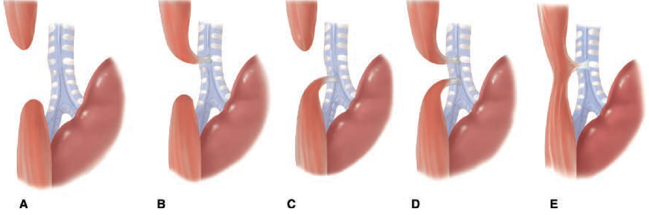
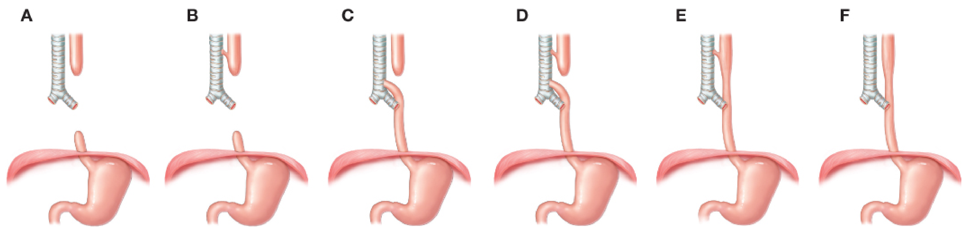
Full Gross Classification (for context)
| Type | Description | Frequency |
|---|---|---|
| A | EA only - no fistula ("pure atresia") | 8% |
| B | EA + proximal TEF only | 1% |
| C | EA + distal TEF only | 86% |
| D | EA + both proximal AND distal TEF | 1% |
| E | TEF only - NO esophageal atresia ("H-type") | 4% |
| F | Congenital esophageal stenosis (no atresia, no TEF) | Rare |
Key Differences: Types C vs D vs E
TYPE C - EA with Distal TEF
(Most common - 85-86% of all cases)
- Esophageal atresia: YES - proximal esophagus ends in a blind pouch
- Fistula location: The distal esophagus connects to the trachea (usually at the carina or just above)
- Proximal fistula: ABSENT
- Stomach gas: PRESENT (air enters stomach via the distal fistula from trachea)
- Clinical features:
- Excessive salivation and drooling at birth
- Choking, gagging, cyanosis with first feed
- Abdominal distension (air passes into stomach through distal fistula)
- Aspiration pneumonia from gastric acid reflux up through the fistula
- X-ray: OG tube coiled in upper pouch; gas present in abdomen (due to distal fistula)
- Prenatal: Polyhydramnios + small/absent stomach bubble
TYPE D - EA with Both Proximal AND Distal TEF
(Rarest combined type - ~1%)
- Esophageal atresia: YES - proximal esophagus ends in a blind pouch
- Fistula location: TWO fistulae - one from the proximal pouch to trachea AND one from the distal esophagus to trachea
- Clinical features:
- All features of Type C PLUS
- Additional proximal fistula makes aspiration even worse
- Saliva directly enters trachea through the proximal fistula
- Higher aspiration risk than Type C
- Abdominal distension present (distal fistula still allows air into stomach)
- Surgical challenge: Both fistulae must be identified and divided - risk of missing the proximal one
TYPE E - TEF without Esophageal Atresia ("H-type")
(4-6% of cases)
- Esophageal atresia: ABSENT - esophagus is completely intact and continuous
- Fistula location: A narrow, oblique fistula connecting an intact trachea to an intact esophagus, usually in the lower cervical/upper thoracic region (runs obliquely from trachea above to esophagus below)
- No blind pouch
- Clinical features - very different from C and D:
- Delayed diagnosis (often missed in newborn period)
- Intermittent symptoms: coughing/choking only with feeds
- Recurrent aspiration pneumonia
- Abdominal distension with crying (air enters esophagus → stomach)
- No excessive drooling (esophagus is patent)
- No OG tube obstruction
- Diagnosis: Contrast esophagogram (tube esophagram) in prone position; bronchoscopy
- Surgery: Right cervical approach (because fistula is high), NOT thoracotomy (unlike C/D)
Summary Comparison Table
| Feature | Type C | Type D | Type E |
|---|---|---|---|
| Esophageal atresia | YES | YES | NO |
| Proximal blind pouch | YES | YES | NO |
| Proximal fistula | No | YES | No |
| Distal fistula | YES | YES | No |
| H-type fistula | No | No | YES |
| Frequency | 86% | 1% | 4-6% |
| Stomach gas on X-ray | YES | YES | YES |
| OG tube coiling | YES | YES | NO |
| Diagnosis age | At birth | At birth | Delayed (weeks-months) |
| Surgical approach | Right thoracotomy | Right thoracotomy | Cervical approach |
| Aspiration risk | High | Very High | Moderate (intermittent) |
Key Memory Aid
- Type C = most Common - proximal pouch, one fistula (distal)
- Type D = Double fistula (proximal + distal) + atresia
- Type E = Esophagus intact - "H-type" - no atresia, fistula only
Sources: Creasy & Resnik's Maternal-Fetal Medicine, p. 520 | Schwartz's Principles of Surgery, 11e, p. 1745 | Fischer's Mastery of Surgery, 8e
Reason of TOF
Tetralogy of Fallot (TOF) results from abnormal anterior and cephalad deviation of the outlet (infundibular) septum during cardiac development, which creates all four defects simultaneously. The exact cause is multifactorial - genetic, environmental, and sporadic.
Searching sources...
Finding Sources
Finding Sources
Finding Sources
Finding Sources
Reading File
Finding Sources
Loading Image
Finding Sources
Causes / Etiology of Tetralogy of Fallot (TOF)
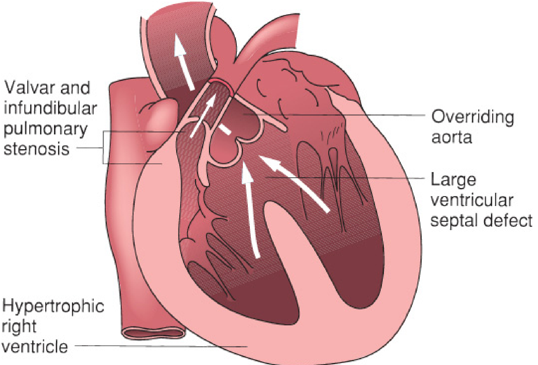
The Core Embryological Reason
TOF arises from a single primary defect in early cardiac development:
Unequal division of the conotruncus with anterior and cephalad (superior) malalignment of the infundibular (outlet) septum during the 5th week of gestation
This one event causes all four components of TOF simultaneously:
How One Defect Creates Four Anomalies
| Event | Result |
|---|---|
| Conotruncus divides unequally | Pulmonary artery becomes narrowed/small |
| Infundibular septum deviates anteriorly | Crowds the RVOT → Pulmonary stenosis |
| Malaligned septum fails to fuse with muscular septum | Large anterior malaligned VSD |
| Aorta is displaced anteriorly and rightward | Overriding aorta (straddles both ventricles) |
| RV works against high resistance (pulmonary stenosis) | RV hypertrophy (secondary/compensatory) |
"The primary morphologic abnormality, anterior and superior displacement of the infundibular septum, results in a malalignment VSD, overriding of the aortic valve, and obstruction of the RVOT. RV hypertrophy is a secondary occurrence." - Mulholland & Greenfield's Surgery, 7e
Neural crest-derived tissue plays a central role - failure of neural crest cell migration into the cardiac outflow tract is the underlying cellular mechanism.
Genetic Causes (25-30% of cases have a genetic basis)
| Genetic Abnormality | Association | Notes |
|---|---|---|
| 22q11.2 deletion (DiGeorge syndrome) | Most common genetic cause (~15%) | CATCH-22 phenotype; also causes pulmonary atresia with TOF |
| Trisomy 21 (Down syndrome) | ~5% of TOF cases | Often with complete AVSD |
| Trisomy 18 | Rare | Poor prognosis |
| JAG1 / NOTCH2 mutations (Alagille) | Occasional | Pulmonary arterial involvement |
| NKX2-5, GATA4, TBX1 mutations | Rare single-gene causes | TBX1 is the main gene deleted in 22q11.2 |
| VEGF gene polymorphisms | Sporadic TOF | Implicated in some studies |
Environmental / Maternal Risk Factors
| Risk Factor | Mechanism |
|---|---|
| Maternal diabetes | Hyperglycemia disrupts cardiac septation |
| Maternal phenylketonuria (PKU) | Phenylalanine teratogenicity |
| Maternal rubella | Direct teratogenic viral damage |
| Maternal alcohol (FAS) | Disrupts neural crest migration |
| Retinoic acid (Vitamin A excess) | Disrupts conotruncal development |
| Thalidomide (historical) | Limb + cardiac teratogen |
| Maternal fever / febrile illness in 1st trimester | Hyperthermia disrupts cardiac development |
| Advanced maternal age | Increases chromosomal anomaly risk |
Summary: Why Does TOF Happen?
CAUSE → Abnormal neural crest cell migration
            ↓
Unequal conotruncal division (Week 5 gestation)
            ↓
Anterior malalignment of infundibular septum
            ↓
┌────────────────────────────────────┐
│ 1. Pulmonary stenosis (RVOTO)      │
│ 2. Large malaligned VSD            │  ← ALL 4 from ONE defect
│ 3. Overriding aorta                │
│ 4. RV hypertrophy (secondary)      │
└────────────────────────────────────┘
            ↓
Right-to-left shunt → CYANOSIS
In ~70-75% of cases, no identifiable genetic cause is found (multifactorial/sporadic). In the rest, chromosomal deletions (especially 22q11.2) or known syndromes are responsible.
- Mulholland & Greenfield's Surgery, 7e, p. 4416-4417
This is a shared conversation. Sign in to Orris to start your own chat.