
I am going to give you question you give answer with easy drawable diagram and clinical 10 marks
I'm ready! Go ahead and share your question. I'll provide a clear answer with an easy-to-draw diagram and clinical points, formatted for a 10-mark answer.
Movement at radioulnar joint
The radioulnar joints (proximal and distal) work together to produce pronation and supination of the forearm. The radius rotates around the ulna - at the proximal joint the radial head spins, and at the distal joint the radius swings around the ulnar head.
Searching sources...
Reading File
Finding Sources
Finding Sources
Reading File
Reading File
Reading File
Loading Image
Loading Image
Finding Sources
Now I have excellent textbook content and diagrams. Let me compose the complete 10-mark answer.
Movement at the Radioulnar Joint
Overview
The radioulnar joints are two pivot (trochoid) joints that act together as a single functional unit to produce pronation and supination of the forearm. The ulna remains stationary while the radius rotates around the ulna.
Types of Radioulnar Joints
| Feature | Proximal RUJ | Distal RUJ |
|---|---|---|
| Location | Below elbow | Above wrist |
| Articulation | Head of radius in radial notch of ulna | Head of ulna in sigmoid notch of radius |
| Joint type | Pivot (trochoid) | Pivot (trochoid) |
| Stabilizer | Annular ligament | TFCC (Triangular Fibrocartilage Complex) |
Movement: PRONATION & SUPINATION
Axis of Movement
Runs obliquely from the centre of the radial head (proximal RUJ) to the fovea of the ulnar head / styloid process of ulna (distal RUJ).
Range
- Supination: 0 - 90°
- Pronation: 0 - 90°
- Total rotation: ~180° (with shoulder adding ~270°)
Easy Drawable Diagram
SUPINATION                        PRONATION
(Palm faces UP)                   (Palm faces DOWN)

PROXIMAL VIEW:                    PROXIMAL VIEW:
  [Radius] [Ulna]                   [Radius crosses
   parallel                          over Ulna]
      ↑                                  ↑
  Annular lig.                      Annular lig.
  holds radius                      holds radius
  in radial notch                   in radial notch


ANTERIOR VIEW (Right forearm):

SUPINATION          PRONATION
  R    U             U
  |    |            / \
  |    |           R   |
  |    |           |   |
  R = Radius, U = Ulna
  (parallel)      (radius crosses over ulna)


AXIS LINE:
  Radial head (above)
       |
       |  (oblique axis)
       |
  Ulnar styloid (below)


DISTAL RUJ CROSS SECTION:

SUPINATION:     NEUTRAL:       PRONATION:
  [R][U]         [R][U]         [U][R]
  side by side   in contact    radius crossed
Muscles Producing Movement
SUPINATION (stronger movement)
| Muscle | Nerve | Note |
|---|---|---|
| Biceps brachii | Musculocutaneous (C5, C6) | Most powerful supinator; best when elbow flexed at 90° |
| Supinator | Posterior interosseous nerve (C6) | Works at all positions |
| Brachioradialis | Radial nerve | Returns from mid-position |
Mnemonic: "SUPinator + BICeps = SUPination" - The Biceps also SUPinates
PRONATION (weaker than supination)
| Muscle | Nerve | Note |
|---|---|---|
| Pronator teres | Median nerve (C6, C7) | Main pronator |
| Pronator quadratus | Anterior interosseous nerve (C8) | Works alone in slow pronation |
| Flexor carpi radialis | Median nerve | Weak accessory |
| Brachioradialis | Radial nerve | Returns from full supination |
Mnemonic: "Pronator Teres + Pronator Quadratus = Pronation"
Connecting Structure: Interosseous Membrane
- A fibrous sheet between the shafts of radius and ulna
- Transmits force from radius to ulna (e.g., falls on outstretched hand)
- Fibers run obliquely: downward and medially from radius to ulna
- Functionally links proximal and distal RUJ - movement of one joint is always accompanied by movement of the other
- Central band is thickest = main load transfer
Easy Diagram: Interosseous Membrane
RADIUS (lateral)          ULNA (medial)
    |    \\\\\\\\\\\\\\\\\\ |
    |     \\\\\\\\\\\\\\\\\ |
    |      \\\\\\\\\\\\\\\\ |  ← Oblique fibers
    |       \\\\\\\\\\\\\\\ |     (radius → ulna,
    |        \\\\\\\\\\\\\\ |      downward + medially)
    |                       |
  [Radial                [Ulnar
   tuberosity]            attachment]
Biomechanics at the Distal RUJ
- In neutral position: styloid process of ulna is aligned with sigmoid notch of radius
- In pronation: ulnar head rotates - styloid process moves to the palmar side; radius migrates proximally (increases ulnar variance)
- In supination: styloid process moves to the dorsal side
- The two distal articular surfaces are in closest contact (congruent) only in neutral/semipronated position
Diagram: Radial head cross-section (Proximal RUJ)
(Based on Thieme Atlas, p. 302)
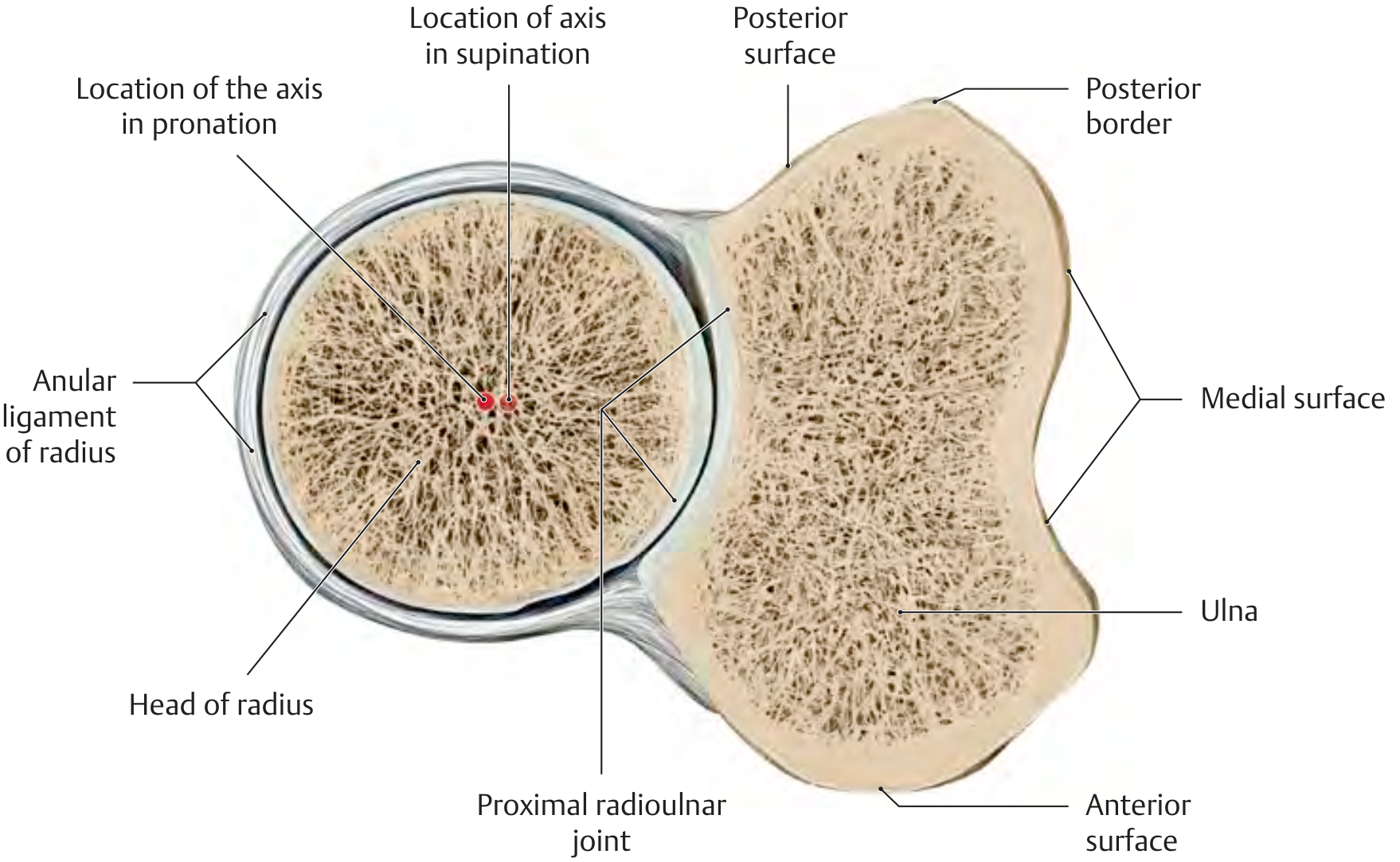
Note: The radial head is slightly oval - the pronation/supination axis shifts ~2 mm radially during pronation. Articular cartilage is thicker on the pronation side (adapts to higher pressure in pronated position).
Diagram: Distal RUJ - Rotation of radius around ulna
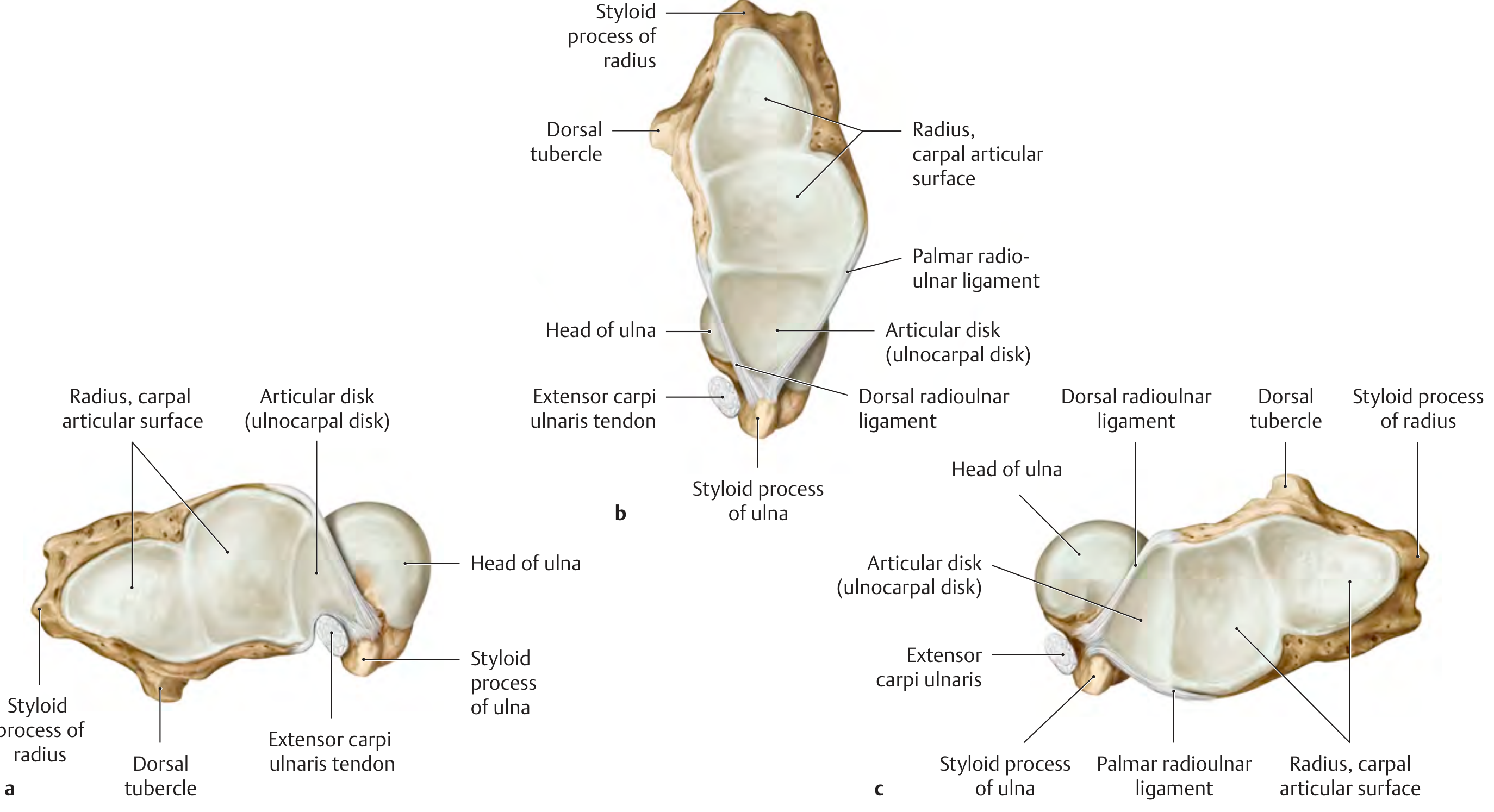
Clinical Points (High Yield)
1. Pulled Elbow (Nursemaid's Elbow)
- Common in children < 5 years
- Mechanism: sudden pull on the hand when the forearm is pronated
- Radial head slips out of the annular ligament
- Child holds arm in slight flexion and pronation, refuses to supinate
- Treatment: hyperpronation or supination + flexion manoeuvre - feel/hear a click
2. Monteggia Fracture-Dislocation
- Fracture of the proximal ulna + dislocation of the radial head from the proximal RUJ
- Disrupts the radioulnar joint
- Must always X-ray the elbow when there is a forearm fracture
- Rule: A line through the radial neck/head should always pass through the capitulum in all views
3. Distal Radioulnar Joint (DRUJ) Disruption
- Seen with distal radius fractures (Galeazzi fracture)
- Signs: pain at DRUJ, prominence of ulnar head (piano key sign), restricted and painful pronation/supination
- Stable DRUJ is essential for painless forearm rotation
4. TFCC (Triangular Fibrocartilage Complex) Injury
- Stabilizes the DRUJ
- Injury causes ulnar-sided wrist pain worse on pronation/supination and grip
- Diagnosed by MRI; treatment ranges from physiotherapy to arthroscopic repair
5. Radioulnar Synostosis
- Bony or fibrous union between radius and ulna
- Forearm is fixed in pronation - loss of supination
- Can be congenital or post-traumatic (after severe forearm fractures, especially if both bones fractured at same level)
6. Superior Radioulnar Joint in Rheumatoid Arthritis
- Synovitis leads to destruction of the annular ligament
- Causes instability of the radial head and loss of rotation
Summary Table
| Feature | Detail |
|---|---|
| Type of movement | Pronation and Supination |
| Joint type | Pivot / Trochoid |
| Axis | Radial head to ulnar styloid/fovea |
| Range | ~90° pronation, ~90° supination |
| Strongest supinator | Biceps brachii |
| Strongest pronator | Pronator teres |
| Proximal stabiliser | Annular ligament |
| Distal stabiliser | TFCC + dorsal/palmar RU ligaments |
| Link between joints | Interosseous membrane |
| Moving bone | Radius (rotates around the ulna) |
| Fixed bone | Ulna (relatively stationary) |
Sources: General Anatomy and Musculoskeletal System, Thieme Atlas of Anatomy (p. 270-302); Imaging Anatomy Text and Atlas Vol. 3, Biomechanics of the Arm and Forearm; Rockwood and Green's Fractures in Adults, 10th ed. 2025
This is a shared conversation. Sign in to Orris to start your own chat.