
Give me the muscle atasment of hand, with a short video
Reading File
Searching the Web
muscle attachments of the hand anatomy video
Finding Sources
Searching the Web
hand intrinsic muscles attachments anatomy YouTube short tutorial
Finding Sources
Reading File
Reading File
Loading Image
Loading Image
Loading Image
Here is a full breakdown of hand muscle attachments with atlas-quality diagrams from the THIEME Atlas of Anatomy.
Muscle Attachments of the Hand
The intrinsic muscles of the hand are divided into three groups: Thenar, Hypothenar, and Metacarpal muscles (lumbricals + interossei).
Thenar Muscles (Base of Thumb)
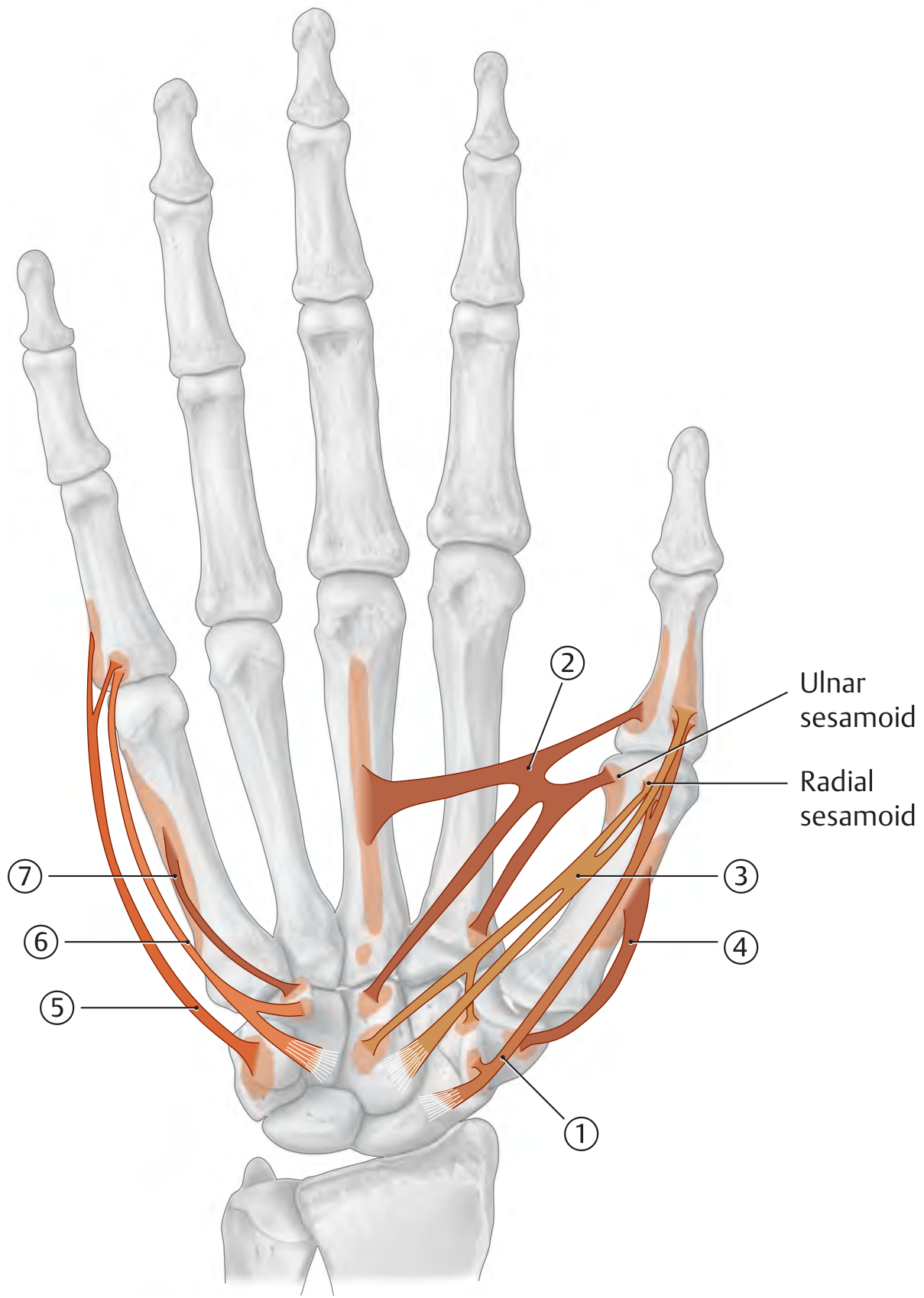
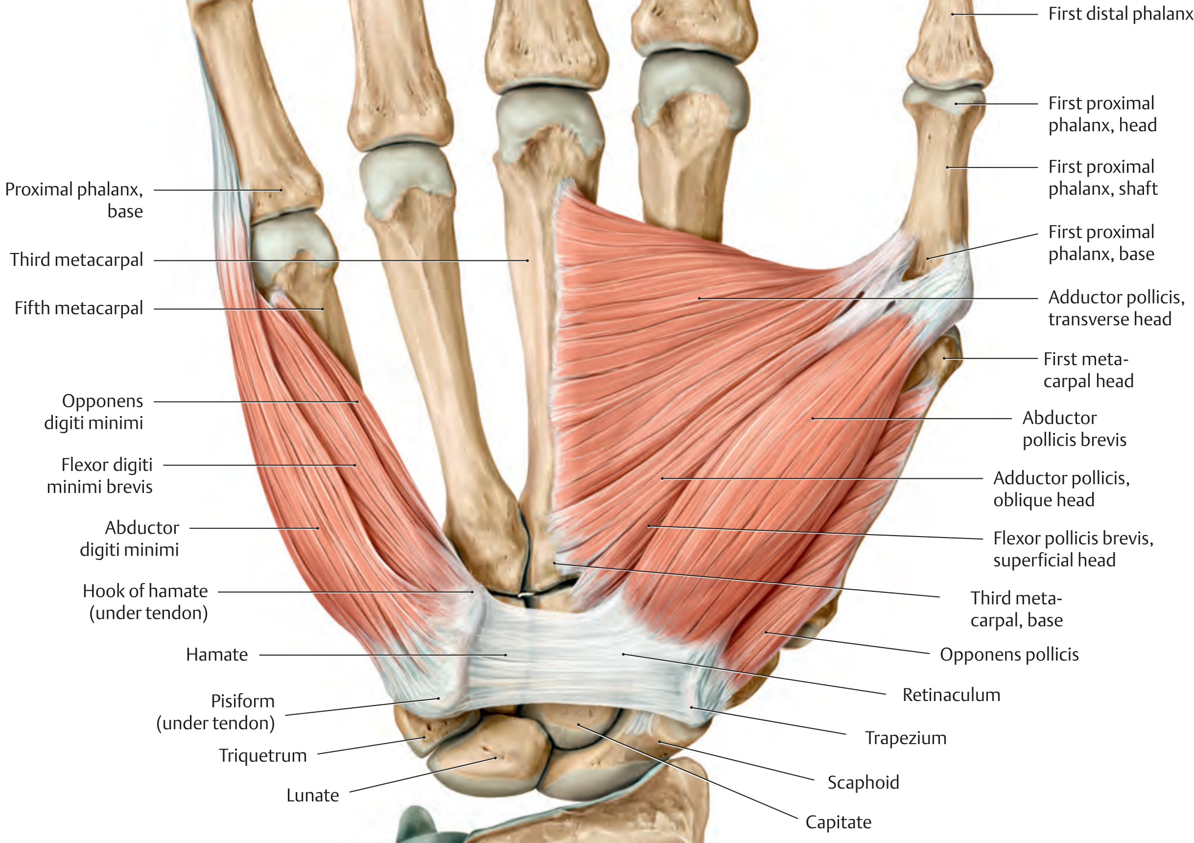
| Muscle | Origin | Insertion | Action | Nerve |
|---|---|---|---|---|
| Abductor pollicis brevis | Scaphoid, trapezium, flexor retinaculum | Base of proximal phalanx of thumb (via radial sesamoid) | Abduction of thumb | Median (C8, T1) |
| Adductor pollicis | Transverse head: palmar surface of 3rd metacarpal; Oblique head: capitate, base of 2nd & 3rd metacarpals | Base of proximal phalanx of thumb (via ulnar sesamoid) | Opposition (CMC), flexion (MCP) | Ulnar (C8, T1) |
| Flexor pollicis brevis | Superficial head: flexor retinaculum; Deep head: capitate, trapezium | Base of proximal phalanx of thumb (via radial sesamoid) | Flexion & opposition (CMC), flexion (MCP) | Superficial: Median; Deep: Ulnar (C8, T1) |
| Opponens pollicis | Trapezium | Radial border of 1st metacarpal | Opposition of thumb (CMC) | Median (C8, T1) |
Hypothenar Muscles (Base of Little Finger)
| Muscle | Origin | Insertion | Action | Nerve |
|---|---|---|---|---|
| Abductor digiti minimi | Pisiform | Ulnar base of proximal phalanx of 5th digit + dorsal digital expansion | Flexion & abduction (MCP); extension (PIP, DIP) | Ulnar (C8, T1) |
| Flexor digiti minimi brevis | Hook of hamate, flexor retinaculum | Base of proximal phalanx of 5th digit | Flexion at MCP of little finger | Ulnar (C8, T1) |
| Opponens digiti minimi | Hook of hamate, flexor retinaculum | Ulnar border of 5th metacarpal | Opposition (draws metacarpal palmarly) | Ulnar (C8, T1) |
| Palmaris brevis | Ulnar border of palmar aponeurosis | Skin of hypothenar eminence | Tightens palmar aponeurosis (protective) | Ulnar (C8, T1) |
Metacarpal Muscles: Lumbricals & Interossei
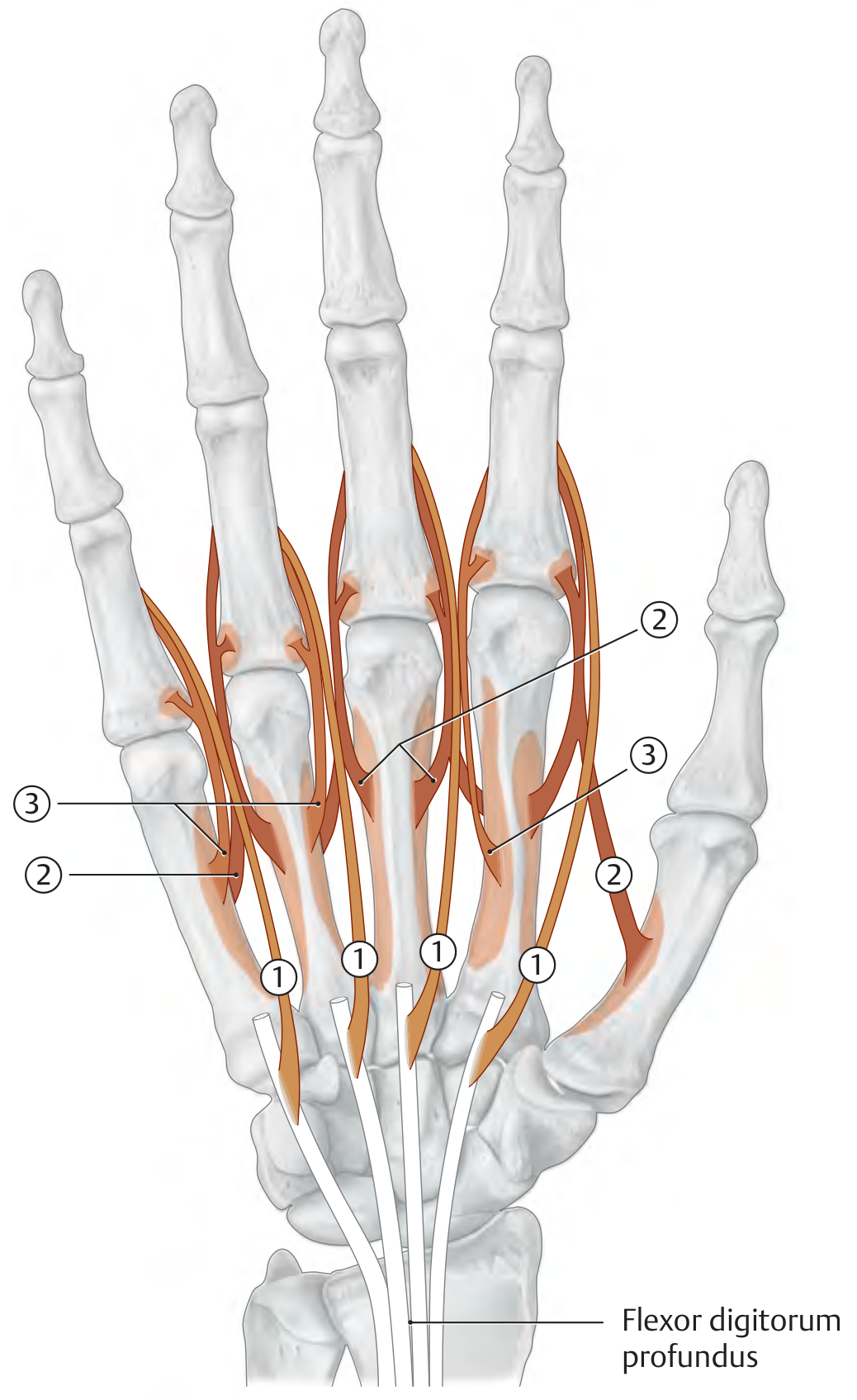
Lumbricals (1st-4th)
- Origin: Radial sides of the flexor digitorum profundus tendons
- Insertion: Dorsal digital expansion (extensor hood) of digits 2-5
- Action: Flexion at MCP joints; extension at PIP and DIP joints
- Nerve: 1st & 2nd (radial two): Median nerve; 3rd & 4th (ulnar two): Ulnar nerve (C8, T1)
Palmar Interossei (1st-3rd or 4th)
- Origin: Palmar surface of 2nd, 4th, and 5th metacarpals (PAD = Palmar ADduction)
- Insertion: Dorsal digital expansion of the same digit
- Action: Adduction of fingers toward the middle finger; MCP flexion + PIP/DIP extension
- Nerve: Ulnar nerve (C8, T1)
Dorsal Interossei (1st-4th)
- Origin: Adjacent sides of two metacarpals (bipennate) (DAB = Dorsal ABduction)
- Insertion: Dorsal digital expansions of digits 2-4
- Action: Abduction of fingers away from the middle finger; MCP flexion + PIP/DIP extension
- Nerve: Ulnar nerve (C8, T1)
Source: THIEME Atlas of General Anatomy and Musculoskeletal System, sections 2.13-2.14
Video Tutorial
Here is a detailed anatomy tutorial covering origin, insertion, and innervation of all hand muscles using 3D models:
This is a shared conversation. Sign in to Orris to start your own chat.