Regulation of tone and posture mbbs 1st year physiology notes for exam pov
Reading File
Finding Sources
Finding Sources
Finding Sources
Finding Sources
Reading File
Finding Sources
Finding Sources
Finding Sources
Finding Sources
Finding Sources
Reading File
Reading File
Loading Image
Loading Image
I now have all the material needed for comprehensive MBBS exam notes. Here are the complete, exam-oriented notes:
Regulation of Tone and Posture
MBBS 1st Year Physiology - Exam Notes
Source: Ganong's Review of Medical Physiology (26th Ed.), Adams & Victor's Principles of Neurology
1. MUSCLE TONE - Definition & Basics
Muscle tone = the resistance of a muscle to passive stretch (also called "tonus").
| State | Description | Cause |
|---|---|---|
| Flaccid | Very little resistance | Severed motor nerve |
| Normal tone | Intermediate resistance | Normal γ-motor discharge |
| Hypertonic/Spastic | High resistance | Hyperactive stretch reflexes |
Key mechanism: Tone is mainly regulated by γ (gamma) motor neurons
- High γ-motor discharge → Hypertonia
- Low γ-motor discharge → Hypotonia
Exam tip: Gamma motor neurons innervate intrafusal (spindle) fibers; alpha motor neurons innervate extrafusal (working) fibers.
2. MUSCLE SPINDLE & STRETCH REFLEX (Basis of Tone)
Muscle Spindle Structure
- Intrafusal fibers - innervated by γ-motor neurons
- Nuclear bag fibers - detect rate of change (dynamic)
- Nuclear chain fibers - detect static stretch
- Ia afferents - primary endings (both bag + chain) → monosynaptic reflex
- II afferents - secondary endings (mainly chain) → polysynaptic reflex
Stretch Reflex Arc (Monosynaptic)
Muscle stretched → Spindle Ia afferent activated → Enters dorsal horn
→ Directly synapses on α-motor neuron (ventral horn)
→ Same muscle contracts (homonymous)
→ Reciprocal inhibition of antagonist (via Ia inhibitory interneuron)
Gamma loop (Fusimotor system):
- γ-motor neuron fires → contracts intrafusal fiber → sensitizes spindle → increases Ia firing → increases α-motor neuron discharge → muscle tone rises
3. INVERSE STRETCH REFLEX (Golgi Tendon Organ Reflex)
- Receptor: Golgi tendon organ (GTO) - in series with muscle fibers (at musculotendinous junction)
- Afferent: Ib afferent fibers
- Effect: When tension is HIGH → GTO fires → inhibits α-motor neurons of same muscle + excites antagonist
Clasp-Knife Effect (clinically important!)
In a spastic limb:
- Moderate passive stretch → muscle contracts (stretch reflex)
- Further stretch → GTO fires → muscle suddenly relaxes
- Resistance followed by sudden give = Clasp-knife effect
Resembles closing a pocket knife - seen in upper motor neuron lesions
4. ROLE OF SUPRASPINAL CENTERS IN TONE & POSTURE
The following descending tracts regulate muscle tone:
| Tract | Origin | Effect on Tone |
|---|---|---|
| Lateral corticospinal | Motor cortex | Inhibits tone (mainly distal) |
| Vestibulospinal | Lateral vestibular nucleus (Deiters') | Facilitates extensor tone |
| Reticulospinal (medullary) | Medullary reticular formation | Inhibits tone |
| Reticulospinal (pontine) | Pontine reticular formation | Facilitates tone |
| Rubrospinal | Red nucleus (midbrain) | Excites flexors, inhibits extensors |
| Tectospinal | Superior colliculus | Postural adjustments (neck/head) |
Balance in normal state: Facilitatory and inhibitory influences are balanced → normal tone.
5. DECEREBRATE vs. DECORTICATE RIGIDITY
This is a high-yield exam topic - memorize the differences!
Decerebrate Rigidity (Lower midbrain/Upper pons transection)
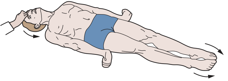
- Level of lesion: Between red nucleus and vestibular nucleus (lower midbrain/upper pons)
- Mechanism: Red nucleus (rubrospinal - flexor facilitating) is disconnected; Vestibulospinal (extensor facilitating) is intact → unopposed extensor facilitation
- Posture:
- All 4 limbs → extended (opisthotonos)
- Neck and back → arched backward
- Upper limbs: extended + pronated + fingers flexed
- Lower limbs: extended + plantar flexed + toes inward
- Abolished by: Cutting dorsal roots (γ-loop dependent)
Decorticate Rigidity (Upper midbrain lesion / Cortical lesion)
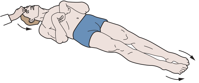
- Level of lesion: Above the red nucleus (rostral to superior colliculus)
- Mechanism: Corticospinal inhibition lost; red nucleus and rubrospinal intact → flexor drive to arms preserved; vestibulospinal still facilitates leg extensors
- Posture:
- Upper limbs: flexed (elbows, wrists)
- Lower limbs: extended
- Head: extended
Memory Trick
"De-CER-ebrate = all Extended" (CER sounds like "stiff") "De-COR-ticate = arms CORE/Curled in" (arms flex inward)
6. SPINAL SHOCK
After sudden complete transection of the spinal cord:
Immediately after transection:
- Complete flaccid paralysis below the lesion
- Loss of ALL reflexes (areflexia) - even stretch reflexes disappear
- Loss of sensation
- Loss of bladder/bowel control
- Hypotension
Recovery (weeks to months):
- Reflexes return - first flexor reflexes then extensor
- Hyperreflexia and spasticity eventually develop
- Babinski sign positive
- Mass reflex may develop
Why does spinal shock occur? Sudden removal of tonic facilitatory impulses (especially from vestibulospinal and reticulospinal tracts) → temporary inexcitability of spinal neurons.
7. POSTURAL REFLEXES
These are reflexes that help maintain body posture:
A. Static Reflexes (Tonic Reflexes)
Maintain body position against gravity - sustained responses.
| Reflex | Receptor | Response |
|---|---|---|
| Tonic neck reflexes | Proprioceptors in neck muscles | Head turned right → right arm extends, left flexes |
| Tonic labyrinthine reflexes | Otolith organs (utricle/saccule) | Regulate limb tone based on head position in space |
| Positive supporting reaction | Pressure receptors in sole + stretch receptors | Contact of sole → limb becomes rigid pillar |
| Negative supporting reaction | Proprio-ceptors | Releases the rigidity of positive supporting reaction |
B. Statokinetic Reflexes (Righting Reflexes)
Dynamic - operate to restore normal posture when it is disturbed.
| Reflex | Purpose |
|---|---|
| Labyrinthine righting reflex | Head rights itself in space (needs intact labyrinths) |
| Neck righting reflex | Once head is righted, neck reflexes right the body |
| Body righting reflex | Asymmetric pressure on body surface rights the trunk |
| Optical righting reflex | Visual input helps orient head in space |
Integration centers for righting reflexes: Midbrain (and higher centers). In decerebrate animals, righting reflexes are lost.
8. VESTIBULAR CONTRIBUTION TO POSTURE
- Lateral vestibular nucleus (Deiters' nucleus) → Vestibulospinal tract → ipsilateral extensor facilitation (anti-gravity muscles)
- Medial vestibular nucleus → Medial vestibulospinal tract → Cervical motor neurons (head/neck posture)
- Input from: Otolith organs (linear acceleration, gravity) + Semicircular canals (angular acceleration)
9. CEREBELLUM & POSTURE
- Anterior lobe (spinocerebellum/paleocerebellum): Receives proprioceptive input; regulates muscle tone - lesion causes hypotonia
- Flocculonodular lobe (vestibulocerebellum/archicerebellum): Equilibrium and vestibular responses - lesion causes truncal ataxia
- Cerebellar lesions cause ipsilateral deficits
10. BASAL GANGLIA & POSTURE
- Regulate background tone for voluntary movements
- Substantia nigra (dopamine) → striatum
- Lesions: Parkinson's disease - cogwheel/lead-pipe rigidity + akinesia
- Huntington's disease - hypotonia + chorea
QUICK REVISION SUMMARY TABLE
| Feature | Decerebrate | Decorticate | Spinal Shock | Cerebellar |
|---|---|---|---|---|
| Arms | Extended | Flexed | Flaccid | Hypotonic |
| Legs | Extended | Extended | Flaccid | Hypotonic |
| Tone | Hypertonic | Hypertonic | Flaccid → Spastic | Hypotonic |
| Level | Lower midbrain/pons | Above red nucleus | Below transection | - |
| Reflexes | Present (exaggerated) | Present | Absent → Exaggerated | Pendular |
HIGH-YIELD EXAM POINTS
- Clasp-knife = GTO (Ib afferents) = upper motor neuron lesion (vs. cogwheel = basal ganglia = Parkinson's)
- Decerebrate = all extended; Decorticate = arms flexed, legs extended
- Decerebrate rigidity is abolished by cutting dorsal roots - proves it depends on the γ-loop/muscle spindle
- Gamma motor neurons set spindle sensitivity → regulates tone
- Vestibulospinal = major extensor facilitator (anti-gravity)
- Medullary reticulospinal = inhibitory; Pontine reticulospinal = facilitatory
- Spinal shock: Flaccid first, then spastic - due to loss of supraspinal facilitation
- Righting reflexes are integrated at the midbrain level
- Anterior lobe of cerebellum damage → hypotonia (not ataxia)
- Positive supporting reaction = limb becomes weight-bearing pillar on foot contact
Source: Ganong's Review of Medical Physiology, 26th Ed. - Ganong's Review of Medical Physiology, p. 241-250
This is a shared conversation. Sign in to Orris to start your own chat.