
Give me difference point btw ring worm. Nummular eczema and psoriasis
Reading File
Now let me search the medical library for all three conditions simultaneously.
Finding Sources
Finding Sources
Finding Sources
Good, I have the file paths. Let me now read the relevant sections from the textbooks in parallel.
Reading File
Reading File
Reading File
Let me get clinical features of ringworm/tinea corporis and psoriasis, and more detailed nummular eczema clinical features.
Finding Sources
Finding Sources
Finding Sources
Excellent - Tintinalli's has a direct comparison table. Let me read that table and also get detailed clinical features from each condition's dedicated section.
Reading File
Reading File
Reading File
I now have enough rich content from authoritative dermatology textbooks to write a comprehensive differential comparison. Let me also fetch a couple of the clinical images to include in the response.
Loading Image
Finding Sources
Finding Sources
I now have comprehensive information from multiple authoritative dermatology textbooks. Here is the complete differential comparison:
Ringworm (Tinea Corporis) vs. Nummular Eczema vs. Psoriasis
These three conditions all produce scaly, coin-shaped or annular plaques and are among the most commonly confused dermatoses in clinical practice. The distinctions below are drawn from Dermatology 2-Volume Set 5e, Fitzpatrick's Dermatology, Tintinalli's Emergency Medicine, and Harrison's Principles of Internal Medicine.
Quick Summary Table
| Feature | Ringworm (Tinea Corporis) | Nummular Eczema | Psoriasis |
|---|---|---|---|
| Cause | Dermatophyte fungus (Trichophyton, Microsporum, Epidermophyton) | Non-infectious; multifactorial (xerosis, allergens, irritants, Staph colonization) | Immune-mediated (T-cell driven); genetic predisposition |
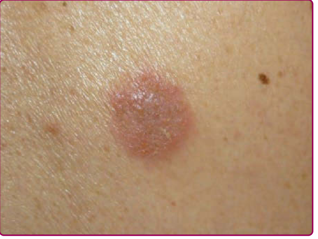
| Primary lesion | Annular/ring-shaped plaque with active scaly border and central clearing | Coin-shaped (nummular = coin) plaque; pinpoint papulovesicles that ooze and crust | Well-demarcated erythematous plaque with thick silvery scale |
| Border | Active, raised, scaly, erythematous rim; center tends to clear as lesion expands | Poorly defined; papulovesicular and exudative at edges, may merge | Very sharply defined, abrupt edge |
| Scale character | Fine, dry scale at the peripheral active border | Crust/serous exudate; weeping, oozing in acute phase; dry and lichenified in chronic | Thick, silver-white (micaceous) scale; layers of scale peel off |
| Shape | Classic ring/annular (can coalesce into gyrate patterns) | Coin-shaped/discoid, rarely annular (central clearing only in chronic cases) | Oval to irregular plaques; not typically annular |
| Central area | Clears as ring expands - central clearing is the hallmark | Remains involved (erythematous, crusted, or lichenified) - no clearing | Uniformly involved plaque, no central clearing |
| Distribution | Trunk, legs, arms, neck; any exposed skin; avoids palms/soles (usually) | Extensor limbs (esp. lower legs), dorsum of hands; lower extremities predominate | Extensor surfaces (elbows, knees), scalp, lumbosacral area, nails |
| Itch | Variable; mild to moderate | Often severe, especially evenings | Variable; chronic itch |
| Special signs | KOH prep: branching septate hyphae; Wood's lamp: some species fluoresce | Pinpoint oozing and crusting is distinctive (Fitzpatrick's sign of nummular eczema) | Auspitz sign (pinpoint bleeding on scale removal), Koebner phenomenon, nail pitting |
| Nail involvement | Onychomycosis if nails affected | Not typical | Nail pitting, onycholysis, oil-drop sign, subungual hyperkeratosis |
| Systemic features | None (except in immunocompromised - deeper invasion) | Associated with dental infection/caries in some patients; no systemic disease | Psoriatic arthritis (up to 30%), metabolic syndrome, cardiovascular risk |
| Seasonality | Year-round; more in warm/humid conditions | Flares in winter (dry skin); troughs in summer | Flares in winter; improves with sunlight |
| Age/demographics | All ages; contact with animals, soil, infected individuals | Bimodal: young men and elderly women; associated with xerosis/dry skin | Onset typically 20s-30s; hereditary predilection |
| Lab/diagnosis | KOH scraping (gold standard): hyphae; fungal culture | Clinical diagnosis; patch testing in recalcitrant cases; lesions are sterile on culture | Clinical diagnosis; biopsy if uncertain |
| Histopathology | Hyphae in stratum corneum; PAS/GMS stain positive | Spongiosis + psoriasiform hyperplasia + scale-crust (plasma and neutrophils in parakeratosis); perivascular lymphocytes and eosinophils | Regular acanthosis, thin suprapapillary plates, elongated rete ridges, Munro microabscesses, dilated papillary capillaries |
| Response to steroids | Worsens (tinea "incognito" - border lost, lesion spreads) | Improves | Usually improves (but rebound on withdrawal) |
| Treatment | Topical antifungals (clotrimazole, terbinafine, ketoconazole); oral if extensive | Emollients + mid-potency topical corticosteroids; tacrolimus; treat underlying infection | Topical corticosteroids, vitamin D analogues, retinoids; phototherapy; biologics (anti-TNF, anti-IL-17, anti-IL-23) for moderate-severe |
| Course | Resolves with treatment; recurs if re-exposed | Often chronic (can persist for decades); intermittent or persistent | Chronic relapsing; rarely self-limited |
Key Distinguishing Points in a Nutshell
Ringworm - The ring tells you it's fungi. Look for an active scaly border with central clearing. Confirmed by KOH. Steroids make it worse. Treat with antifungals.
Nummular Eczema - The coin is solid and weeping, not a ring. Lesion center stays involved and crusted. No hyphae on KOH. Sterile culture. Worsens in winter/dry conditions. Treat with moisturizers and steroids.
Psoriasis - Look for thick silvery scale, very sharp borders, extensor surface preference, nail changes, and the Auspitz sign. Associated systemic disease (arthritis). Has a hereditary component. Needs long-term management.
Clinical Image: Nummular Eczema
The Most Clinically Useful Differentiators
- KOH scraping - positive hyphae = ringworm, period. Always do this first if uncertain.
- Central clearing - present in ringworm, absent in nummular eczema (and variable in psoriasis).
- Scale color/texture - silvery/thick = psoriasis; fine peripheral scale = ringworm; serous crust/ooze = nummular eczema.
- Nail changes - pitting, oil-drop, onycholysis = psoriasis.
- Response to steroids - nummular eczema and psoriasis improve; ringworm worsens (or mask as tinea incognito).
- Distribution - extensor elbows/knees/scalp = psoriasis; lower extremities = nummular eczema; any skin surface with active border = ringworm.
This is a shared conversation. Sign in to Orris to start your own chat.