
Explain me 10 common heart diseases with ECG changes and ECG images of those diseases
Reading File
Finding Sources
Searching Images
ECG electrocardiogram myocardial infarction ST elevation
Finding Sources
Searching Images
atrial fibrillation ECG irregularly irregular rhythm
Searching Images
heart block complete AV block ECG third degree
Searching Images
ventricular tachycardia ECG wide complex tachycardia
Searching Images
Wolff-Parkinson-White WPW syndrome ECG delta wave short PR
Searching Images
left bundle branch block LBBB ECG broad QRS
Searching Images
hypertrophic cardiomyopathy ECG left ventricular hypertrophy
Searching Images
pericarditis ECG saddle-shaped ST elevation diffuse
Searching Images
long QT syndrome ECG prolonged QT interval torsades de pointes
Searching Images
Brugada syndrome ECG coved ST elevation V1 V2 V3
Searching Images
pulmonary embolism ECG S1Q3T3 right heart strain sinus tachycardia
I now have excellent images and data for all 10 conditions. Let me compile the comprehensive answer.Here is a comprehensive guide to 10 common heart diseases with their ECG changes and representative ECG images.
10 Common Heart Diseases: ECG Changes & Images
1. ST-Elevation Myocardial Infarction (STEMI)
Pathophysiology: Complete occlusion of a coronary artery causing transmural myocardial ischemia and injury.
Key ECG Changes:
- ST-segment elevation ≥1 mm in ≥2 contiguous limb leads, or ≥2 mm in ≥2 contiguous precordial leads
- Hyperacute (peaked) T-waves — earliest sign, preceding ST elevation
- Reciprocal ST depression in leads facing the opposite wall
- Pathological Q waves — develop hours to days later (necrosis marker)
- T-wave inversion — repolarization abnormality in evolving/resolved STEMI
- Lead localization: Anterior (V1–V4 → LAD), Inferior (II, III, aVF → RCA), Lateral (I, aVL, V5–V6 → LCx)
- Tombstone morphology in massive proximal LAD occlusion
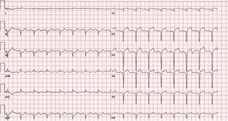
Anterior STEMI: Massive convex ST elevation across V1–V6 with hyperacute T-waves, consistent with proximal LAD occlusion.
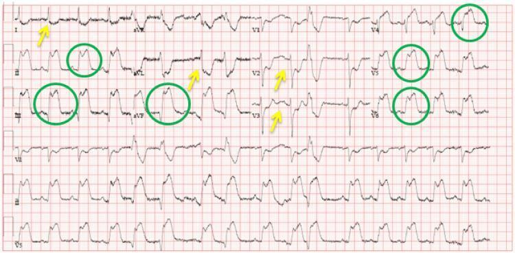
Inferolateral STEMI: ST elevation in II, III, aVF and V4–V6 with reciprocal depression in I, aVL, and anterior leads.
2. Atrial Fibrillation (AF)
Pathophysiology: Chaotic, disorganized atrial electrical activity from multiple re-entrant circuits, leading to loss of coordinated atrial contraction.
Key ECG Changes:
- Absent P waves — replaced by fibrillatory (f) waves, best seen in V1
- Irregularly irregular R-R intervals — pathognomonic finding
- Narrow QRS complexes (unless aberrant conduction or pre-existing BBB)
- Ventricular rate varies: controlled (<100 bpm), rapid response (>100 bpm)
- Fine vs. coarse fibrillatory baseline — coarser waves often suggest valvular cause
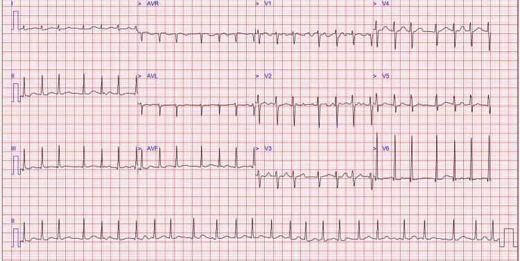
AF with rapid ventricular response: Absent P waves, fibrillatory baseline prominent in V1, irregularly irregular QRS complexes.
3. Complete (Third-Degree) AV Block
Pathophysiology: Complete failure of conduction between atria and ventricles; atria and ventricles beat independently — AV dissociation.
Key ECG Changes:
- Complete AV dissociation — no relationship between P waves and QRS complexes
- P waves march through at their own regular rate (faster than QRS)
- Escape rhythm — either:
- Junctional: narrow QRS (~40–60 bpm)
- Ventricular: wide QRS (<40 bpm, more dangerous)
- Fixed R-R intervals (regular escape rhythm)
- PR interval varies — no consistent PR relationship
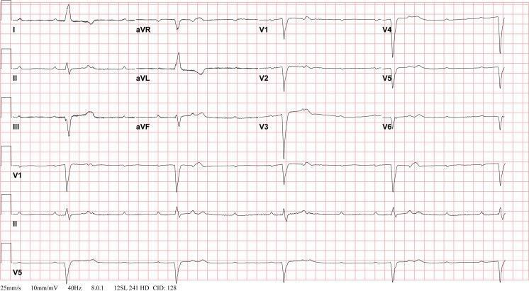
Third-degree heart block: Independent atrial and ventricular activity. Wide QRS escape rhythm at ~30 bpm, P waves unrelated to QRS complexes.
4. Ventricular Tachycardia (VT)
Pathophysiology: Rapid, potentially life-threatening rhythm originating below the bundle of His, often from a re-entrant circuit in scarred ventricular myocardium.
Key ECG Changes:
- Wide QRS complexes (>120 ms), rate typically 100–250 bpm
- AV dissociation — P waves unrelated to QRS (when visible)
- Fusion beats and capture beats — pathognomonic of VT
- Concordance across precordial leads (all positive or all negative)
- Brugada/Josephson criteria help differentiate from SVT with aberrancy
- Monomorphic VT — uniform QRS morphology (structural heart disease)
- Polymorphic VT — varying QRS (ischemia, long QT, CPVT)
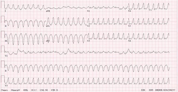
Monomorphic VT: Wide-complex tachycardia with positive concordance across precordial leads, no discernible P waves, superior axis.
5. Wolff-Parkinson-White (WPW) Syndrome
Pathophysiology: An accessory pathway (bundle of Kent) bypasses the AV node, causing ventricular pre-excitation. Risk of rapid AF conducting via accessory pathway causing VF.
Key ECG Changes:
- Short PR interval (<120 ms) — AV node bypass
- Delta wave — slurred, slow upstroke at the beginning of QRS
- Wide QRS (>120 ms) — due to fusion of normal + pre-excited conduction
- Discordant ST-T changes — secondary to abnormal depolarization
- "Pseudo-infarction" — negative delta waves in inferior leads can mimic Q waves
- Accessory pathway localization by delta wave polarity in specific leads
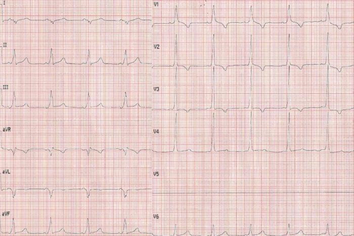
WPW syndrome: Short PR interval, delta waves visible in II, III, aVF, and V2–V6 causing QRS widening — the classic pre-excitation triad.
6. Left Bundle Branch Block (LBBB)
Pathophysiology: Failure of conduction through the left bundle branch, causing delayed and abnormal left ventricular depolarization. Often signals underlying structural disease (cardiomyopathy, ischemia, hypertension).
Key ECG Changes:
- Wide QRS (≥120 ms)
- Broad, notched "M-shaped" R waves in lateral leads (I, aVL, V5, V6)
- Deep QS or rS complexes in right precordial leads (V1–V3)
- Absence of septal Q waves in I, aVL, V5, V6
- Discordant ST-T changes — ST/T in opposite direction to QRS
- New LBBB in the setting of chest pain is treated as STEMI equivalent (Sgarbossa criteria)
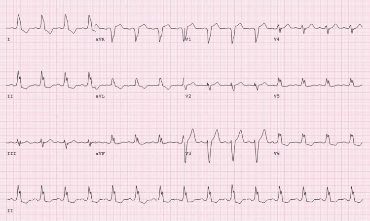
LBBB: Wide QRS, deep S-waves in V1–V3, broad monophasic R-waves in I, V5–V6, and discordant ST-T changes.
7. Hypertrophic Cardiomyopathy (HCM)
Pathophysiology: Genetic sarcomere protein mutations causing asymmetric (typically septal) hypertrophy, myofiber disarray, and diastolic dysfunction. A leading cause of sudden cardiac death in young athletes.
Key ECG Changes:
- Left ventricular hypertrophy (LVH) — high-voltage QRS (Sokolow-Lyon: S in V1 + R in V5/V6 ≥35 mm)
- Deep, wide Q waves in lateral leads (I, aVL, V5–V6) and inferior leads — "septal Q waves"
- ST-segment depression and T-wave inversion in lateral leads — strain pattern
- Apical HCM (Yamaguchi): Giant T-wave inversions (≥10 mm) in precordial leads V2–V5
- Left axis deviation common
- Atrial fibrillation may develop with LA enlargement
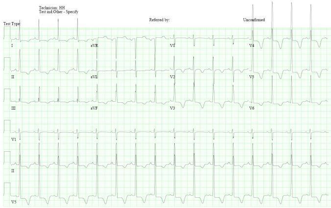
Apical HCM (Yamaguchi syndrome): High-voltage QRS with giant symmetric T-wave inversions in V2–V5, characteristic of apical hypertrophy.
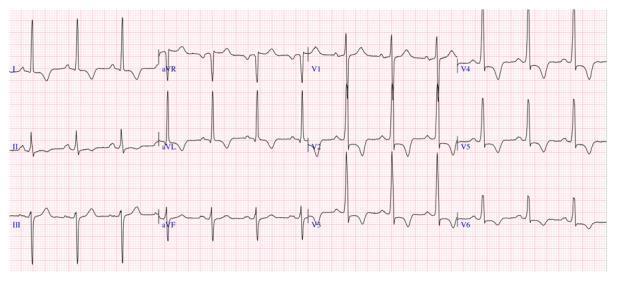
Septal HCM: High R-wave amplitude in V2–V4, ST depression and deep T-wave inversions in V2–V6 and lateral leads, positive T in aVR (risk marker).
8. Acute Pericarditis
Pathophysiology: Inflammation of the pericardium (viral, autoimmune, post-MI/Dressler's, uremic, idiopathic). The inflammatory process affects the epicardium, producing diffuse ST changes.
Key ECG Changes (4 Stages):
- Stage 1: Diffuse, concave (saddle-shaped) ST elevation in nearly all leads except aVR and V1; PR depression in most leads, PR elevation in aVR
- Spodick's sign: Downward sloping TP segment (highly specific)
- Stage 2: ST normalization, PR depression persists
- Stage 3: T-wave inversions (diffuse)
- Stage 4: ECG normalizes
- Differentiator from STEMI: No reciprocal changes in the opposite territory (except aVR), no Q waves, concave vs. convex ST elevation
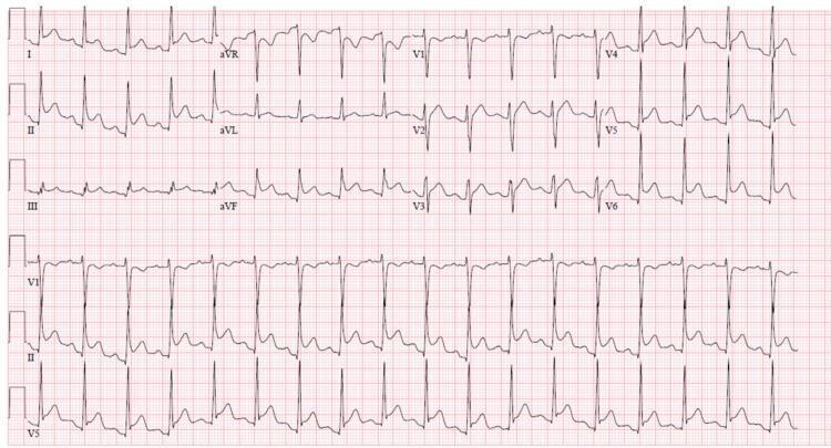
Acute pericarditis: Diffuse saddle-shaped ST elevation in I, II, III, aVF, V2–V6; PR depression in II (most visible); reciprocal ST depression and PR elevation in aVR. Spodick's sign present.
9. Long QT Syndrome (LQTS)
Pathophysiology: Prolonged ventricular repolarization due to ion channel mutations (congenital: LQT1–LQT15) or acquired causes (electrolyte disturbances, drugs). Predisposes to Torsades de Pointes (TdP) → ventricular fibrillation → sudden death.
Key ECG Changes:
- Prolonged QTc interval: Males >440 ms, Females >460 ms; >500 ms is high risk
- QTc formula: QT ÷ √RR interval (Bazett formula)
- T-wave morphology subtypes:
- LQT1: Broad-based T waves
- LQT2: Bifid (notched) T waves
- LQT3: Long isoelectric ST segment with late narrow T wave
- T-wave alternans — beat-to-beat variation in T morphology (pre-TdP sign)
- Torsades de Pointes: Polymorphic VT with "twisting of the points" around isoelectric line, triggered by R-on-T phenomenon
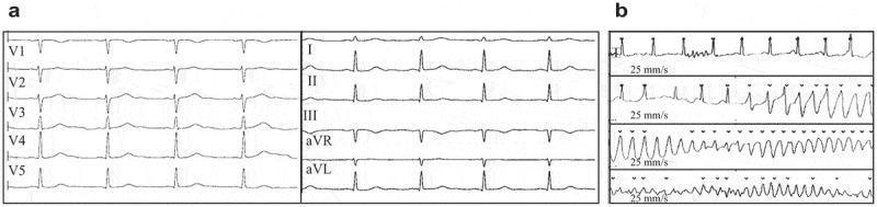
LQTS with TdP: Panel (a) shows markedly prolonged QTc (478 ms). Panel (b) shows the characteristic "twisting" polymorphic VT pattern of Torsades de Pointes.
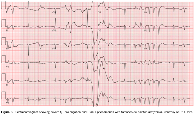
Severe QT prolongation → R-on-T phenomenon (PVC landing on T wave) → triggering polymorphic VT (TdP).
10. Brugada Syndrome
Pathophysiology: Genetic sodium channelopathy (SCN5A mutation) causing abnormal repolarization in the right ventricle outflow tract (RVOT). Associated with risk of polymorphic VT/VF and sudden cardiac death, particularly in young males during sleep/rest.
Key ECG Changes:
- Type 1 (Diagnostic): Coved ST-segment elevation ≥2 mm with convex (downward sloping) morphology in V1–V2, terminating in negative (inverted) T-wave — "coved pattern"
- Type 2: Saddle-back ST elevation ≥0.5 mm in V1–V2 (no longer diagnostic alone)
- Type 3: Saddle-back elevation <2 mm (non-diagnostic)
- Pattern may be dynamic — fever, vagotonia, certain drugs (flecainide, ajmaline) unmask it
- Right precordial leads best seen with V1–V2 placed in 2nd/3rd intercostal space
- Incomplete RBBB morphology often co-exists
- No QT prolongation (distinguishes from LQTS)
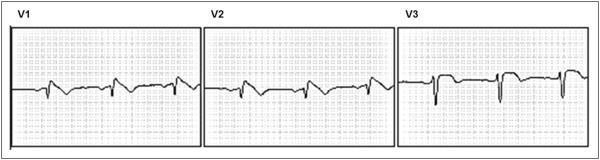
Brugada Type 1: Classic coved ST elevation ≥2 mm in V1–V2 with convex morphology descending into inverted T-wave — pathognomonic for Brugada syndrome.
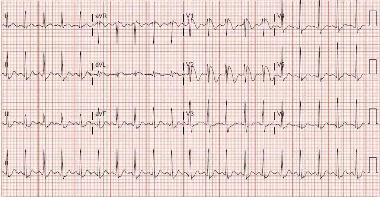
Brugada Type 1 on 12-lead: Coved ST elevation isolated to V1–V2, with normal ST segments in all other leads — sinus tachycardia.
Quick Reference Summary Table
| # | Disease | Core ECG Finding | Key Differentiator |
|---|---|---|---|
| 1 | STEMI | Convex ST elevation + reciprocal depression | Localizes to coronary territory; Q waves follow |
| 2 | Atrial Fibrillation | No P waves, irregularly irregular | Fibrillatory baseline in V1 |
| 3 | Complete AV Block | AV dissociation, escape rhythm | P waves unrelated to QRS |
| 4 | Ventricular Tachycardia | Wide QRS, fast, AV dissociation | Fusion/capture beats; concordance |
| 5 | WPW Syndrome | Short PR + delta wave + wide QRS | Pre-excitation triad; pseudo-Q waves |
| 6 | LBBB | Wide QRS, M-shaped R in lateral, QS in V1–V3 | New LBBB = STEMI equivalent |
| 7 | HCM | LVH + deep lateral Q waves; giant T inversions (apical) | Yamaguchi pattern: giant T inversions V2–V5 |
| 8 | Pericarditis | Diffuse saddle-shaped ST↑, PR depression | Spodick's sign; no reciprocal changes |
| 9 | Long QT Syndrome | QTc >450–500 ms, TdP | T-wave alternans; twisting VT morphology |
| 10 | Brugada Syndrome | Coved ST elevation V1–V2 | Type 1 only is diagnostic; no QT prolongation |
Clinical note: ECG interpretation always requires correlation with clinical history, symptoms, and hemodynamic status. Apparent "normal" ECGs do not exclude significant cardiac disease, and dynamic ECG changes over serial tracings are often more informative than a single recording.The guide above covers 10 major heart diseases with detailed ECG changes and real ECG images from peer-reviewed medical literature. Here's a brief overview of what was covered:
- STEMI — Convex ST elevation localizing to a coronary territory, with reciprocal changes and evolving Q waves
- Atrial Fibrillation — Absent P waves replaced by fibrillatory baseline; irregularly irregular rhythm
- Complete AV Block — Full AV dissociation with independent atrial and ventricular pacemakers
- Ventricular Tachycardia — Wide-complex tachycardia with AV dissociation and concordance
- WPW Syndrome — Short PR + delta wave + wide QRS (pre-excitation triad)
- LBBB — Wide QRS with M-shaped lateral R waves and deep right precordial QS complexes
- Hypertrophic Cardiomyopathy — LVH criteria + lateral Q waves or giant T-wave inversions (apical variant)
- Acute Pericarditis — Diffuse saddle-shaped ST elevation + PR depression (Spodick's sign)
- Long QT Syndrome — Prolonged QTc > 450–500 ms, T-wave alternans, Torsades de Pointes
- Brugada Syndrome — Coved ST elevation ≥2 mm in V1–V2 with inverted T-wave (Type 1 pattern)
This is a shared conversation. Sign in to Orris to start your own chat.