
Relation for epidural fat ligamentum flavum diramater in endoscopic dissectomy
endoscopic discectomy epidural fat ligamentum flavum dura mater anatomy
Relationship of Epidural Fat, Ligamentum Flavum, and Dura Mater in Endoscopic Discectomy
1. Spatial Architecture of the Posterior Spinal Canal
Spinous process / Lamina
        ↓
Ligamentum Flavum (posterior wall of epidural space)
        ↓
Posterior Epidural Fat (cushion between LF and dura)
        ↓
Dura Mater (outer meningeal layer)
        ↓
Arachnoid Mater → Subarachnoid Space (CSF) → Pia Mater
        ↓
Spinal Cord / Cauda Equina
2. Ligamentum Flavum
- It extends from the foramen magnum to the sacral hiatus
- It is actually two paired ligaments (right and left) that join in the midline with a ventral opening
- It is not uniform in thickness - it is largest in the lumbar region (where discectomy is most common)
- Immediately posterior to the LF are the lamina and spinous processes
- When hypertrophied, it can compress the dural sac and nerve roots
- The lateral portion is first detached from the caudal edge of the superior lamina and the cephalad edge of the inferior lamina using a curette
- A blunt dissector lifts the edge so it can be excised with a Kerrison rongeur
- En bloc removal in one piece is the goal - avoids repeated nibbling that risks dural injury
- The rongeur is kept parallel to the nerve root to avoid root damage
3. Epidural Fat
- Posteriorly: the dura mater is in contact with the posterior epidural fat, which separates the dura from the ligamenta flava, posterior joints, and laminae - Grainger & Allison's Diagnostic Radiology
- In the thoracic region this fat layer can be up to 5 mm thick (must not be confused with epidural lipomatosis)
- In the lumbar region, fat forms multiple, metameric, discrete collections in both anterior and posterior aspects of the epidural space - Barash Clinical Anesthesia (9e)
- The cervical level contains no epidural fat - making cervical procedures riskier for dural injury
- Epidural fat is not removed during discectomy (Campbell's Operative Orthopaedics, 15e, Step 15)
- It is a useful landmark on MRI - the bright T1 signal fat stripe posterior to the dura confirms intact epidural anatomy - Neuroanatomy through Clinical Cases (3e)
4. Dura Mater
- The dura is the outermost meningeal covering; deep to the epidural fat
- The epidural space surrounds the dura anteriorly, laterally, and posteriorly
- Anterior: dura is in contact with the posterior longitudinal ligament (PLL) almost the entire spine length
- Lateral: dura closely contacts the medial border of the pedicles; above/below the pedicles it contacts epidural fat continuing into the intervertebral foramina
- Posterior: dura contacts posterior epidural fat, which separates it from the ligamentum flavum
- After LF removal, the dural sac and traversing nerve root become visible - the epidural fat marks the safe plane
- Step 14 (Campbell's): a gentle extradural exploration is made beneath the nerve with a 90-degree blunt hook, taking care not to tear the dura - the small operating corridor and magnification can make the edge of the dural sac appear to be the nerve root
- Step 19: the surgeon confirms the nerve root and dural sac are free of any disc protrusions before closure
5. Surgical Sequence Summary (Endoscopic / Tubular Discectomy)
| Step | Structure Encountered | Key Action |
|---|---|---|
| Skin → fascia | Paraspinal muscles | Dilators placed, tubular retractor docked on lamina |
| Lamina/LF junction | Ligamentum flavum | Curette detaches LF from lamina edges |
| LF removal | LF with Kerrison rongeur | En bloc excision, rongeur parallel to nerve |
| Post-LF removal | Posterior epidural fat | Identifies safe working plane; NOT removed |
| Fat plane | Dura mater exposed | Gentle dissection; blunt hook exploration |
| Dura → nerve root | Traversing nerve root | Mobilized medially with nerve hook |
| Under nerve root | Herniated disc | Annulotomy + pituitary rongeur removal |
6. Clinical Significance
- Dural tear risk: The posterior epidural fat is the only buffer between the LF and dura. When the LF is thickened and adherent, or when epidural fat is minimal, dural injury risk rises sharply.
- Identifying the correct plane: Seeing posterior epidural fat after LF removal confirms the surgeon is in the correct extradural plane - the dura lies just deep to it.
- Epidural fat as a tamponade: Fat left in situ contributes to hemostasis in the epidural space.
- MRI correlation: Preoperatively, loss of posterior epidural fat stripe on sagittal T1 MRI signals LF hypertrophy, adhesions, or severe stenosis - warning of difficult dissection.
- Campbell's Operative Orthopaedics, 15e (2026), Steps 11-19 of Tubular Retractor Discectomy
- Miller's Anesthesia (10e) - Epidural Space Anatomy, p. 6017-6018
- Barash Clinical Anesthesia (9e) - Epidural Space, p. 2795
- Neuroanatomy through Clinical Cases, 3e - Nerve Roots in Relation to Vertebral Bones, p. 344-345
- Grainger & Allison's Diagnostic Radiology - Neural Structures, p. 1221
Create a visual diagram poster of the posterior spinal canal layers and their relationships in discectomy
Medical educational poster showing a detailed cross-sectional anatomical diagram of the posterior spinal canal layers as encountered during endoscopic discectomy. The diagram should show a sagittal/axial cross-section of the lumbar spine with clearly labeled layers from posterior (top) to anterior (bottom): 1. Spinous Process and Lamina (bone, shown in beige/ivory) 2. Ligamentum Flavum - yellow elastic ligament (shown in yellow-gold), labeled as "posterior wall of epidural space" 3. Posterior Epidural Fat - soft tissue cushion (shown in orange-yellow), labeled "buffer zone, NOT removed during discectomy" 4. Dura Mater - thick fibrous membrane (shown in blue), labeled "outermost meningeal layer" 5. Epidural Space with Batson's venous plexus (shown in red vessels) 6. Arachnoid Mater (shown in light blue) 7. Subarachnoid Space with CSF (shown in pale blue) 8. Pia Mater closely investing the spinal cord (shown in pink) 9. Spinal Cord / Cauda Equina (shown in light pink/cream) Include a side panel showing the surgical sequence steps: Step 1: Dock tubular retractor on lamina Step 2: Curette detaches ligamentum flavum from lamina Step 3: Kerrison rongeur - en bloc LF removal Step 4: Posterior epidural fat visible (safe plane landmark) Step 5: Dura mater exposed - blunt dissection only Step 6: Nerve root mobilized medially Step 7: Disc herniation excised Include color-coded arrows showing the surgical direction (posterior to anterior). Add a small inset showing the MRI T1 appearance with bright fat stripe. Title at top: "Posterior Spinal Canal Layers in Endoscopic Discectomy". Professional medical illustration style, clean white background, bold clear labels.
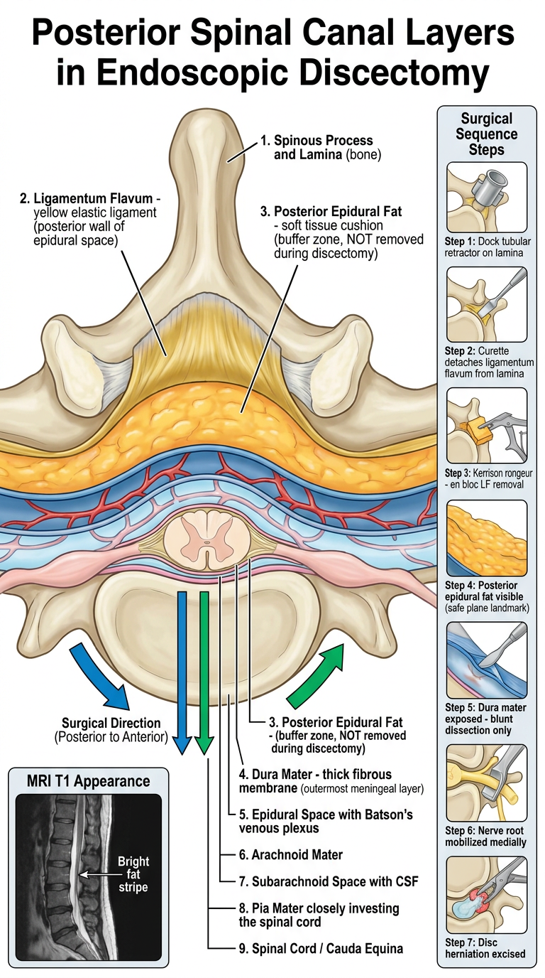
- Cross-sectional anatomy from posterior to anterior - bone (lamina/spinous process) → ligamentum flavum (yellow) → posterior epidural fat (orange-yellow buffer) → dura mater (blue) → arachnoid → CSF/subarachnoid space → pia mater → spinal cord/cauda equina
- Epidural space contents including the Batson venous plexus
- Surgical sequence panel - the 7 steps from retractor docking through disc excision
- Color-coded surgical direction arrows showing the posterior-to-anterior approach
- MRI T1 inset showing the bright fat stripe that serves as the preoperative landmark
Endoscopic images of various structure during endoscopic dissectomy
endoscopic discectomy intraoperative images ligamentum flavum epidural fat dura mater nerve root disc herniation
percutaneous endoscopic interlaminar discectomy intraoperative endoscope view epidural fat nerve root dura mater site:pmc.ncbi.nlm.nih.gov OR site:researchgate.net
Endoscopic Intraoperative Images During Discectomy
IMAGE SET 1 - Full Endoscopic Interlaminar Discectomy (A-H surgical sequence)

| Panel | Structure | What You See |
|---|---|---|
| A | Ligamentum Flavum | Yellowish-white fibrous tissue being grasped and excised by endoscopic punch/scissors. The LF is the first neural canal structure encountered after bone. Firm, elastic texture. |
| B | Epidural Space / Epidural Fat | Lobulated, yellowish-pink fat globules filling the posterior epidural space. This is the critical "safe plane" landmark - its appearance confirms you are extradural. |
| C | Bone / Lamina | Triangular endoscopic instrument (Kerrison rongeur) biting at the lamina edge. Background shows bleeding and muscle/fat mix. |
| D | Dura Mater + Epidural Fat Interface | Glistening white-blue dural membrane becoming visible beneath epidural fat. Surrounding red tissues are epidural vessels. This is the danger zone - blunt dissection only. |
| E | Dura Mater / Thecal Sac | Smooth, shiny, taut white membrane = dural sac. The pulsatile nature (with irrigation flow) confirms intact dura with CSF inside. |
| F | Traversing Nerve Root + Dura | Star (★) = thecal sac/dura; Arrow = traversing nerve root emerging from it. The nerve has a redder, cord-like appearance distinct from the white dura. |
| G | Nerve Root Mobilization | Blunt probe/nerve hook mobilizing the nerve root medially to expose underlying disc. Small instrument tip visible. |
| H | Herniated Disc + Post-decompression | After nerve mobilization - red epidural tissue and white fibrous disc/annular material visible. Pulsatile movement of neural structures confirms adequate decompression. |
IMAGE SET 2 - Percutaneous Endoscopic Interlaminar Discectomy + 3D Spine Reconstruction
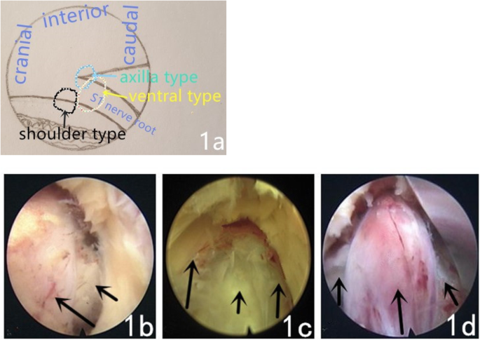
| Panel | What You See |
|---|---|
| 1a | Schematic diagram showing disc herniation types relative to the S1 nerve root: shoulder type (black circle, lateral to root), axilla type (cyan dashed, medial to root axilla), ventral type (yellow, anterior to root). Orientation: cranial (top-left), caudal (bottom-right), interior (top). |
| 1b | Intraoperative endoscopic view - ligamentum flavum being dissected (arrows point to its edges). The yellow-white fibrous tissue is the flavum being mobilized. |
| 1c | Epidural space after flavectomy - arrows show the yellow-orange glistening epidural fat, the key landmark confirming extradural access. |
| 1d | Nerve root exposed - arrows indicate the nerve root (pink-white cord-like structure) after mobilization, with surrounding dura visible. The nerve is clearly distinguishable from the thecal sac. |
Key Endoscopic Appearance Summary
| Structure | Endoscopic Appearance | Clinical Significance |
|---|---|---|
| Ligamentum flavum | Yellow-white, firm, fibrous; does not pulsate | First structure to excise; en bloc removal preferred |
| Posterior epidural fat | Lobular, yellowish-orange, soft, floats with irrigation | Safe plane - confirms extradural position |
| Dura mater | Shiny, taut, blue-white membrane; pulsates with heartbeat | Do NOT incise - blunt instruments only |
| Nerve root | Pinkish-red cord; more vascular than dura; moves with probing | Mobilize medially before disc access |
| Disc material | Dull white/grey, avascular, fibrous; does not pulsate | Excise with pituitary rongeur |
| Epidural veins | Red, bleed readily, surround fat and nerve | Bipolar cautery; one-sided contact only near nerve |
Surgical Video Reference
Yes please
- Identifying epidural fat as the landmark for the spinal canal floor
- Distinguishing the traversing nerve root from surrounding fat
- Visualizing the annulus/disc in real time
- The technique of palpating vs. directly visualizing neural structures under continuous irrigation