
Discuss role of usg in Airway assessment. Q
Reading File
Finding Sources
Finding Sources
Searching PubMed
"airway ultrasound"[Title] AND assessment
Reading File
Reading File
Finding Sources
Finding Sources
Finding Sources
Finding Sources
Reading File
Reading File
Reading File
Reading File
Excellent image found - this maps out the whole POCUS airway assessment approach. Now I have enough to write a comprehensive, well-organized answer.
Finding Sources
Searching the Web
point of care ultrasound airway assessment difficult intubation prediction parameters 2024
I have enough material now. Here's a comprehensive answer.
Role of Ultrasound (USG) in Airway Assessment
Point-of-care ultrasound (POCUS) has become a valuable bedside adjunct to traditional airway examination (Mallampati, thyromental distance, upper lip bite test), which have only modest sensitivity for predicting a difficult airway - "the absence of these findings is predictive of relative ease of intubation... [but] any patient's airway may be found to be surprisingly difficult in spite of assuring bedside airway screening tests" (Morgan and Mikhail's Clinical Anesthesiology, p. 571-572). Ultrasound adds objective, reproducible anatomical data that can be gathered rapidly at the bedside without radiation.
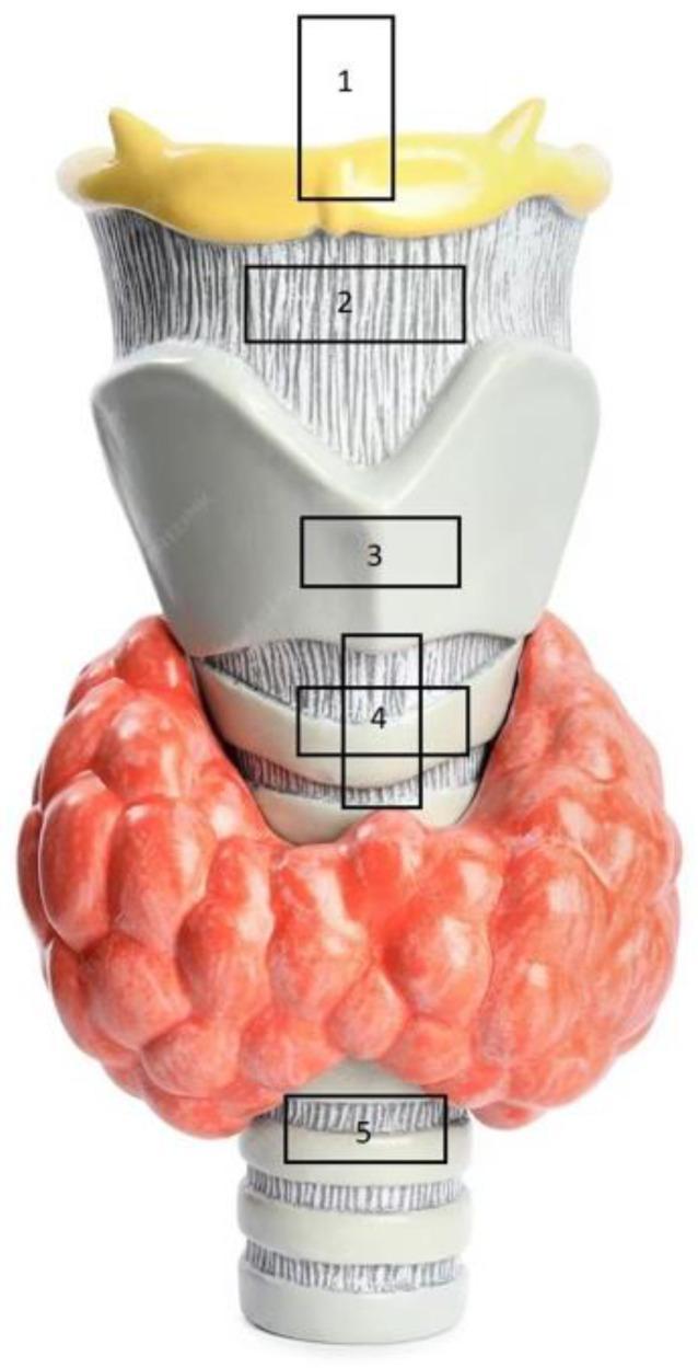
1. Preoperative prediction of a difficult airway
Using a linear high-frequency probe scanned from the submental region down to the suprasternal notch, several sonographic parameters correlate with difficult laryngoscopy/intubation:
- Tongue thickness / tongue base measurement - increased tongue thickness is an established predictor of airway difficulty, similar in concept to Mallampati grading (Miller's Anesthesia).
- Distance from skin to epiglottis (DSE) and hyomental distance - increased soft-tissue depth correlates with harder glottic visualization.
- Anterior neck soft-tissue thickness at the level of the hyoid, thyrohyoid membrane, and vocal cords - quantified sonographically, this predicts difficult laryngoscopy particularly in obese patients (Barash, Cullen & Stoelting's Clinical Anesthesia, referencing Ezri et al., Anaesthesia 2003).
- These measures are especially useful in obesity, where redundant pharyngeal tissue and increased neck circumference make bedside tests less reliable, and in patients who cannot cooperate with standard clinical tests (altered consciousness, cervical collar, trauma).
A 2025 systematic review and meta-analysis in Anaesthesia (Soni et al., PMID 40891437) and a 2023 meta-analysis in the Journal of Cardiothoracic and Vascular Anesthesia (Benavides-Zora et al., PMID 37012134) both evaluated the diagnostic performance of these airway ultrasound parameters for predicting difficult laryngoscopy - useful if you want the most current pooled sensitivity/specificity data; worth flagging since this is an active, evolving evidence base.
2. Localizing the cricothyroid membrane (CTM)
This is one of the most well-validated uses. Digital palpation of the CTM is unreliable, especially in obese, edematous, or distorted necks. Ultrasound (sagittal or transverse view over the anterior neck) allows accurate, rapid identification of the CTM:
- Improves speed and success of emergency cricothyroidotomy / front-of-neck access (FONA) in a "can't intubate, can't oxygenate" scenario (ROSEN's Emergency Medicine; Barash's Clinical Anesthesia; Roberts and Hedges' Clinical Procedures).
- Studies cited in Barash's Clinical Anesthesia (Kristensen & Teoh) and Roberts and Hedges' show ultrasound-guided marking of the CTM is more accurate than palpation alone, particularly in obese patients and pregnant women in labor.
- This preprocedural marking is now built into difficult-airway algorithms as part of the "preintubation difficult airway assessment" (ROSEN's Emergency Medicine, Difficult Cricothyrotomy: SMART section).
3. Confirming endotracheal tube (ETT) placement
- Real-time transverse tracheal ultrasound during intubation can visualize the tube passing through the trachea (versus esophageal intubation), seen as increased echogenicity/movement just distal to the tracheal cartilage (Morgan and Mikhail's Clinical Anesthesiology, Fig 19-7).
- Particularly useful when capnography is unreliable - e.g., low cardiac output states, cardiac arrest, or neonates - where ultrasound confirmation of tube position has been reviewed as an adjunct (Barash's Clinical Anesthesia; Tintinalli's Emergency Medicine; Rosen's Emergency Medicine references on neonatal ETT confirmation).
- Can also estimate correct ETT/tracheal tube sizing by measuring subglottic/tracheal diameter, especially useful in pediatric airway planning (Miller's Anesthesia).
4. Guiding surgical airway and tracheostomy procedures
- POCUS assists in determining tracheal size, localizing tracheal rings, and guiding percutaneous tracheostomy, reducing complications from blind puncture (Miller's Anesthesia, "Ultrasound for Procedural Guidance").
- In trauma and emergency settings, ultrasound-guided rapid four-step cricothyrotomy technique has been simplified by pre-marking the CTM with ultrasound before the skin incision (Roberts and Hedges' Clinical Procedures).
5. Adjunct assessments relevant to airway/anesthetic risk
- Gastric ultrasound (antral cross-sectional area) to assess fasting status/aspiration risk before induction - not airway anatomy itself but part of the broader "airway-adjacent" POCUS bundle used by anesthesiologists (Miller's Anesthesia).
- Lung ultrasound post-intubation to confirm bilateral ventilation (lung sliding) and exclude pneumothorax or mainstem intubation.
Summary of clinical value
| Application | Benefit |
|---|---|
| Predicting difficult laryngoscopy | Objective anterior neck/tongue measurements, useful in obesity |
| Cricothyroid membrane localization | Faster, more accurate FONA than palpation |
| ETT placement confirmation | Reliable when capnography is compromised |
| Tube/tracheal sizing | Guides pediatric tube selection |
| Percutaneous tracheostomy guidance | Reduces procedural complications |
Limitations
Ultrasound airway assessment is operator-dependent, requires training, and normative cutoff values vary between studies - current meta-analyses still report heterogeneity in diagnostic accuracy across different sonographic parameters, so it is used as an adjunct to, not a replacement for, standard clinical airway examination and universal difficult-airway preparedness.
Recent evidence flag: the 2025 Anaesthesia systematic review (PMID 40891437) and 2023 JCVA meta-analysis (PMID 37012134) represent the most current pooled data on diagnostic accuracy of these parameters - if precise sensitivity/specificity figures are needed for exam purposes, I can pull the exact numbers from these papers on request.
This is a shared conversation. Sign in to Orris to start your own chat.