
Explain the anatomical snuffbox with its clinical importance and clinical anatomy with simple line diagrams for four marks.
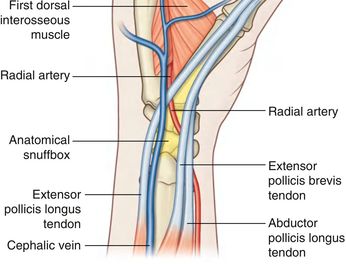
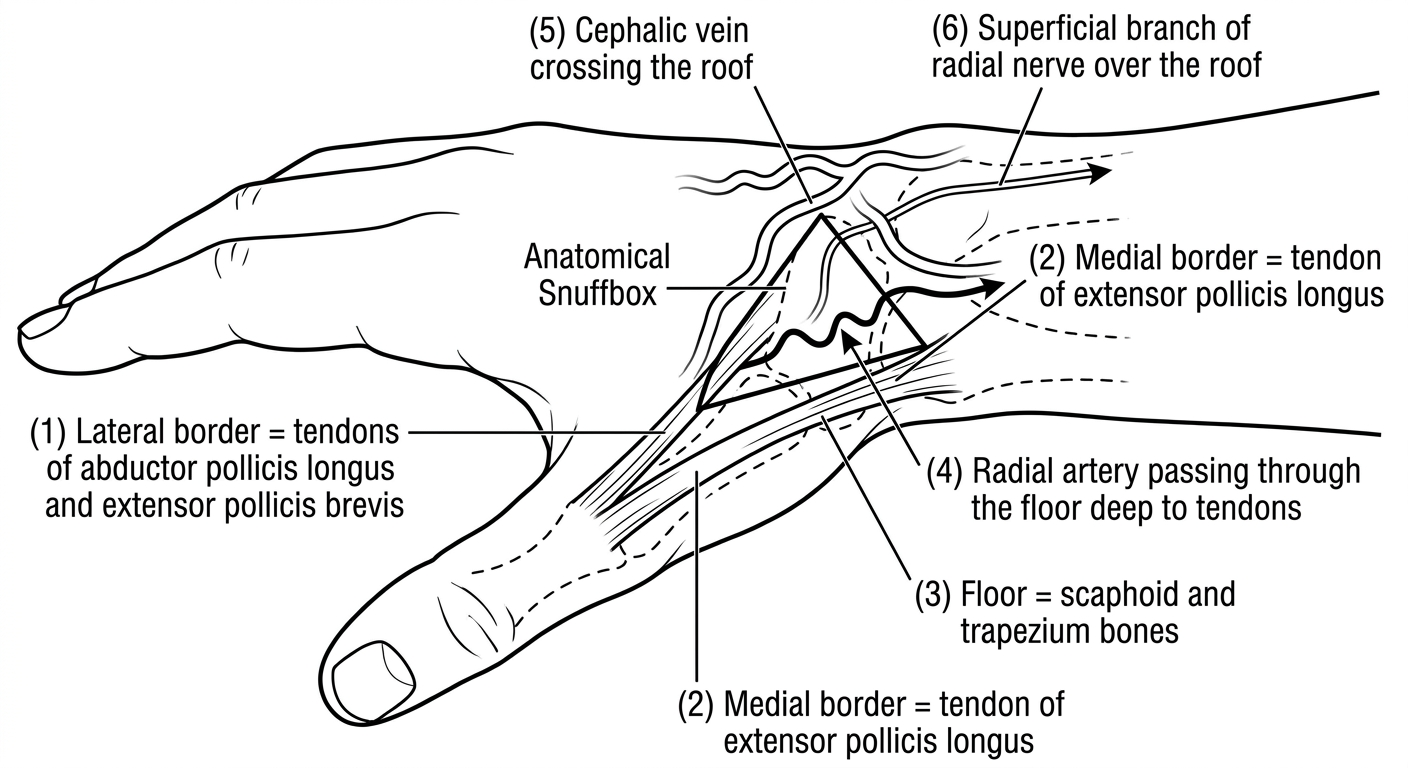
Simple labeled line diagram of the anatomical snuffbox of the hand showing: dorsolateral view of the wrist and thumb in extension, with clear labels for: (1) lateral border = tendons of abductor pollicis longus and extensor pollicis brevis, (2) medial border = tendon of extensor pollicis longus, (3) floor = scaphoid and trapezium bones, (4) radial artery passing through the floor deep to tendons, (5) cephalic vein crossing the roof, (6) superficial branch of radial nerve over the roof, (7) the triangular depression labeled "Anatomical Snuffbox". Clean anatomical illustration style, black and white, educational medical diagram quality.
Anatomical Snuffbox
Definition
Boundaries (Clinical Anatomy)
| Structure | What forms it |
|---|---|
| Lateral border (anterior wall) | Tendons of abductor pollicis longus (APL) and extensor pollicis brevis (EPB) |
| Medial border (posterior wall) | Tendon of extensor pollicis longus (EPL) |
| Floor | Scaphoid and trapezium bones; distal tendons of extensor carpi radialis longus (ECRL) and extensor carpi radialis brevis (ECRB) |
| Base | At the wrist (proximal) |
| Apex | Points toward the thumb (distal) |
Contents / Roof Structures
- Radial artery - passes obliquely through the snuffbox deep to the extensor tendons, lying directly on the scaphoid and trapezium
- Cephalic vein - crosses the roof (superficial to tendons), originating from the dorsal venous arch
- Superficial branch of radial nerve - passes subcutaneously over the roof
Diagrams
Clinical Importance
1. Scaphoid Fracture
- The floor of the snuffbox overlies the scaphoid bone directly.
- Tenderness in the anatomical snuffbox after a fall on an outstretched hand (FOOSH) is the classic sign of a scaphoid fracture - even when initial X-rays appear normal.
- With the hand in ulnar deviation, the scaphoid moves distally and becomes palpable within the snuffbox, allowing direct assessment.
- Scaphoid fractures are dangerous because the proximal scaphoid has a precarious blood supply entering distally, risking avascular necrosis if missed.
2. Radial Pulse Palpation
- The radial artery pulse can be felt in the floor of the snuffbox (in addition to the classic wrist position). This is useful when the wrist pulse is absent or difficult to palpate.
3. Cephalic Vein Access
- The cephalic vein in the snuffbox is a reliable site for intravenous cannulation, especially when antecubital veins are inaccessible (e.g., in difficult IV access or emergencies).
4. Radial Nerve Injury
- Terminal branches of the superficial branch of the radial nerve cross the snuffbox roof.
- These can be injured by tight wristbands, handcuffs, or direct trauma, causing dorsolateral hand numbness ("handcuff neuropathy").
- They can be palpated by rolling against the EPL tendon - a useful clinical test.
5. Surgical Access
- The snuffbox is used as an approach for radial artery cannulation (intra-arterial monitoring) and as an access site for transradial cardiac catheterization.
Memory Aid
"LAME" for the lateral border - Lateral = APL + EPB; Medial = EPLFloor = Scaphoid + Trapezium (press here for scaphoid fracture)Artery deep, vein and nerve superficial