
*A 12-year-old boy presents with a 24-hour history of periumbilical pain that has migrated to the right iliac fossa. He has nausea, vomiting, and tenderness in the right lower quadrant. A provisional diagnosis of acute appendicitis is made, and he is taken for surgery. Intraoperatively, the appendix appears normal. Further exploration reveals a 4-cm inflamed outpouching arising from the antimesenteric border of the ileum, about 60 cm proximal to the ileocecal junction.*
Reading File
This is a classic Meckel's diverticulum presentation. Let me search the medical library thoroughly.
Searching sources...
Finding Sources
Finding Sources
Excellent results. Let me now read the most authoritative sections in parallel.
Reading File
Reading File
Reading File
Reading File
Now let me get the surgical management section and fetch the pathology image, plus look at Robbins for the pathology details.
Reading File
Loading Image
Finding Sources
I now have comprehensive, authoritative information from multiple textbooks. Here is the full clinical analysis:
Meckel's Diverticulum - Complete Clinical Analysis
Diagnosis
This boy has Meckel's diverticulitis. The intraoperative findings are pathognomonic:
- Outpouching from the antimesenteric border of the ileum
- Located ~60 cm proximal to the ileocecal junction
- Inflamed (diverticulitis)
- Normal appendix
This is the most common congenital anomaly of the gastrointestinal tract.
Embryology
Meckel's diverticulum results from incomplete closure (failed involution) of the omphalomesenteric (vitelline) duct - the structure that connects the midgut to the yolk sac during fetal development. It is a true diverticulum, meaning it contains all three layers of the bowel wall (mucosa, muscularis, serosa) and has its own blood supply (a branch of the superior mesenteric artery).
- Robbins, Cotran & Kumar Pathologic Basis of Disease, p. 1492
- Sabiston Textbook of Surgery, p. 2535
The "Rule of 2s"
A classic mnemonic summarizing its features:
| Feature | Value |
|---|---|
| Prevalence in population | ~2% |
| Distance from ileocecal valve | ~2 feet (60 cm) |
| Length | ~2 inches (5 cm) |
| Male:female ratio | 2:1 |
| Age most often symptomatic | By age 2 years |
| Lifetime risk of symptoms | ~2% (only ~4% are ever symptomatic) |
This patient's diverticulum is 4 cm long and located 60 cm proximal to the ileocecal junction - perfectly consistent with the rule of 2s.
- Bailey and Love's Short Practice of Surgery, 28th Ed, p. 1333
- Robbins, Cotran & Kumar, p. 1492-1503
Gross Pathology
This is what a Meckel's diverticulum looks like on gross pathology (from Robbins):
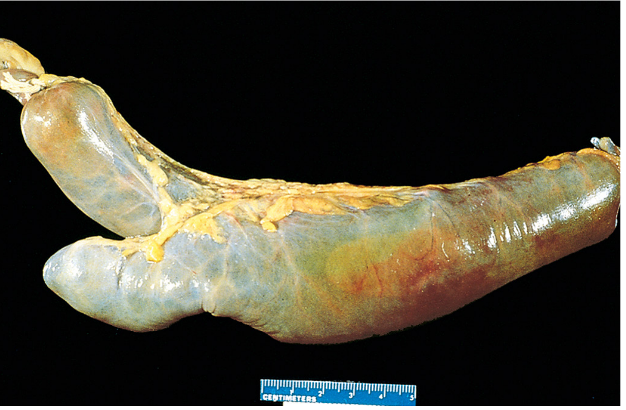
Meckel's diverticulum: the blind pouch is located on the antimesenteric side of the small bowel
Heterotopic Mucosa
In approximately 20-50% of cases, the diverticulum contains ectopic (heterotopic) tissue:
- Gastric mucosa - most common (the parietal cells secrete HCl, causing acid-induced peptic ulceration of the adjacent ileal mucosa)
- Pancreatic mucosa - second most common
- Rarely: colonic mucosa
The mucus-secreting cells of ectopic gastric mucosa are the basis for the Meckel's scan (Tc-99m pertechnetate scintigraphy), described below.
- Sabiston, p. 2535
- Robbins, p. 1503
Clinical Presentations / Complications
| Complication | Details |
|---|---|
| Diverticulitis | Clinically indistinguishable from appendicitis (as in this case) - presents with periumbilical pain migrating to RLQ, nausea, vomiting |
| Haemorrhage | Most common symptomatic presentation in children <2 years; caused by peptic ulceration from ectopic gastric mucosa; presents as painless dark rectal bleeding or melaena |
| Intestinal obstruction | Via: (1) volvulus around a fibrous band connecting diverticulum apex to umbilicus, (2) intussusception (Meckel's acts as lead point - ileoileal or ileocolic), (3) Littre's hernia (incarceration in inguinal hernia) |
| Perforation | Follows diverticulitis; may resemble perforated duodenal ulcer |
| Chronic ulceration | Periumbilical pain (midgut origin) |
| Neoplasm | Rare (0.5-3.2%); NETs most common (33-44%), followed by leiomyosarcoma, adenocarcinoma, GIST |
Key surgical teaching point: When a normal appendix is found during exploration for suspected appendicitis, the distal ileum must be examined for an inflamed Meckel's diverticulum - exactly as occurred in this case.
- Bailey and Love's, p. 1333-1334
- Sabiston, p. 2540-2544
Diagnosis (Preoperative, if not explored surgically)
-
Plain abdominal X-ray, CT, ultrasound: rarely diagnostic
-
Tc-99m pertechnetate scintigraphy (Meckel's scan): Investigation of choice in children
- Pertechnetate is taken up by mucus-secreting cells of ectopic gastric mucosa
- Sensitivity ~85%, specificity ~95%, accuracy ~90% in children
- Sensitivity drops to ~63% in adults (less ectopic gastric tissue)
- Cimetidine pre-treatment enhances sensitivity (reduces secretion, increases radionuclide retention)
-
Mesenteric arteriography: useful in acute haemorrhage
-
Double-balloon endoscopy, barium contrast: for false-negative scans
-
Sabiston, p. 2553-2555
Surgical Management
Symptomatic Meckel's diverticulum requires prompt surgery:
- Diverticulectomy - for non-bleeding Meckel's diverticulitis (hand-sewn or stapled across the base in a diagonal/transverse line to prevent stenosis)
- Segmental ileal resection - required for haemorrhage (bleeding ulcer is in the adjacent ileum, not the diverticulum itself); also required for complications such as volvulus/gangrene
- Laparoscopic vs. open: retrospective studies show equivalent outcomes
Incidentally found (asymptomatic) Meckel's diverticulum:
-
In children: generally recommended to resect
-
In adults: controversial; resection considered if risk factors are present:
- Age <50 years
- Male sex
- Length >2 cm
- Presence of ectopic tissue or palpable abnormalities
-
Sabiston, p. 2560-2568
-
Bailey and Love's, p. 1333-1334
Summary for This Case
| Feature | This Patient |
|---|---|
| Age | 12 years (pediatric - higher risk if symptomatic) |
| Diagnosis | Meckel's diverticulitis mimicking acute appendicitis |
| Finding | 4-cm diverticulum, antimesenteric border, 60 cm from IC junction - classic |
| Management | Diverticulectomy (non-bleeding, so simple excision at the base is appropriate) |
| Underlying mechanism | Failed involution of omphalomesenteric duct |
| Potential ectopic tissue | Ectopic gastric or pancreatic mucosa possible (~20-50%) |
This is a shared conversation. Sign in to Orris to start your own chat.