
Action potential
I'll look this up in the medical library for a thorough answer.
Reading File
Now let me search the medical library for action potential content in parallel.
Finding Sources
Finding Sources
Excellent. Now let me read the most relevant sections - Guyton (physiology gold standard) and Neuroscience: Exploring the Brain, in parallel.
Reading File
Reading File
Let me now fetch the key images and read the Costanzo section on cardiac action potential and Guyton's refractory period section in parallel.
Loading Image
Loading Image
Reading File
Reading File
Let me fetch the Costanzo conductance image and also get the cardiac action potential section and the conduction velocity section.
Loading Image
Reading File
Reading File
Excellent - I have comprehensive material from Guyton, Costanzo, and Neuroscience: Exploring the Brain. Now let me fetch the Na channel states image and get the conduction section.
Loading Image
Reading File
I now have all the material needed for a comprehensive answer. Let me compile it.
Action Potential
An action potential is a rapid, transient reversal of the membrane potential that travels along the surface of excitable cells (neurons, muscle fibers) to transmit information over distance. It lasts roughly 1-2 milliseconds and is the fundamental unit of communication in the nervous system.
1. Resting Membrane Potential
Before an action potential can occur, the cell sits at its resting membrane potential, approximately -65 to -70 mV (inside negative). This resting state is maintained because:
- K⁺ conductance is high - K⁺ leaks out down its concentration gradient, driving the potential toward the K⁺ equilibrium potential (~-85 mV)
- Na⁺ conductance is very low - Na⁺ cannot flow in despite strong electrochemical driving force
- The Na⁺/K⁺-ATPase actively maintains the concentration gradients
The cell is said to be "polarized" at rest.
2. Phases of the Action Potential
Here is the classic Guyton waveform with all phases labeled:
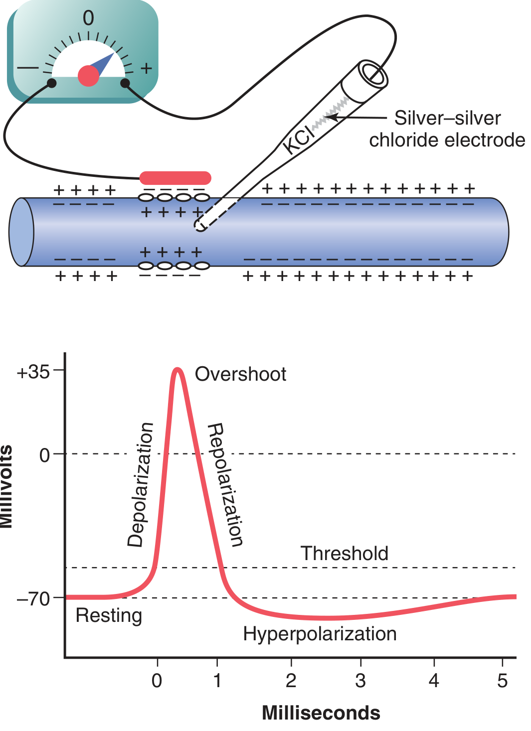
(Guyton and Hall Textbook of Medical Physiology)
Phase 1 - Resting Stage (-70 mV)
The membrane is polarized. Na⁺ channels are closed but available (activation gate closed, inactivation gate open). K⁺ channels are open.
Phase 2 - Threshold and Depolarization (Upstroke / Rising Phase)
A depolarizing stimulus (synaptic input, sensory, or current spread from a neighboring site) brings the membrane to threshold (~-55 to -60 mV). At this critical level, enough voltage-gated Na⁺ channels open that the inward Na⁺ current overwhelms the resting K⁺ current - creating a positive feedback loop (the Hodgkin cycle):
Depolarization → more Na⁺ channels open → more Na⁺ influx → more depolarization
The membrane potential shoots upward rapidly.
Phase 3 - Overshoot
In large myelinated fibers, the potential overshoots 0 mV to reach approximately +35 to +40 mV. The membrane interior is now transiently positive. The membrane potential approaches (but does not fully reach) the Na⁺ equilibrium potential (~+65 mV).
Phase 4 - Repolarization (Falling Phase)
Two events combine to bring the potential back down:
- Na⁺ channel inactivation - the slow inactivation gate closes (even though the activation gate stays open). Na⁺ conductance falls sharply.
- Delayed K⁺ channel opening - voltage-gated K⁺ channels (triggered by depolarization ~1 ms earlier) finally open. K⁺ rushes out, driving the potential back toward the K⁺ equilibrium potential.
Phase 5 - Undershoot (Afterhyperpolarization)
The K⁺ channels remain open slightly longer than needed. The membrane hyperpolarizes briefly below the resting potential, approaching E_K (~-85 mV). This corresponds to the relative refractory period.
3. Ionic Conductance Changes
The graph below shows the precise timing of Na⁺ and K⁺ conductance changes alongside the membrane potential:
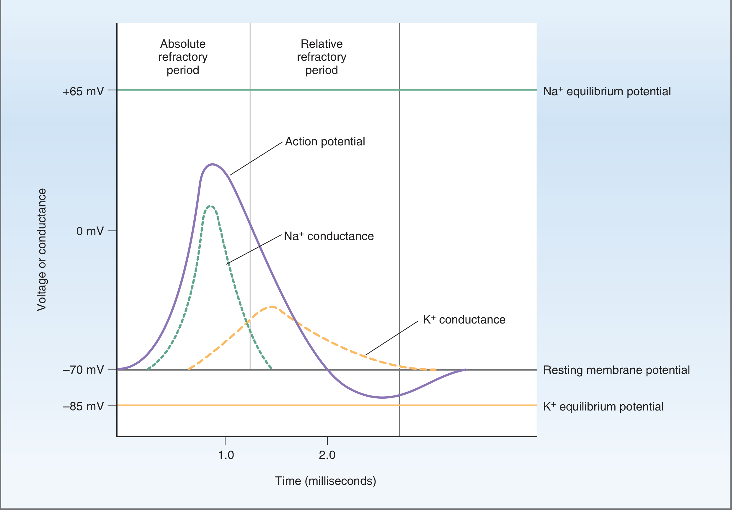
(Costanzo Physiology, 7th Edition)
Key points:
- Na⁺ conductance peaks before K⁺ conductance
- K⁺ conductance is delayed and broader, causing the undershoot
- Resting membrane potential = -70 mV; Na⁺ equilibrium = +65 mV; K⁺ equilibrium = -85 mV
4. The Voltage-Gated Na⁺ Channel: Three States
The Na⁺ channel has two gates whose timing is the key to the action potential:
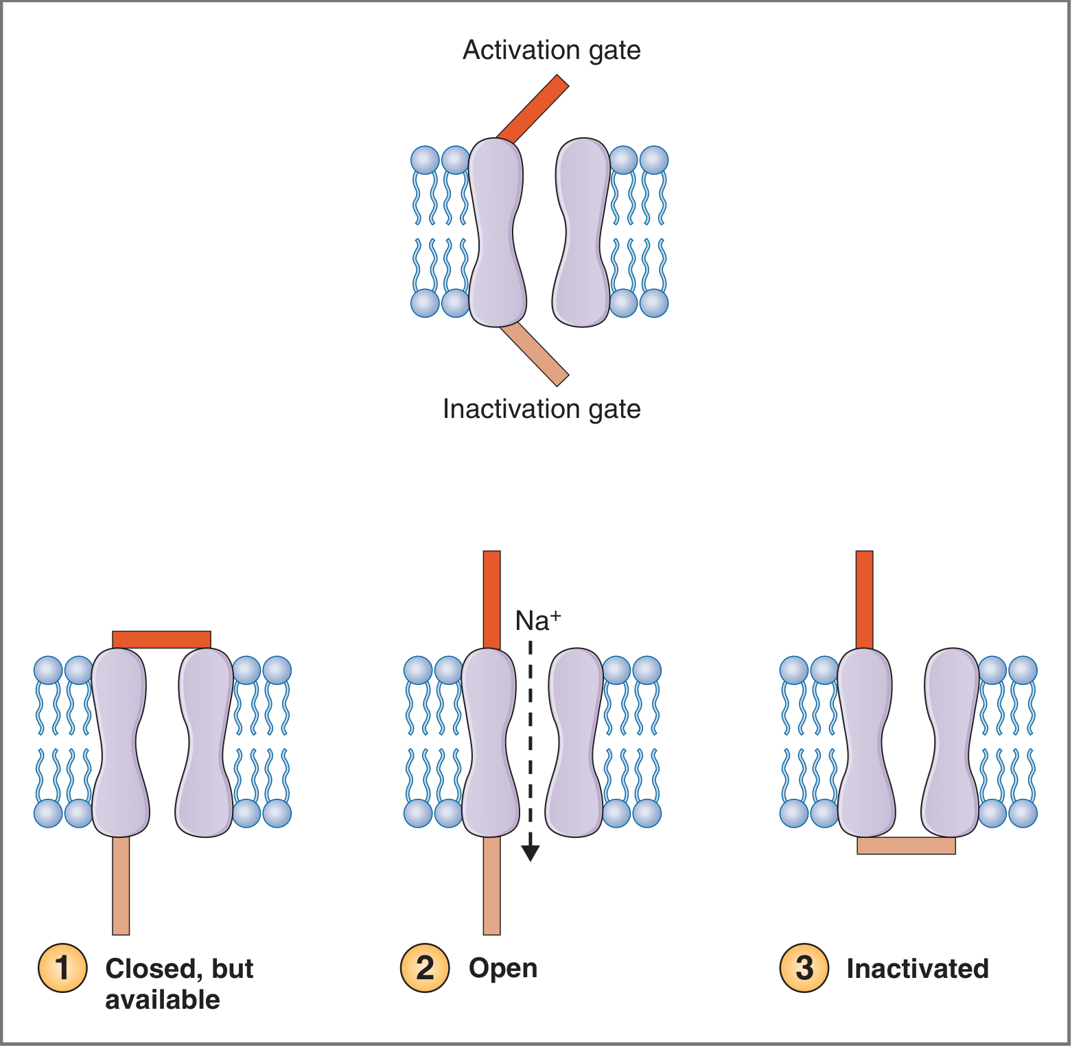
(Costanzo Physiology, 7th Edition)
| State | Activation Gate | Inactivation Gate | Na⁺ Flow | When |
|---|---|---|---|---|
| Closed, available | Closed | Open | None | Resting membrane potential |
| Open | Open | Open | Inward rush | Upstroke |
| Inactivated | Open | Closed | None | Peak/falling phase |
Recovery to the "closed, available" state requires repolarization back to near resting potential, which causes the inactivation gate to reopen.
5. Refractory Periods
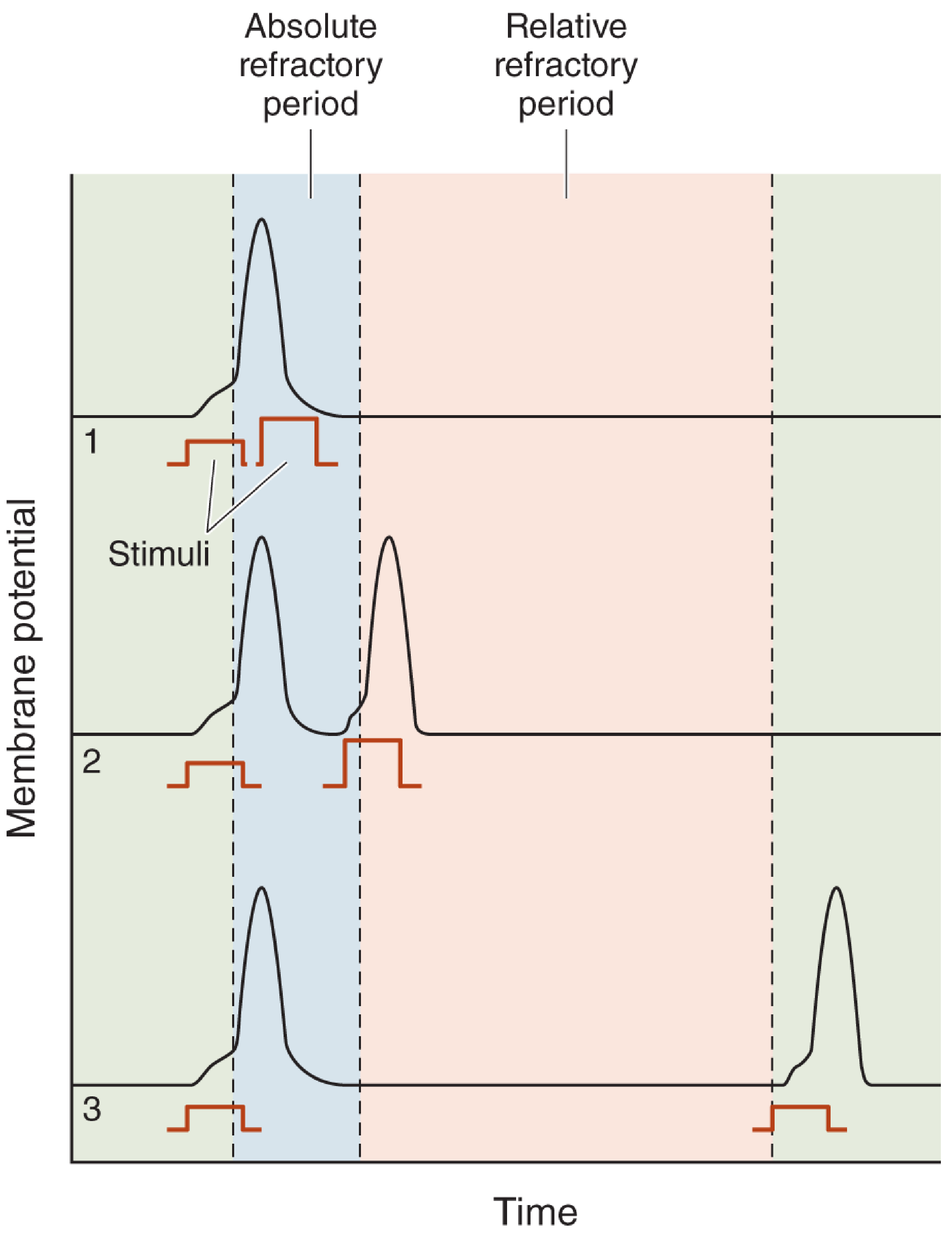
(Neuroscience: Exploring the Brain, 5th Edition)
- Absolute refractory period (~1 ms in large myelinated fibers): Na⁺ channels are inactivated. No stimulus of any strength can trigger a new action potential. This sets the maximum firing frequency (~1000 impulses/second).
- Relative refractory period (2-4 ms, during hyperpolarization): K⁺ channels are still open; the membrane is below resting potential. A new action potential can be triggered but requires a larger-than-normal stimulus.
The refractory period ensures unidirectional conduction - action potentials cannot travel backward through recently depolarized membrane.
6. Key Properties (All-or-None Law)
As described in Costanzo Physiology:
- All-or-none: The action potential either fires fully or not at all - there is no "partial" action potential. Stimulus strength is encoded in frequency, not amplitude.
- Stereotypical size and shape: Every normal action potential in a given cell type reaches the same peak voltage and has the same shape.
- Nondecremental propagation: Each successive patch of membrane is brought to threshold by local currents, regenerating the action potential at full amplitude at every point along the fiber.
7. Conduction Along the Axon
Action potentials spread by local circuit currents: the depolarized patch of membrane drives current into adjacent resting membrane, depolarizing it to threshold and triggering the next action potential.
Factors increasing conduction velocity:
- Larger axon diameter - lower internal resistance, faster current spread
- Myelination - myelin insulates internodal segments, restricting current to Nodes of Ranvier only (saltatory conduction, from Latin saltare = to jump). Myelinated A-α fibers conduct at up to 120 m/s; unmyelinated C fibers conduct at <2 m/s.
8. Pharmacological Relevance
| Drug/Toxin | Mechanism | Effect |
|---|---|---|
| Tetrodotoxin (TTX) | Blocks voltage-gated Na⁺ channels (puffer fish toxin) | Blocks action potential generation |
| Local anesthetics (lidocaine, procaine) | Block Na⁺ channel activation gates | Abolish nerve conduction |
| Tetraethylammonium (TEA) | Blocks voltage-gated K⁺ channels | Prolongs action potential, impairs repolarization |
| High extracellular Ca²⁺ | Stabilizes membrane, reduces Na⁺ permeability | Raises threshold, harder to fire |
Sources:
- Guyton and Hall Textbook of Medical Physiology, Chapter 5
- Costanzo Physiology, 7th Edition, Chapter 1
- Neuroscience: Exploring the Brain, 5th Edition, Chapter 4
For note short
Action Potential - Short Notes
Definition
Rapid reversal of membrane potential in excitable cells. Lasts ~1-2 ms.
Resting Membrane Potential
- -70 mV (inside negative)
- High K⁺ permeability, low Na⁺ permeability
- Maintained by Na⁺/K⁺-ATPase
Phases
| Phase | Voltage | Ion Event |
|---|---|---|
| Resting | -70 mV | K⁺ channels open |
| Depolarization | -70 → +35 mV | Na⁺ channels open → Na⁺ rushes IN |
| Overshoot | > 0 mV | Peak Na⁺ influx |
| Repolarization | +35 → -70 mV | Na⁺ channels inactivate + K⁺ channels open → K⁺ rushes OUT |
| Hyperpolarization (undershoot) | < -70 mV | K⁺ channels slow to close |
Threshold = ~-55 mV (must be reached to fire)
Na⁺ Channel States
- Closed/Available → resting (activation gate closed, inactivation gate open)
- Open → upstroke (both gates open)
- Inactivated → peak/falling phase (inactivation gate closes)
Refractory Periods
- Absolute (~1 ms): Na⁺ channels inactivated → NO AP possible at any stimulus strength
- Relative (2-4 ms): Hyperpolarization → AP possible only with stronger stimulus
Key Properties
- All-or-none law - fires fully or not at all
- Strength coded by frequency, not amplitude
- Nondecremental - same amplitude throughout propagation
Conduction Velocity
- ↑ axon diameter → ↑ velocity
- Myelination → saltatory conduction (jumps node to node) → fastest (~120 m/s)
- Unmyelinated C fibers → slowest (<2 m/s)
Key Drugs
| Drug | Action |
|---|---|
| Tetrodotoxin (TTX) | Blocks Na⁺ channels → no AP |
| Lidocaine | Blocks Na⁺ channels → local anesthesia |
| TEA | Blocks K⁺ channels → prolonged AP |
This is a shared conversation. Sign in to Orris to start your own chat.