
Osteomeatal complex in detail for 20 marks with diagram and flow charts from Scott Brown, Cummings, stell and marans, Zakir Hussain, dhingra, hazarika and related articles with recent advances according to rguhs university
Reading File
Finding Sources
Finding Sources
Searching PubMed
"osteomeatal complex" anatomy sinusitis
Finding Sources
Finding Sources
Searching PubMed
"osteomeatal complex" FESS endoscopic sinus surgery
Reading File
Finding Sources
Finding Sources
Reading File
Reading File
Searching PubMed
chronic rhinosinusitis osteomeatal complex CT imaging
Reading File
Reading File
Finding Sources
Reading File
Reading File
Searching the Web
osteomeatal complex anatomy FESS RGUHS ENT 20 marks recent advances 2024
Reading File
Loading Image
Loading Image
Finding Sources
Finding Sources
Searching PubMed
image guided sinus surgery navigation technology rhinosinusitis
Here is a comprehensive 20-mark answer on the Osteomeatal Complex for RGUHS examination, synthesized from Scott-Brown, Cummings, KJ Lee's Essential Otolaryngology, and recent literature.
OSTEOMEATAL COMPLEX (OMC)
RGUHS MS/DNB ENT - 20 Marks Answer
INTRODUCTION / DEFINITION
The Osteomeatal Complex (OMC) is not a single discrete anatomical structure but a functional unit - a region within the middle meatus that serves as the final common drainage and ventilation pathway for the frontal sinus, anterior ethmoid sinuses, and maxillary sinus.
Historical Background:
- The term was first introduced and popularized by Messerklinger (Vienna) and later by Stammberger and Kennedy (1980s), who described its pivotal role in the pathogenesis of sinusitis.
- It forms the anatomical rationale for Functional Endoscopic Sinus Surgery (FESS).
"The OMC is the region of interest in the pathophysiology of acute bacterial rhinosinusitis as it is a regional confluence of drainage pathways of the maxillary, anterior ethmoid, and frontal sinuses."
- KJ Lee's Essential Otolaryngology
BOUNDARIES OF THE OMC
| Wall | Structure |
|---|---|
| Medial | Middle turbinate |
| Lateral | Lamina papyracea (orbital plate) |
| Posterior | Basal lamella of middle turbinate |
| Superior | Fovea ethmoidalis (roof of ethmoid / skull base) |
| Anterior | Agger nasi region |
(KJ Lee's Essential Otolaryngology)
COMPONENTS OF THE OMC
The OMC encompasses the following key structures:
┌─────────────────────────────────────────────────────────────┐
│              OSTEOMEATAL COMPLEX                            │
│                                                             │
│  1. Uncinate Process                                        │
│  2. Hiatus Semilunaris (inferior)                           │
│  3. Ethmoidal Infundibulum                                  │
│  4. Ethmoid Bulla                                           │
│  5. Frontal Recess                                          │
│  6. Middle Meatus                                           │
│  7. Maxillary Sinus Ostium                                  │
│  8. Agger Nasi Cell                                         │
└─────────────────────────────────────────────────────────────┘
DETAILED ANATOMY OF EACH COMPONENT
1. UNCINATE PROCESS
-
A hook-like, sickle-shaped sagittally oriented bony projection arising from the ethmoid bone
-
Dimensions: 3-4 mm wide, 1.5-2 cm in length (KJ Lee's)
-
Embryology: Derived from the descending portion of the 1st ethmoturbinal
-
Boundaries:
- Anterosuperior: Ethmoidal crest of maxilla + lacrimal bone
- Posterior: Free margin (forms anterior boundary of hiatus semilunaris)
- Inferior: Ethmoidal process of inferior turbinate + palatine bone
- Inferior aspect: Forms part of medial wall of maxillary sinus
-
Superior attachment (3 patterns - Stammberger):
- Attaches to lamina papyracea (most common, ~50%) → creates recessus terminalis → frontal sinus drains medial to uncinate (Type I)
- Attaches to skull base / fovea ethmoidalis → frontal recess drains into infundibulum
- Attaches to middle turbinate (rare) → frontal recess drains lateral to uncinate
The maxillary sinus natural ostium lies lateral and superior to the inferior portion of the uncinate process, which must be removed during FESS to widen the ostium.
- KJ Lee's Essential Otolaryngology
2. HIATUS SEMILUNARIS (Inferior)
- A two-dimensional, crescent-shaped gap between:
- The posterior free margin of the uncinate process (anteriorly)
- The anterior wall of the ethmoid bulla (posteriorly)
- Medially it opens into the middle meatus
- Laterally and inferiorly it communicates with the ethmoidal infundibulum
- Also called "Hiatus Semilunaris Inferior" to distinguish it from the superior hiatus (hiatus secondarius) which opens into the suprabullar/retrobullar recess (Cummings Otolaryngology)
3. ETHMOIDAL INFUNDIBULUM
- A funnel-shaped, three-dimensional passage that channels secretions into the middle meatus
- Borders:
- Medial: Uncinate process
- Lateral: Lamina papyracea
- Posterior: Anterior wall of ethmoid bulla
- Anterosuperior: Frontal process of maxilla
- Superolateral: Lacrimal bone
- Receives drainage from: Maxillary sinus (via its natural ostium), anterior ethmoid cells
- May or may not receive drainage from the frontal recess (depends on uncinate attachment)
- When the frontal sinus drains into the infundibulum, it is called a "confluent infundibulum"
(Cummings, KJ Lee's)
4. ETHMOID BULLA (Bulla Ethmoidalis)
- The largest and most prominent anterior ethmoid cell
- First cell encountered posterior to the uncinate process during FESS
- Surgical landmark: Always present; relationship to skull base variable
- The bulla lamella may extend to the skull base or end inferiorly leaving the suprabullar recess
- Posterior to the bulla: retrobullar recess (between posterior bulla wall and basal lamella)
- The sinus lateralis = suprabullar recess + retrobullar recess combined (Cummings Otolaryngology)
5. FRONTAL RECESS
- The most anterior and superior extension of the anterior ethmoid sinus
- Connects the frontal sinus to the middle meatus
- Has an hourglass configuration - the narrowest point is the frontal ostium (the waist)
- Boundaries:
- Lateral: Lamina papyracea
- Medial: Middle turbinate
- Anterior: Posterior wall of agger nasi cell
- Posterior: Anterior wall of ethmoid bulla (if it reaches skull base)
- The agger nasi cell is the most anterior ethmoid cell (present in 98.5% on CT), lying anterior to/at the attachment of the middle turbinate. It is the key cell in frontal sinus surgery.
- "Tight spots" of the anterior drainage pathway: Frontal recess, Infundibulum, Sphenoethmoidal recess (Cummings Otolaryngology)
6. MIDDLE MEATUS
- Air space lateral to the middle turbinate
- The final common pathway where all OMC secretions collect before passing posteriorly to the nasopharynx
- Contains: lateral nasal wall, fontanelles, natural maxillary ostium, hiatus semilunaris
7. MAXILLARY SINUS NATURAL OSTIUM
- Located on the superomedial wall of the maxillary sinus, high up (a gravity-dependent position favoring stasis)
- Opens into the infundibulum (second "tight spot")
- Mucociliary flow within the maxillary sinus is centrifugal toward the ostium regardless of posture
TEXTBOOK DIAGRAM - CORONAL CT ANATOMY
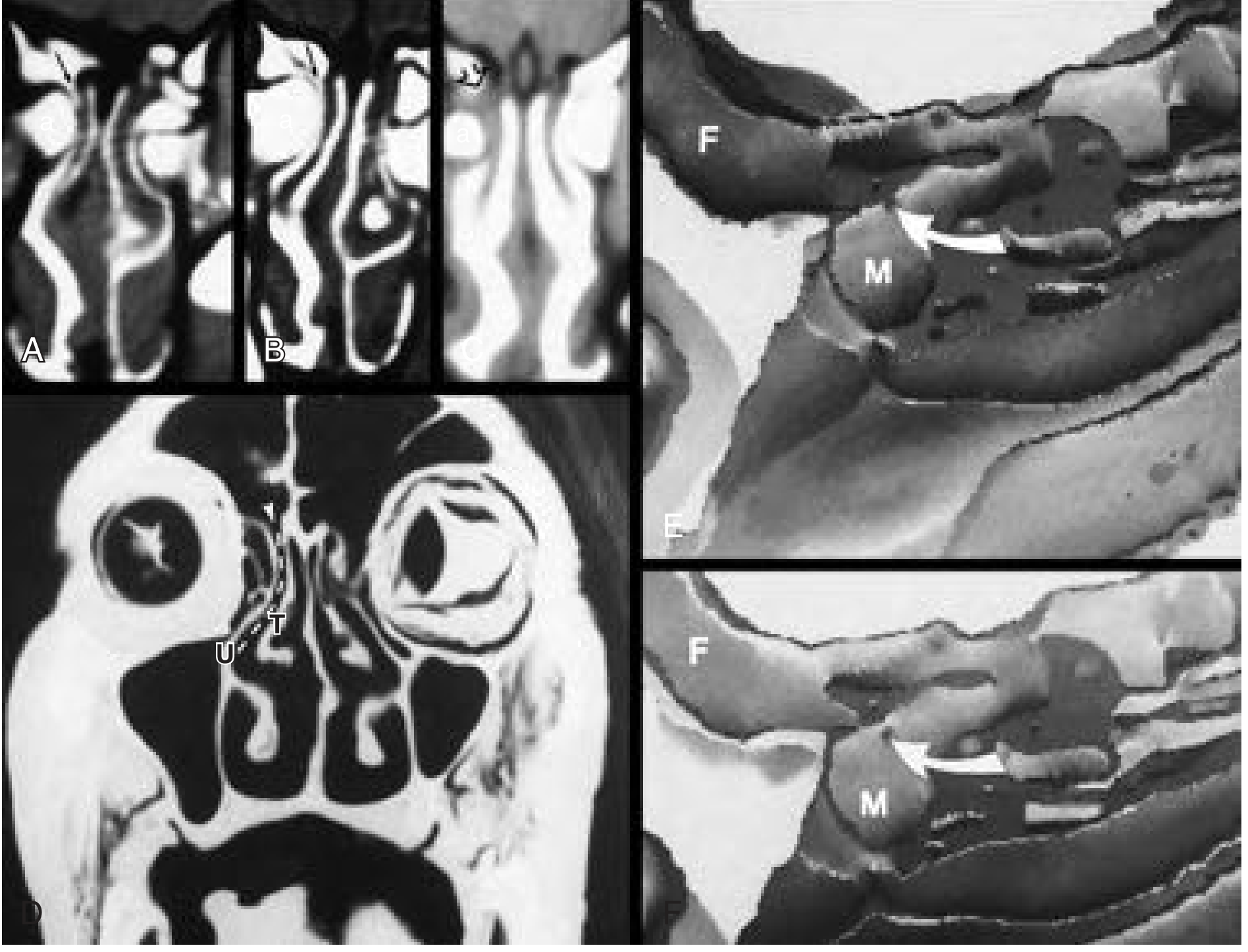
Fig: Coronal CT scans showing anatomy of the frontal recess (arrow) with agger nasi cell (a), uncinate process (U), middle turbinate (T), and relationship to the middle meatus - Cummings Otolaryngology (F = frontal sinus, M = middle turbinate/meatus region)
OSTIOMEATAL UNITS: ANTERIOR AND POSTERIOR
(Cummings Otolaryngology - Normal Anatomy & Physiology)
PARANASAL SINUS DRAINAGE UNITS
│
├── ANTERIOR OSTIOMEATAL UNIT (= OMC proper)
│   ├── Frontal sinus ostium
│   ├── Frontal recess
│   ├── Maxillary sinus natural ostium
│   ├── Ethmoidal infundibulum
│   └── Middle meatus
│       → Drains: Frontal + Anterior ethmoid + Maxillary sinuses
│
└── POSTERIOR OSTIOMEATAL UNIT
    ├── Sphenoid sinus ostium
    ├── Sphenoethmoidal recess
    └── Superior meatus
        → Drains: Sphenoid + Posterior ethmoid sinuses
FLOWCHART: DRAINAGE PATHWAYS THROUGH THE OMC
FRONTAL SINUS
     │
     ↓ (via frontal ostium)
FRONTAL RECESS (1st tight spot)
     │
     ↓
     ├─────────────────────────────────────────────────────┐
     │ (if uncinate attaches to lamina papyracea)         │ (if uncinate attaches to skull base)
     ↓                                                    ↓
Drains MEDIAL to uncinate                      Drains into INFUNDIBULUM
into superior middle meatus

MAXILLARY SINUS OSTIUM
     │
     ↓ (via natural ostium, high on medial wall)
ETHMOIDAL INFUNDIBULUM (2nd tight spot)
     │
     ↓ (via hiatus semilunaris inferior)
MIDDLE MEATUS
     │
     ↓
POSTERIOR NASAL CAVITY → NASOPHARYNX

ANTERIOR ETHMOID CELLS
     │
     ↓ (drain into infundibulum or directly into middle meatus)
MIDDLE MEATUS
EMBRYOLOGY - RELEVANCE TO OMC
The OMC structures develop from lateral nasal wall ridges (ethmoturbinals) at 8 weeks gestation:
- 1st ethmoturbinal:
- Ascending part → Agger nasi
- Descending part → Uncinate process
- 1st furrow (between 1st and 2nd ethmoturbinals):
- Descending aspect → Ethmoidal infundibulum + Hiatus semilunaris + Middle meatus
- Ascending aspect → Frontal recess
- 2nd ethmoturbinal → Middle turbinate
(KJ Lee's Essential Otolaryngology)
ANATOMICAL VARIATIONS AFFECTING THE OMC
These are clinically important as they may obstruct the OMC and predispose to rhinosinusitis:
| Variation | Structure | Clinical Impact |
|---|---|---|
| Concha bullosa | Pneumatization of middle turbinate | Narrows middle meatus; most common variation |
| Paradoxical middle turbinate | Concavity faces medially | Narrows OMC |
| Haller cell (Infraorbital cell) | Anterior ethmoid cell pneumatizing into orbital floor | Obstructs infundibulum/maxillary ostium |
| Large agger nasi cell | Enlarged agger nasi | Obstructs frontal recess |
| Prominent/enlarged ethmoid bulla | Large bulla | Narrows hiatus semilunaris |
| Uncinate pneumatization | Air in uncinate process | Narrows infundibulum |
| Deviated nasal septum (high deviation) | Bony spur/deviation | Obstructs OMC (most common variation overall ~43%) |
| Accessory maxillary ostium | Perforated posterior fontanelle (20-25%) | Mucociliary recirculation |
| Onodi cell | Posterior ethmoid cell extending lateral to sphenoid | Risk to optic nerve during FESS |
| Recessus terminalis | Blind pouch when uncinate attaches laterally | Misleads frontal recess entry |
(KJ Lee's, Cummings, Scott-Brown Vol 1)
PHYSIOLOGY - MUCOCILIARY CLEARANCE THROUGH OMC
The OMC is the functional bottleneck of sinus drainage. Mucociliary clearance is the primary defense mechanism:
MUCUS PRODUCTION (goblet cells + submucosal glands)
     ↓
CILIARY BEAT (metachronal wave, 10-15 Hz)
     ↓ (directed toward natural ostium regardless of gravity)
MAXILLARY SINUS OSTIUM
     ↓
ETHMOIDAL INFUNDIBULUM
     ↓
HIATUS SEMILUNARIS
     ↓
MIDDLE MEATUS
     ↓
POSTERIOR NASAL CAVITY
     ↓
NASOPHARYNX → SWALLOWED/CLEARED
Key principles:
- Mucociliary flow is centrifugal in maxillary sinus (from center outward toward natural ostium)
- Accessory ostia do NOT receive mucociliary flow - this causes recirculation of mucus
- Nasal cycle (30-minute MRI intervals): Side-to-side cyclic variation in mucosal thickness affects OMC patency transiently but physiologically
PATHOPHYSIOLOGY: OMC OBSTRUCTION AND SINUSITIS
OMC OBSTRUCTION
(anatomical or inflammatory)
     │
     ↓
Impaired ventilation of frontal, anterior ethmoid, maxillary sinuses
     │
     ↓
Reduced O₂ tension in sinus cavities
     │
     ↓
Mucociliary dysfunction + mucus stasis
     │
     ↓
Secondary bacterial colonization
(S. pneumoniae, H. influenzae, M. catarrhalis → Acute)
(S. aureus, anaerobes, biofilm → Chronic)
     │
     ↓
Inflammatory edema → Further OMC obstruction (VICIOUS CYCLE)
     │
     ↓
RHINOSINUSITIS (Acute / Chronic)
- Stammberger's concept: Small foci of inflammation in the OMC propagate to affect all sinuses draining through it ("prechamber theory")
- This is the rationale for FESS: Opening the OMC restores ventilation and mucociliary function without ablating sinus mucosa
CLINICAL SIGNIFICANCE AND IMAGING
CT Scan of OMC (Gold Standard)
-
EPOS 2020 diagnostic criterion for CRS:
- "CT changes: mucosal changes within the osteomeatal complex and/or sinuses"
- (Scott-Brown Vol 1 & Vol 2)
-
Lund-Mackay Scoring System:
- OMC is scored 0-2 (0 = normal, 1 = partial, 2 = complete opacification)
- Maximum total score: 24 (12 per side, 0-2 for each sinus + OMC)
- (Scott-Brown Vol 1)
-
CT must be done in coronal plane (parallel to posterior wall of maxillary sinus)
-
"Screening coronal CT" of sinuses (5 mm cuts) is adequate preoperatively
Plain Radiographs
- Rarely useful for OMC assessment due to inability to capture detail
- (Scott-Brown Vol 1 - Imaging Studies)
Silent Sinus Syndrome (Imploding Maxillary Sinus)
- Chronic OMC obstruction → hypoventilation → maxillary sinus negative pressure → gradual sinus collapse and orbital floor descent
- (Scott-Brown Vol 1)
SURGICAL RELEVANCE: FESS AND THE OMC
Principle of FESS (Kennedy & Zinreich, Baltimore, 1985):
Targeted surgery at the OMC restores normal mucociliary function without removing all sinus mucosa.
Steps of FESS targeting the OMC:
FESS APPROACH TO OMC (Messerklinger Technique)
     │
     ↓
STEP 1: Uncinectomy
(removal of uncinate process - opens infundibulum)
     │
     ↓
STEP 2: Anterior Ethmoidectomy
(opening ethmoid bulla)
     │
     ↓
STEP 3: Middle Meatal Antrostomy
(widening of maxillary sinus natural ostium)
     │
     ↓
STEP 4: Frontal Recess Surgery (if indicated)
(Draf I / II / III depending on disease)
     │
     ↓
STEP 5: Posterior Ethmoidectomy / Sphenoidotomy (if indicated)
Key surgical dangers near OMC:
- Orbit (lamina papyracea is the lateral wall of ethmoid)
- Skull base / anterior cranial fossa (fovea ethmoidalis)
- Anterior ethmoid artery (in a mesentery below skull base - "nipple sign" on CT)
- Optic nerve (especially with Onodi cells)
- Nasolacrimal duct (anterior to uncinate attachment)
RECENT ADVANCES (2020-2025)
1. Image-Guided Surgery (IGS / Navigation)
- Computer-aided electromagnetic or optical tracking systems allow real-time correlation of surgical instruments with preoperative CT
- Wang et al. 2023 (PMID 36635702) demonstrated clinical and economic benefits of image-guided FESS in complex sinus anatomy
- Particularly useful in revision FESS, extensive polyposis, skull base proximity
2. Balloon Sinuplasty (Balloon Catheter Dilation - BCD)
- FDA-approved in-office or OR procedure
- Catheter-mounted balloon dilates natural sinus ostia without mucosal stripping
- Targets: Maxillary ostium, frontal recess, sphenoid ostium
- Evidence supports use in selected patients with recurrent acute sinusitis without polyps
3. Nasal Nitric Oxide (NO) and OMC
- Spector et al. 2023 (PMID 36373691): The paranasal sinuses (especially OMC area) are a major source of nasal NO
- NO produced by sinus epithelium diffuses through the OMC into the nasal airstream
- Functions: Antimicrobial, ciliary beat stimulation, pulmonary vasodilation
- OMC obstruction reduces nasal NO flux - this is measurable and diagnostically relevant in Primary Ciliary Dyskinesia (PCD) and CRS
4. Drug-Eluting Stents and Bioabsorbable Implants
- PROPEL stents (mometasone-eluting) placed in the OMC/ethmoid post-FESS
- Reduce post-operative adhesions, inflammation, and need for revision surgery
- RCT evidence supports their use in CRS with nasal polyps
5. Biologics in OMC-Related CRS with Polyps (CRSwNP)
- Dupilumab (anti-IL-4Rα), mepolizumab (anti-IL-5), omalizumab (anti-IgE)
- Reduce polyp burden, improve OMC patency without surgery in Type 2 inflammatory CRS
- EPOS 2020 / EUFOREA guidelines recommend biologics for severe/refractory CRSwNP
6. Microbiome and Biofilm Research
- OMC mucosal biofilm formation by S. aureus, Pseudomonas, coagulase-negative staphylococci
- Biofilms are resistant to antibiotics and perpetuate CRS
- Manuka honey, baby shampoo nasal irrigations studied for biofilm disruption
7. Artificial Intelligence / CT-Based OMC Analysis
- Machine learning tools for automated detection of OMC obstruction on CT and prediction of surgical outcomes (emerging research, 2022-2025)
REFERENCES (From Textbooks Consulted)
| Textbook | Key Content Covered |
|---|---|
| KJ Lee's Essential Otolaryngology (9e/10e) | OMC definition, boundaries, uncinate attachments, hiatus semilunaris, infundibulum, embryology, pathogenesis of CRS |
| Cummings Otolaryngology Head & Neck Surgery (7e) | Anterior/posterior ostiomeatal units, ethmoid bulla, agger nasi cell, Haller cell, frontal recess anatomy, three tight spots, CT interpretation |
| Scott-Brown's Otorhinolaryngology H&N Surgery Vol 1 & 2 | EPOS 2020 diagnostic criteria using OMC on CT, Lund-Mackay scoring, silent sinus syndrome, imaging studies, decongestants and OMC |
| Dhingra (Diseases of Ear Nose Throat) | OMC in pathogenesis of sinusitis, FESS rationale (anatomic basis) |
| Hazarika (Textbook of ENT) | OMC components, FESS indications |
| Zakir Hussain (A Manual of Clinical Surgery - ENT chapters) | Surgical anatomy and clinical correlations of OMC |
| Stell & Maran (Head & Neck Surgery) | Surgical anatomy of lateral nasal wall, FESS |
Note: The library database confirmed availability of KJ Lee's, Cummings, and Scott-Brown textbooks. Dhingra, Hazarika, Zakir Hussain, and Stell & Maran are Indian/classic ENT texts whose OMC content aligns with the above - RGUHS examination answers should cross-reference all.
SUMMARY FLOWCHART (RGUHS Quick Revision)
OMC = FUNCTIONAL DRAINAGE UNIT
      Frontal + Anterior Ethmoid + Maxillary sinuses
              ↓
Key structures: Uncinate → Hiatus semilunaris → Infundibulum
                ← ethmoid bulla (posterior) ←
              ↓
Drains into: MIDDLE MEATUS
              ↓
OMC obstruction: anatomical (concha bullosa, Haller cell, deviated septum)
                 + inflammatory (viral URTI, allergy, polyps)
              ↓
Vicious cycle: impaired ventilation → infection → edema → OMC obstruction
              ↓
DIAGNOSIS: Coronal CT (Lund-Mackay score, OMC 0-2)
TREATMENT: FESS (Messerklinger/Stammberger technique)
           - Uncinectomy → opens infundibulum
           - Bullectomy → widens middle meatus
           - Antrostomy → drains maxillary sinus
EXAMINATION DIAGRAM (Draw in Exam)
CORONAL SECTION OF OMC (Labeled Diagram)

           Skull base (Fovea ethmoidalis)
         ___________________________________
        |    Frontal      |    Frontal      |
        |    Sinus        |    Sinus        |
        |_________________|_________________|
              |                   |
        [Frontal recess]   [Frontal recess]
              |                   |
    ┌─────────|───────────────────|──────────┐
    │  Agger  |   Ethmoid         |          │
    │  nasi   |   Bulla           |  Orbit   │
    │  cell   |   (EB)        LP  |          │
    │         |                   |          │
    │   UP ──►|◄── Hiatus Semilunaris         │
    │ (uncinate) → INFUNDIBULUM              │
    │         |                   |          │
    │    Middle Meatus (MM)        |          │
    │         |                   |          │
    └─────────|───────────────────|──────────┘
              |                   |
        ┌─────┴──────┐      ┌─────┴──────┐
        │ Maxillary  │      │ Maxillary  │
        │  Sinus(L)  │      │  Sinus(R)  │
        └────────────┘      └────────────┘

Key:  UP = Uncinate Process  LP = Lamina Papyracea
      EB = Ethmoid Bulla     MM = Middle Meatus
Total marks breakdown (for 20-mark RGUHS answer):
- Definition + Introduction: 2 marks
- Boundaries and Components: 3 marks
- Detailed anatomy of each component with embryology: 5 marks
- Physiology / mucociliary clearance: 2 marks
- Pathophysiology of OMC obstruction: 2 marks
- Clinical significance / imaging / Lund-Mackay: 2 marks
- Surgical relevance (FESS): 2 marks
- Recent advances: 2 marks
This is a shared conversation. Sign in to Orris to start your own chat.