Here is everything — all 20 MCQs, full cheatsheet, hints, and explanations — right here in chat.
⚡ RAPID REVISION CHEATSHEET — PAGE 1 of 2
Development of Urinary Bladder & Urethra
Batterjee Medical College Jeddah — Embryology Block
🧫 Cloaca Division (Weeks 4–7)
| Structure | Derived Part |
|---|
| Divider | Urorectal septum (MESODERMAL) |
| Anterior cloaca | Urogenital sinus |
| Posterior cloaca | Primitive rectum |
| Cloacal membrane (anterior) | Urogenital membrane |
| Cloacal membrane (posterior) | Anal membrane |
🫧 Urogenital Sinus — Subdivisions
- Above mesonephric duct openings = Vesicourethral canal
- Below mesonephric duct openings = Definitive urogenital sinus
- Cranial = pelvic part
- Caudal = phallic part
- Allantois opens at cranial end of vesicourethral canal
🔺 Urinary Bladder Origins
| Layer | Origin |
|---|
| Epithelium (whole bladder) | Endoderm (vesicourethral canal) |
| Trigone epithelium | MESODERM (absorbed mesonephric ducts) → later replaced by endoderm |
| Muscle + serosa | Splanchnopleuric intraembryonic mesoderm |
| Bladder onset | Week 5 IUL |
🔗 Urachus & Anomalies
| Anomaly | What's Open | Presentation |
|---|
| Urachal fistula | Entire lumen | Urine from umbilicus |
| Urachal cyst | Middle segment only | Cystic swelling, NO urine, NO communication |
| Urachal sinus | Upper part (near umbilicus) | Drains at umbilicus only |
| Patent urachus | Full, continuous with bladder | Urine from umbilicus |
| Adult name | — | Median umbilical ligament |
⚡ Median = urachus (1 structure, midline) | Medial = obliterated umbilical arteries (2 structures) | Lateral fold = inferior epigastric vessels
🚨 Exstrophy of Bladder
- Cause: Failure of lateral body wall folds to close in midline (pelvic/hypogastric region)
- Result: Posterior bladder wall exposed, no anterior abdominal wall between umbilicus and genital tubercle
- Always with: Epispadias (dorsal) — NEVER hypospadias (ventral)
- Incidence: 2 per 10,000 live births
⚡ RAPID REVISION CHEATSHEET — PAGE 2 of 2
Male & Female Urethra + Adrenal Glands
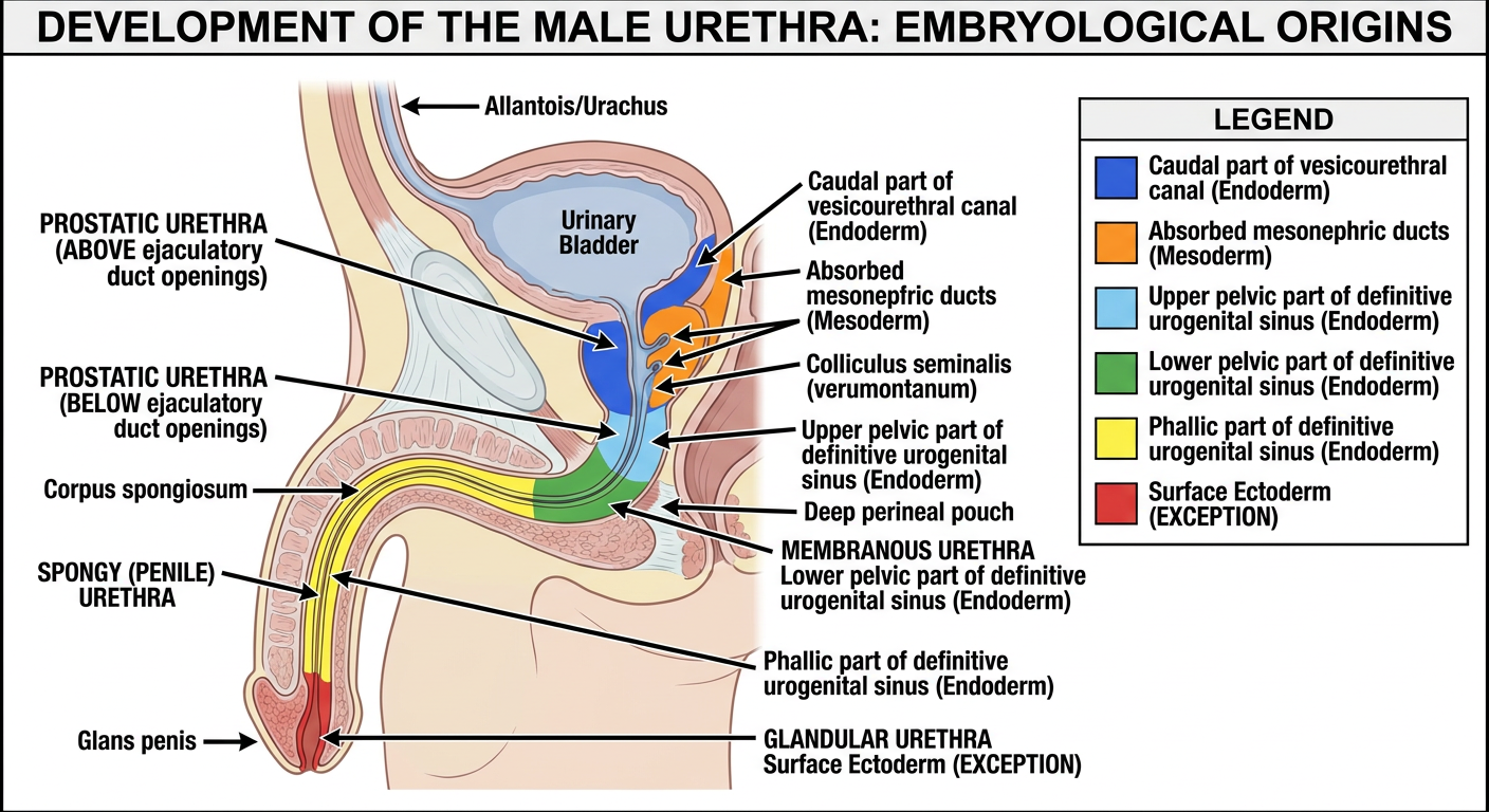
♂ Male Urethra — Origins Map
| Part | Source |
|---|
| Prostatic urethra ABOVE ejac. ducts | Caudal vesicourethral canal; posterior wall = mesoderm |
| Prostatic urethra BELOW ejac. ducts | Upper pelvic part of definitive UG sinus |
| Membranous urethra | Lower pelvic part of definitive UG sinus (in deep perineal pouch) |
| Spongy urethra | Phallic part of definitive UG sinus |
| Glandular urethra (glans penis) | Surface ECTODERM ← only exception |
⚡ All urethra = endoderm. The ONE exception = glandular (terminal) part = ectoderm
♀ Female Urethra
| Part | Source |
|---|
| Major part | Caudal vesicourethral canal (endoderm) |
| Terminal part | Pelvic part of definitive UG sinus (endoderm) |
| Phallic part forms | Vestibule of vagina |
| Dorsal wall | Mesoderm (absorbed mesonephric ducts) — exception |
| Equivalent in male | Prostatic urethra ABOVE colliculus seminalis |
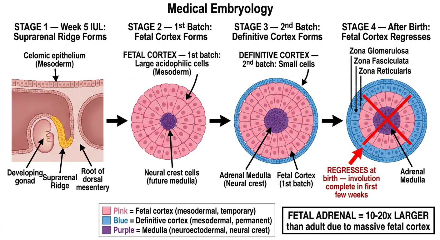
🔴 Adrenal Gland — Origins (Most Tested)
| Part | Origin | Type |
|---|
| Cortex | Celomic epithelium | MESODERM |
| Medulla | Neural crest (sympathochromaffin cells) | NEUROECTODERM |
Cortex Development — Two Waves:
- 1st batch = Large ACIDOPHILIC cells = Fetal cortex → surrounds medulla → REGRESSES after birth
- 2nd batch = Small cells = Definitive cortex → surrounds fetal cortex → persists for life
Medulla Development:
- Neural crest cells migrate and enter fetal cortex from the MEDIAL side
- Cells = sympathochromaffin cells (similar to postganglionic sympathetic neurons)
- Onset = Week 5 IUL
- Fetal adrenal gland = 10–20× LARGER than adult adrenal gland
🧬 Master Quick-Reference Table
| Structure | Origin |
|---|
| UB epithelium | Endoderm |
| UB trigone | Mesoderm |
| UB muscle/serosa | Splanchnopleuric mesoderm |
| Urorectal septum | Mesoderm |
| Adrenal cortex | Mesoderm (celomic epi) |
| Adrenal medulla | Neuroectoderm (neural crest) |
| Glandular urethra | Ectoderm (surface) |
| All other urethra | Endoderm |
🧠 Memory Mnemonics
"MEN Cause Trigone" → Mesonephric duct = Mesoderm = Trigone
"CAM" → Cortex = celomic (Mesoderm); medullA = neural crest (ectoderm)
"GLANDS = GLANS" → terminal urethra in GLANS penis = ectoderm
"Median = Midline = Mine alone" → Median umbilical lig. = urachus (one structure)
⚡ THE 8 GOLDEN TRICKY POINTS
- Trigone = mesodermal (NOT endodermal) — #1 most repeated trap
- Urorectal septum = mesodermal (not ectodermal)
- Adrenal cortex = mesoderm; medulla = neuroectoderm
- Fetal adrenal = 10–20× larger than adult
- Female urethra = prostatic urethra above colliculus seminalis
- Glandular urethra = ectoderm (only exception)
- Urachal fistula = urine from umbilicus; urachal cyst = closed, no urine
- Fetal cortex regresses at birth; definitive cortex persists
📝 ALL 20 HIGH-YIELD MCQs
With Tricky Options · Hints · Full Explanations · Past Year Frequency
PART 1: DEVELOPMENT OF THE URINARY BLADDER
MCQ 1 🔥 CRITICAL — Repeated 3× (2022, 2024)
The cloaca is divided into the urogenital sinus and primitive rectum by which structure?
- A. Urorectal septum ✅
- B. Cloacal membrane (tricky — students confuse "divides cloaca" with "gets divided")
- C. Paramesonephric duct
- D. Urogenital fold
✓ Answer: A. Urorectal septum
💡 Hint: "URO-RECTAL" septum — the name tells you it divides the UROgenital sinus from the RECTum. The cloacal membrane is what GETS divided into two parts (urogenital and anal membranes) — it does not do the dividing.
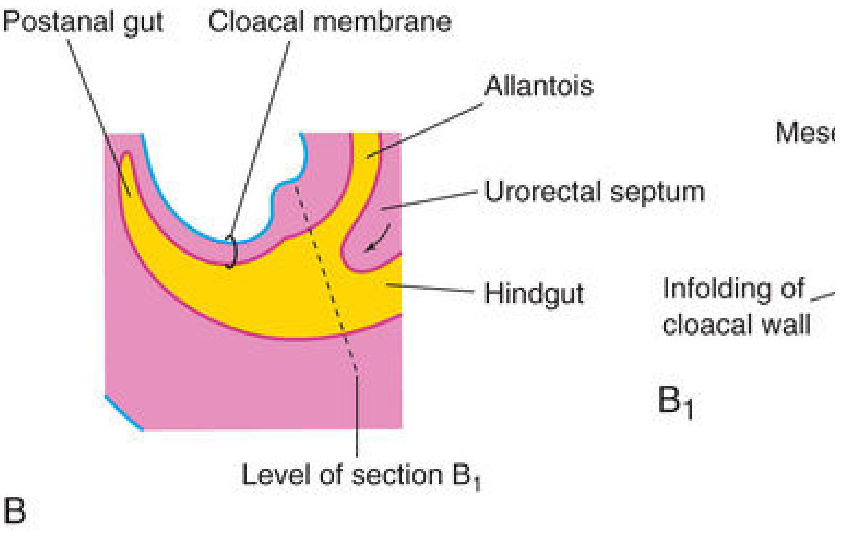
Explanation: The urorectal septum is a wedge of mesoderm growing caudally between the allantois (ventral) and hindgut (dorsal). By week 7 it divides the cloaca into: (1) anterior urogenital sinus and (2) posterior primitive rectum. The cloacal membrane is simultaneously divided into the anterior urogenital membrane and posterior anal membrane.
Why B is wrong: The cloacal membrane is at the caudal end of the embryo — it gets divided BY the urorectal septum; it does not do the dividing. This is the #1 trap because students hear "divides the cloaca" and think of the membrane.
Fig. 11.25B — Urorectal septum (yellow wedge) dividing cloaca. Note the allantois (ventral) and hindgut (dorsal). Source: The Developing Human, Moore 10th Ed.
MCQ 2 🔥 CRITICAL — Appeared 2023
The urorectal septum that divides the cloaca is derived from which embryological layer?
- A. Endoderm (tricky — endodermal signaling initiates it, but the septum itself isn't endoderm)
- B. Mesoderm ✅
- C. Neural crest
- D. Surface ectoderm (tricky)
✓ Answer: B. Mesoderm
💡 Hint: The septum is a "wedge of mesenchyme" — mesenchyme is always mesodermally derived. WALLS and DIVIDERS in the embryo are usually mesodermal (like the muscular coat of gut). Endodermal β-catenin signaling INITIATES its formation, but the structure itself is mesodermal.
Explanation: Both Langman's and Moore's explicitly call it a "wedge of mesenchyme." Mesenchyme = embryonic connective tissue from mesoderm. Endodermal β-catenin is required for its FORMATION (signaling molecule), but the septum itself is mesodermal. Neural crest forms adrenal medulla, melanocytes, peripheral ganglia.
MCQ 3 🔶 HIGH — Appeared 2023
The vesicourethral canal is the portion of the urogenital sinus located:
- A. Below the openings of mesonephric ducts (tricky — that's the definitive UG sinus)
- B. Above the openings of mesonephric ducts ✅
- C. Between the two ureteric bud openings (tricky — that describes the trigone)
- D. Caudal to the phallic part
✓ Answer: B. Above the openings of mesonephric ducts
💡 Hint: The mesonephric ducts act as a dividing line: ABOVE = vesicourethral canal (bladder territory), BELOW = definitive urogenital sinus (urethra territory).
Explanation: Once mesonephric ducts open into the primitive urogenital sinus, they create a landmark. Above = vesicourethral canal → (1) upper dilated part = urinary bladder + (2) lower narrow part = primitive urethra. Below = definitive UG sinus → cranial pelvic + caudal phallic parts.
MCQ 4 🔥 #1 MOST REPEATED — Appeared 2021, 2022, 2023, 2024
The epithelial lining of the trigone of the urinary bladder is derived from:
- A. Endoderm of urogenital sinus (classic wrong answer — students extrapolate "bladder = endoderm" to trigone)
- B. Mesoderm (absorbed mesonephric ducts) → later replaced by endoderm ✅
- C. Paramesonephric (Müllerian) ducts
- D. Splanchnopleuric mesoderm (tricky — that's for muscle/serosa, not epithelium)
✓ Answer: B. Mesoderm (mesonephric ducts)
💡 THE GOLDEN RULE: Everything in the bladder is endodermal EXCEPT the TRIGONE, which starts MESODERMAL. Langman's states: "the mucosa of the trigone is mesodermal. With time, the mesodermal lining of the trigone is replaced by endodermal epithelium."
Explanation: The trigone forms from the absorbed portions of the mesonephric ducts (Wolffian ducts) that get incorporated into the posterior wall of the vesicourethral canal. Both mesonephric ducts and ureters originate from mesoderm, so the trigone is mesodermal. This mesodermal lining is eventually replaced by endodermal epithelium — so the adult bladder epithelium is entirely endodermal — but embryologically the trigone starts as mesodermal.
Why A is wrong: Students know the bladder is mostly endodermal and extrapolate this to the trigone. The trigone is the exception. This is the most repeated MCQ trap in this topic.
MCQ 5 🔶 HIGH — Appeared 2022
The muscular and serous coats of the urinary bladder wall are derived from:
- A. Endoderm of vesicourethral canal (tricky — that gives epithelium only)
- B. Mesonephric ducts (tricky — that gives trigone epithelium)
- C. Splanchnopleuric intraembryonic mesoderm ✅
- D. Somatic mesoderm (tricky — somatic mesoderm gives body wall structures)
✓ Answer: C. Splanchnopleuric intraembryonic mesoderm
💡 Hint: Rule — SPLANCHNO (visceral) mesoderm → forms SMOOTH MUSCLE + SEROSA of ALL hollow viscera (gut, bladder, bronchi). Same principle as gut development: endoderm = mucosa; splanchnic mesoderm = submucosa + muscularis + serosa.
Explanation: The intraembryonic mesoderm surrounding the endodermal gut tube is splanchnopleuric (visceral) mesoderm. It gives rise to smooth muscle, connective tissue, and serosa of all hollow viscera including the urinary bladder. Somatic mesoderm forms body wall structures (muscles, bones, dermis of body wall).
MCQ 6 🔥 CRITICAL — EXACT CASE IN YOUR LECTURE — Appeared 2024
A newborn male presents with continuous leakage of urine from the umbilicus shortly after birth. Ultrasound shows a tubular connection between the apex of the urinary bladder and the umbilicus. Which embryological structure failed to obliterate?
- A. Mesonephric duct (tricky — this was absorbed into the bladder wall)
- B. Urachus (allantois) ✅
- C. Ureteric bud (forms ureter, not related to umbilicus)
- D. Müllerian duct (tricky — forms female reproductive tract)
✓ Answer: B. Urachus (allantois)
💡 Hint: The ONLY structure connecting the bladder APEX to the UMBILICUS is the urachus. Patent urachus = urachal fistula = urine drains from navel. This is the exact case in your lecture PDF page 26.
Explanation: The urachus is the adult remnant of the allantois — normally obliterates to form the median umbilical ligament (bladder apex → umbilicus). If the entire urachus fails to obliterate = urachal fistula = full patency = urine from umbilicus. Tubular connection apex to umbilicus = patent urachus.
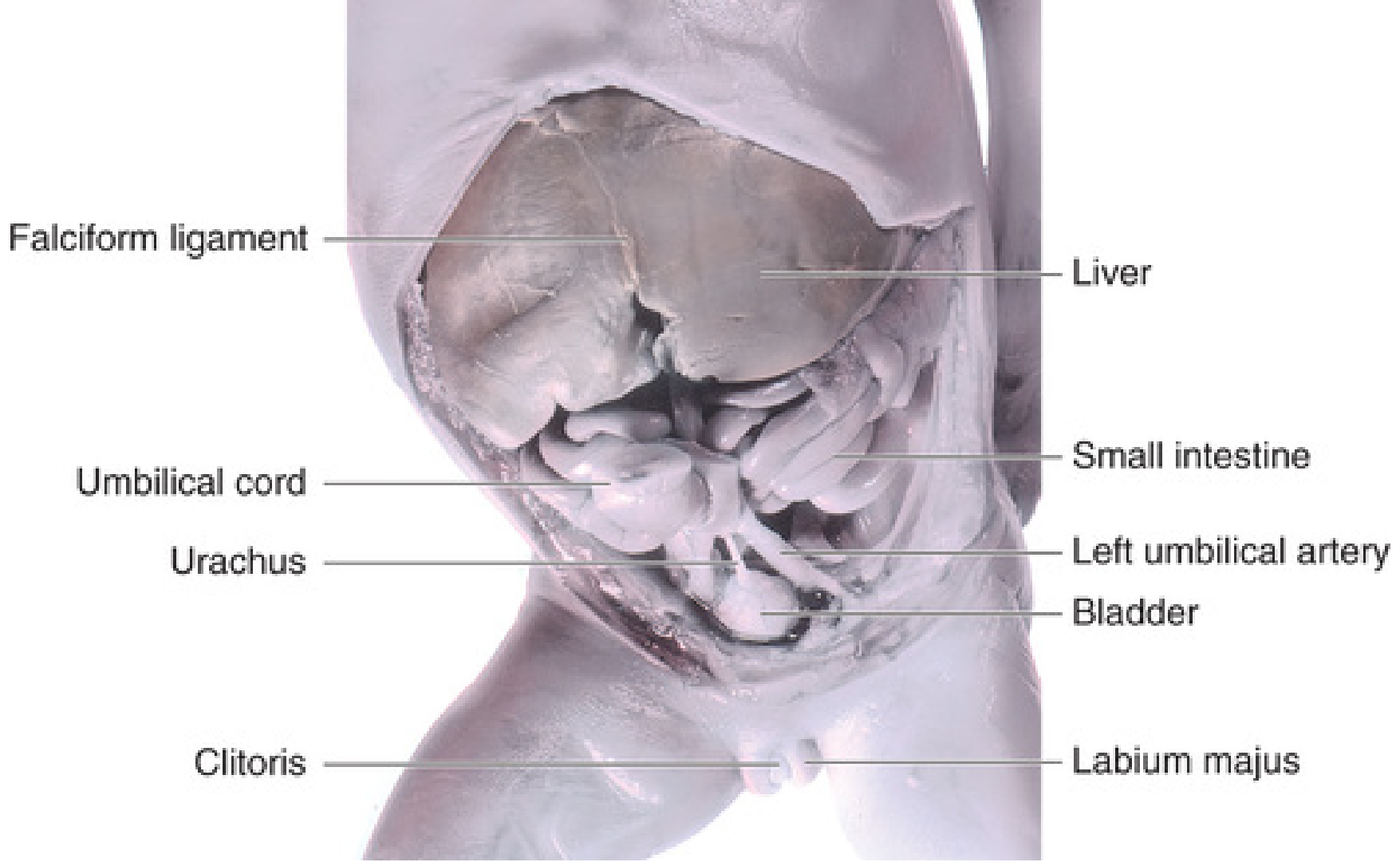
Fig. 12.21 — 18-week female fetus dissection showing urachus connecting bladder apex to umbilical cord. Source: The Developing Human, Moore 10th Ed.
MCQ 7 🔶 HIGH — Appeared 2021, 2023 — Naming trap
The urachus in the adult is represented by which structure?
- A. Lateral umbilical ligament (= inferior epigastric vessels — body wall)
- B. Median umbilical ligament ✅
- C. Medial umbilical ligament (tricky — = obliterated umbilical ARTERIES, not urachus)
- D. Round ligament of uterus
✓ Answer: B. Median umbilical ligament
💡 Naming Guide:
- MEDIAN umbilical ligament = urachus = 1 structure, midline
- MEDIAL umbilical ligament (×2, paired) = obliterated umbilical arteries
- Lateral umbilical fold = inferior epigastric vessels
"MEDIAN = single = midline = urachus"
Explanation: Classic naming trap. Median (1 structure, midline) = urachus. Medial (paired, bilateral) = obliterated umbilical arteries. The round ligament of uterus = gubernaculum/ovarian ligament remnant.
MCQ 8 🔶 HIGH — Appeared 2022, 2024 — Clinical
A 3-year-old child has a cystic swelling midway between the umbilicus and pubic symphysis. It does NOT communicate with the bladder or the umbilicus. What is the diagnosis?
- A. Urachal fistula (entire urachus open — communicates both ends)
- B. Urachal cyst ✅
- C. Urachal sinus (tricky — upper part open, drains at umbilicus only)
- D. Exstrophy of bladder (exposed bladder mucosa — different presentation)
✓ Answer: B. Urachal cyst
💡 Urachal Anomaly Distinguisher:
- Fistula = ENTIRE urachus open = urine from umbilicus
- Cyst = MIDDLE segment only = cystic swelling, NO communication with bladder or umbilicus
- Sinus = UPPER part (near umbilicus) = drains at umbilicus
- Patent urachus = full patency, continuous with bladder
Explanation: A urachal cyst results when only the middle segment of the urachus remains patent. Secretory activity of its epithelial lining produces a cystic dilatation. Neither end is open → no communication with bladder or umbilicus → presents as midline infraumbilical mass. Can become infected.
MCQ 9 🔶 HIGH — Appeared 2021
In exstrophy of the urinary bladder, the primary developmental failure involves:
- A. Incomplete closure of neural tube (= neural tube defects — different entity)
- B. Failure of urorectal septum to descend (tricky — that causes anorectal malformations)
- C. Failure of lateral body wall folds to close in the midline in the pelvic region ✅
- D. Agenesis of the mesonephric duct
✓ Answer: C. Failure of lateral body wall folds to close in midline (pelvic/hypogastric region)
💡 Hint: Exstrophy = exposed bladder = FRONT WALL missing. The anterior abdominal wall fails to form in the hypogastric area (between umbilicus and genital tubercle). ALWAYS associated with EPISPADIAS (dorsal), NEVER hypospadias (ventral). Incidence: 2/10,000.
Explanation: Bladder exstrophy = failure of lateral body wall folds to fuse in the midline in the pelvic region → absent anterior abdominal wall muscles + absent anterior bladder wall → posterior bladder mucosa exposed. Ureteric orifices are visible on the exposed bladder. Always with epispadias (Langman's: incidence 2/10,000).
PART 2: DEVELOPMENT OF THE URETHRA
MCQ 10 🔥 CRITICAL — Appeared 2022, 2023, 2024 — Repeated 3×
The glandular (terminal) part of the male urethra within the glans penis is derived from:
- A. Phallic part of urogenital sinus (endoderm) (tricky — forms most of spongy urethra, NOT glandular part)
- B. Vesicourethral canal (tricky)
- C. Surface ectoderm ✅
- D. Mesonephric duct
✓ Answer: C. Surface ectoderm
💡 THE ONE EXCEPTION: All of the male urethra is endodermal (from UG sinus) EXCEPT the terminal/glandular part in the glans penis, which forms from a cord of SURFACE ECTODERM that grows inward, canalizes, and joins the endodermal spongy urethra. This is the single most important urethra exception.
Explanation: As urogenital folds fuse in the midline, the phallic part of the definitive UG sinus (endoderm) forms most of the spongy urethra. However, the terminal portion within the glans penis forms from a cord of surface ectoderm that grows inward from the tip of the glans, canalizes, and meets the endodermal urethra. Ectodermal origin here is unique and regularly tested.
MCQ 11 🔶 HIGH — Appeared 2023
The membranous urethra in the male develops from which part of the urogenital sinus?
- A. Caudal vesicourethral canal (tricky — that gives prostatic urethra above ejac. ducts)
- B. Upper pelvic part of definitive UG sinus (tricky — that gives prostatic urethra BELOW ejac. ducts)
- C. Lower pelvic part of definitive UG sinus ✅
- D. Phallic part of definitive UG sinus (tricky — that gives spongy urethra)
✓ Answer: C. Lower pelvic part of definitive UG sinus
💡 Male Urethra Map (memorize this):
- Prostatic (above ejac. ducts) → caudal vesicourethral canal
- Prostatic (below ejac. ducts) → UPPER pelvic part of definitive UG sinus
- Membranous → LOWER pelvic part of definitive UG sinus (deep perineal pouch)
- Spongy → phallic part of definitive UG sinus
- Glandular → surface ectoderm
Explanation: The membranous urethra — shortest, narrowest, in the deep perineal pouch between layers of the urogenital diaphragm — develops from the lower pelvic part of the definitive UG sinus. The pelvic part has two subdivisions: upper (prostatic below ejac. ducts) and lower (membranous). It is surrounded by the external urethral sphincter.
MCQ 12 🔶 HIGH — Appeared 2021
The posterior wall of the prostatic urethra ABOVE the openings of the ejaculatory ducts has which embryological origin?
- A. Endoderm of vesicourethral canal (tricky — that gives the main lining)
- B. Absorbed mesonephric ducts (mesoderm) ✅
- C. Paramesonephric duct
- D. Neural crest
✓ Answer: B. Absorbed mesonephric ducts (mesoderm)
💡 Hint: Just like the trigone of the bladder, the POSTERIOR WALL of the prostatic urethra above the ejaculatory duct openings is mesodermal because of the same mesonephric duct absorption. This is a direct extension of the trigone concept.
Explanation: The parts of the mesonephric ducts distal to the ureteric buds are absorbed into the vesicourethral canal wall. This contributes to the posterior wall of the proximal prostatic urethra — making it mesodermal. The colliculus seminalis (verumontanum) marks where the ejaculatory ducts open — above this = mesodermal posterior wall; below this = the definitive UG sinus source.
MCQ 13 🔶 HIGH — Appeared 2022
The female urethra is embryologically equivalent to which part of the male urethra?
- A. Entire prostatic + membranous urethra (tricky — too much)
- B. Prostatic urethra above the colliculus seminalis (verumontanum) ✅
- C. Spongy (penile) urethra
- D. Membranous urethra only (tricky)
✓ Answer: B. Prostatic urethra above the colliculus seminalis
💡 Hint: The female urethra is SHORT because it ONLY corresponds to the top part of the male prostatic urethra (above where ejaculatory ducts open). Below that point, the male has more urethra; the female doesn't — her phallic part of UG sinus forms the vestibule instead.
Explanation: Directly stated in the lecture: "The female urethra corresponds to the prostatic part of the male urethra ABOVE the colliculus seminalis." Above the colliculus = same embryological source as female urethra. Below the colliculus, the male urethra continues; the female's does not — the phallic part of definitive UG sinus forms the vestibule of vagina in females.
MCQ 14 🟢 MEDIUM — Appeared 2021
The phallic part of the definitive urogenital sinus in the female gives rise to:
- A. Clitoris (tricky — clitoris comes from genital tubercle, like penis in male)
- B. Vestibule of vagina ✅
- C. Labia minora (from urogenital folds)
- D. Lower 1/3 of vagina (from sinovaginal bulbs — a different structure)
✓ Answer: B. Vestibule of vagina
💡 Hint: In MALES — phallic part of definitive UG sinus = spongy (penile) urethra. In FEMALES — the same phallic part = vestibule of vagina (the space between labia minora where urethra and vagina open). Clitoris comes from genital tubercle (analogous to penis).
Explanation: The lecture states: "The phallic part of the definitive urogenital sinus forms the vestibule of vagina into which the urethra opens." The vestibule receives the urethra, vaginal orifice, and Bartholin gland ducts. Clitoris = genital tubercle. Labia minora = urogenital folds.
PART 3: DEVELOPMENT OF THE ADRENAL GLANDS
MCQ 15 🔥 CRITICAL — Appears in EVERY Batterjee Past Exam
The adrenal medulla develops from which embryological origin?
- A. Celomic epithelium (mesoderm) (tricky — that's the CORTEX, not medulla)
- B. Neural crest (neuroectoderm) ✅
- C. Suprarenal ridge mesoderm (tricky — that's the cortex again)
- D. Lateral plate mesoderm
✓ Answer: B. Neural crest (neuroectoderm)
💡 The Most Tested Adrenal Fact:
- CORTEX = Celomic epithelium = MESODERM
- MEDULLA = Migrating neural crest = NEUROECTODERM
The medulla cells are sympathochromaffin cells (like postganglionic sympathetic neurons) — confirming neural crest origin.
Explanation: The adrenal gland has TWO completely different embryological origins:
- Cortex: Celomic epithelium → mesodermal. Suprarenal ridge between the developing gonad and root of dorsal mesentery.
- Medulla: Neural crest cells (sympathochromaffin cells) → neuroectodermal. These migrate from neural crest, enter the fetal cortex from the MEDIAL side, and differentiate into chromaffin cells (secreting adrenaline/noradrenaline). They are analogous to postganglionic neurons of sympathetic ganglia. Onset = Week 5 IUL.
MCQ 16 🔶 HIGH — Appeared 2022
The fetal cortex of the adrenal gland is composed of large acidophilic cells. After birth, what happens to the fetal cortex?
- A. It regresses (involution complete in first few weeks of life) ✅
- B. It differentiates into zona glomerulosa (tricky — definitive cortex forms the zones, not fetal cortex)
- C. It persists as the definitive cortex throughout life (tricky)
- D. It becomes the adrenal medulla after birth
✓ Answer: A. It regresses after birth
💡 Hint — Two Batches:
- FIRST batch = large acidophilic cells = FETAL cortex = TEMPORARY, regresses after birth
- SECOND batch = small cells = DEFINITIVE cortex = PERMANENT, persists into adult life
"Big cells = temporary; small cells = permanent"
Explanation: Two successive waves of celomic epithelial proliferation:
- First batch — Large acidophilic cells → fetal cortex → surrounds medulla → regresses after birth (active during fetal life producing DHEA-S for placental estrogen synthesis)
- Second batch — Small cells → definitive cortex → surrounds fetal cortex → differentiates into zona glomerulosa, fasciculata, and reticularis
MCQ 17 🟢 MEDIUM — Appeared 2023
Neural crest cells forming the adrenal medulla enter the fetal cortex from which direction?
- A. Lateral side (tricky)
- B. Medial side ✅
- C. Cranial pole
- D. Caudal pole
✓ Answer: B. Medial side
💡 Hint: Neural crest cells migrate from the neural tube (midline/paravertebral). They approach the developing adrenal from its MEDIAL aspect. "MEDial = MEDulla-forming cells enter from the MEsial side."
Explanation: The sympathochromaffin cells (neural crest-derived) migrate from the neural crest and enter the fetal cortex from the medial side to form the adrenal medulla at the center. The lecture explicitly states: "migrate from neural crest and enter the fetal cortex from the medial side." This is consistent with the neural crest's paravertebral (medial) location.
MCQ 18 🔥 CRITICAL — Clinical — Appeared 2022, 2024
A female neonate is born with ambiguous genitalia. Karyotype is 46,XX. The most likely enzymatic deficiency is:
- A. 21-hydroxylase deficiency ✅
- B. 5α-reductase deficiency (tricky — causes 46,XY pseudohermaphroditism, opposite sex)
- C. 17α-hydroxylase deficiency (tricky — causes mineralocorticoid excess + ambiguous genitalia in males)
- D. 11β-hydroxylase deficiency (2nd most common CAH, less than 5%)
✓ Answer: A. 21-hydroxylase deficiency
💡 Hint: 46,XX + ambiguous genitalia = female pseudohermaphroditism = CAH = 21-hydroxylase deficiency (90–95% of all CAH cases). The lecture specifically names 21-hydroxylase as the "most common" cause. 5α-reductase deficiency = 46,XY appears female (opposite situation).
Explanation: CAH most commonly caused by 21-hydroxylase deficiency (CYP21A2 gene mutation). Blocks cortisol synthesis → ACTH rises → adrenal cortex hyperplasia → excess androgens. In 46,XX female: virilization → clitoral enlargement, labial fusion, ambiguous genitalia = female pseudohermaphroditism. In 46,XY male: precocious puberty = adrenogenital syndrome.
MCQ 19 🟢 MEDIUM — Appeared 2021
Ectopic adrenal tissue is most commonly found in which location?
- A. Deep within the renal capsule OR right lobe of liver ✅
- B. Along the aorta (para-aortic region) (tricky — para-aortic chromaffin tissue is medullary type, different entity)
- C. In the testis/ovary (can occur in CAH but less commonly emphasized in the lecture)
- D. In the retroperitoneum
✓ Answer: A. Renal capsule OR right lobe of liver
💡 Hint: The adrenal cortex develops between the developing gonad and root of dorsal mesentery — close to the kidney and liver. The lecture's exact words: "fused to kidney deep in its capsule or in the right lobe of the liver."
Explanation: Ectopic adrenal tissue (usually cortical) arises because the suprarenal ridge is anatomically adjacent to the kidney and liver during development. Para-aortic chromaffin bodies (organs of Zuckerkandl) are ectopic medullary-type tissue — a different entity and related to pheochromocytoma precursors.
MCQ 20 🟢 MEDIUM — Appeared 2023
Compared to the adult adrenal gland, the fetal adrenal gland is:
- A. 2–5 times larger (too small)
- B. 5–10 times larger (too small)
- C. 10–20 times larger ✅
- D. The same size
✓ Answer: C. 10–20 times larger
💡 Hint: Direct quote from lecture AND Langman's: "The fetal adrenal gland is 10–20 times larger than adult adrenal gland." It's large because of the massive FETAL CORTEX producing DHEA-S for placental estrogen. After birth, fetal cortex regresses → gland shrinks to adult size.
Explanation: The fetal adrenal is disproportionately large due to the prominent fetal cortex (large acidophilic cells). This fetal cortex produces DHEA-S → converted by placenta to estrogen (estradiol, estriol). After birth, fetal cortex involutes rapidly → gland shrinks to adult proportions. The definitive cortex (small cells) takes over.
📊 PAST YEAR FREQUENCY SUMMARY
| Rank | Topic | Times Appeared | Years |
|---|
| 🔥1 | Trigone epithelium = mesodermal | 4× | 2021, 2022, 2023, 2024 |
| 🔥2 | Adrenal medulla = neural crest | 4× | Every exam |
| 🔥3 | Urachus / urine from umbilicus | 3× | 2021, 2022, 2024 |
| 🔥4 | Glandular urethra = ectoderm | 3× | 2022, 2023, 2024 |
| 🔶5 | Median vs medial umbilical ligament | 2× | 2021, 2023 |
| 🔶6 | CAH / 21-hydroxylase deficiency | 2× | 2022, 2024 |
| 🔶7 | Urachal cyst vs sinus vs fistula | 2× | 2022, 2024 |
| 🔶8 | Exstrophy of bladder mechanism | 1× | 2021 |
| 🟢9 | Fetal adrenal size (10–20×) | 1× | 2023 |
| 🟢10 | Fetal cortex regresses at birth | 1× | 2022 |
Sources: Langman's Medical Embryology (14th Ed.) · The Developing Human — Moore (10th Ed.) · Vishram Singh Embryology (3rd Ed.) · Batterjee Medical College Jeddah Lecture Notes