
Anatomy of female reproductive system
Reading File
Reading File
Reading File
Female reproductive anatomy includes:
-
External genitalia (vulva)
Mons pubis, labia majora and minora, clitoris, vestibule, hymen, vestibular bulbs, and greater vestibular (Bartholin) glands. -
Vagina
A fibromuscular canal extending from the vulva to the cervix. It has anterior, posterior, and lateral fornices around the vaginal portion of the cervix. -
Uterus
A pear-shaped pelvic organ with fundus, body, isthmus, and cervix. Its wall has three layers: perimetrium, myometrium, and endometrium. Major supports include the pelvic diaphragm, cardinal ligaments, uterosacral ligaments, and round ligaments. -
Uterine (fallopian) tubes
Four parts: infundibulum with fimbriae, ampulla, isthmus, and intramural/uterine part. Fertilization most commonly occurs in the ampulla. -
Ovaries
Paired gonads located on the lateral pelvic wall. They produce ova and hormones. Key attachments are the ovarian ligament, suspensory ligament of the ovary, and mesovarium. -
Blood supply, lymphatics, and nerves
Includes ovarian and uterine vessels, internal iliac branches, para-aortic and pelvic lymph-node drainage, and autonomic plus pudendal nerve supply. -
Pelvic relations and applied anatomy
Relations to bladder, ureters, rectum, peritoneal pouches, and clinically relevant sites such as the pouch of Douglas.
Which part would you like in detail: external genitalia, internal organs, uterine supports, vessels/nerves, or applied anatomy?
Yes with introduction and definition
Reading File
Finding Sources
Finding Sources
Finding Sources
Searching Images
labeled diagram female reproductive system internal organs uterus ovaries uterine tubes vagina
Searching PubMed
female reproductive anatomy
Searching the Web
female reproductive system anatomy uterus ovaries uterine tubes vagina anatomy
Reading File
Reading File
Reading Web Page
https://www.ncbi.nlm.nih.gov/sites/books/NBK554601
Anatomy of the Female Reproductive System
Introduction
The female reproductive system consists of organs that produce ova (female gametes), secrete sex hormones, receive sperm, provide the site for fertilization and implantation, support fetal development, and enable childbirth. It is divided into external genital organs and internal genital organs.
Definition
The female reproductive system is the group of external and internal organs concerned with reproduction. The external genitalia are collectively called the vulva, while the internal genital organs include the vagina, cervix, uterus, uterine tubes, and ovaries. NCBI anatomy reference
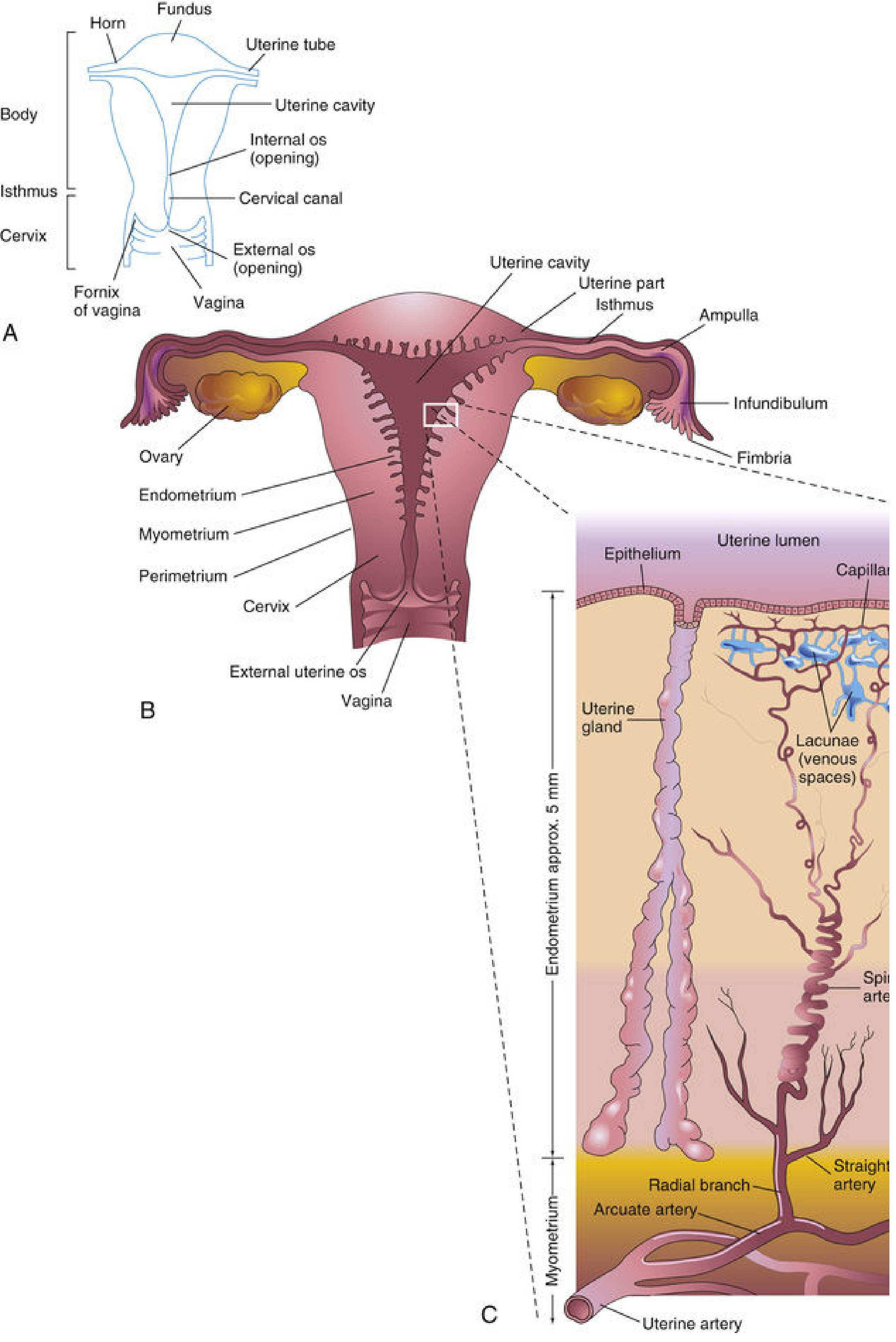
Diagram showing the parts of the uterus, vagina, uterine tubes, and ovaries. The Developing Human: Clinically Oriented Embryology, p. 92.
Classification
1. External genital organs
The external female genitalia are called the vulva. They include:
- Mons pubis
- Labia majora
- Labia minora
- Clitoris
- Vestibule of vagina
- External urethral opening
- Vaginal opening or introitus
- Hymen
- Greater vestibular glands (Bartholin glands)
- Paraurethral glands (Skene glands)
The vulva protects the openings of the urinary and reproductive tracts and contributes to sexual arousal.
2. Internal genital organs
A. Vagina
The vagina is a fibromuscular canal extending from the vulva to the cervix.
- Length: approximately 7-9 cm anteriorly and 9-11 cm posteriorly
- It lies posterior to the urethra and urinary bladder and anterior to the rectum.
- Its upper end surrounds the vaginal part of the cervix, forming anterior, posterior, and two lateral fornices.
- Functions:
- Receives penis and semen during sexual intercourse
- Passage for menstrual flow
- Forms the lower part of the birth canal
B. Uterus
The uterus is a hollow, muscular, pear-shaped organ situated in the pelvis between the urinary bladder and rectum.
Parts
- Fundus: Dome-shaped upper part above the openings of the uterine tubes
- Body: Main central part
- Isthmus: Narrow segment between body and cervix
- Cervix: Lower cylindrical part projecting into the vagina
Normal position
- Usually anteverted and anteflexed:
- The uterus bends forward over the bladder.
- The body is bent forward in relation to the cervix.
Layers of uterine wall
- Perimetrium: Outer serous covering
- Myometrium: Thick smooth-muscle layer, important in labor
- Endometrium: Inner mucosal lining that undergoes cyclical menstrual changes and receives the implanted embryo
Functions
- Menstruation
- Implantation and nourishment of the embryo
- Growth and protection of the fetus
- Expulsion of the fetus during childbirth
C. Uterine tubes
The uterine tubes, also called fallopian tubes or oviducts, extend laterally from the upper part of the uterus toward the ovaries. Each is about 10-12 cm long.
Parts of each uterine tube
- Infundibulum: Funnel-shaped lateral end
- Fimbriae: Finger-like projections that help collect the ovum
- Ampulla: Widest and longest part
- Isthmus: Narrow medial part
- Intramural or uterine part: Part within the uterine wall
Importance: Fertilization usually occurs in the ampulla. NCBI tubal anatomy
D. Ovaries
The ovaries are paired female gonads, almond-shaped and located on the lateral wall of the pelvis.
Functions
- Produce ova
- Secrete hormones, mainly estrogen and progesterone
Attachments
- Ovarian ligament: Connects ovary to uterus
- Suspensory ligament of ovary: Connects ovary to the lateral pelvic wall and contains ovarian vessels
- Mesovarium: Short peritoneal fold attaching the ovary to the broad ligament
Supporting Ligaments of the Uterus
The uterus is supported mainly by pelvic floor muscles and fascial ligaments:
- Pelvic diaphragm, especially levator ani muscles
- Transverse cervical or cardinal ligaments
- Uterosacral ligaments
- Pubocervical ligaments
- Round ligaments of uterus, which help maintain anteversion
- Broad ligaments, which are peritoneal folds but do not provide major mechanical support
Blood Supply
| Organ | Main arterial supply | Venous drainage |
|---|---|---|
| Ovary | Ovarian artery from abdominal aorta | Right ovarian vein to inferior vena cava; left ovarian vein to left renal vein |
| Uterus | Uterine artery, branch of internal iliac artery | Uterine venous plexus to internal iliac veins |
| Vagina | Vaginal artery and branches of uterine artery | Vaginal venous plexus to internal iliac veins |
| Vulva | Internal pudendal artery | Internal pudendal veins |
The uterine artery crosses superior to the ureter near the cervix. This is clinically remembered as: “water under the bridge” - the ureter passes beneath the uterine artery.
Lymphatic Drainage
- Ovaries and uterine tubes: Para-aortic (lumbar) lymph nodes
- Body of uterus: External iliac lymph nodes
- Fundus of uterus: May drain along the round ligament to superficial inguinal nodes
- Cervix: Internal iliac and sacral lymph nodes
- Upper vagina: Internal iliac nodes
- Lower vagina and vulva: Superficial inguinal lymph nodes
Nerve Supply
- Internal reproductive organs receive autonomic fibers through the inferior hypogastric plexus.
- The vulva and lower vagina receive somatic sensation mainly through the pudendal nerve.
- Pain from the upper uterus and ovaries may be referred to lower abdominal or lumbar regions; pain from the lower vagina and vulva is generally more localized.
Applied Anatomy
- Ectopic pregnancy most often occurs in the ampulla of the uterine tube.
- Pelvic inflammatory disease may damage the uterine tubes and cause infertility or ectopic pregnancy.
- The pouch of Douglas or rectouterine pouch is the lowest part of the peritoneal cavity in a standing female and can collect fluid or blood.
- During hysterectomy, the ureter is at risk because it passes close to and beneath the uterine artery.
- Weakness of the pelvic supports can result in uterine prolapse.
The main anatomical components are the vulva externally and the vagina, cervix, uterus, uterine tubes, and ovaries internally, as outlined in Sabiston Textbook of Surgery, “Female Reproductive and Pelvic Anatomy.”
Uterine cancer diagnostic evaluations with purpose
Reading File
Reading File
Finding Sources
Finding Sources
Finding Sources
Searching the Web
NCI endometrial cancer diagnosis tests biopsy transvaginal ultrasound staging
Finding Sources
Searching PubMed
endometrial cancer diagnosis
Reading Web Page
https://www.cancer.gov/types/uterine/hp/endometrial-treatmen…
Uterine Cancer: Diagnostic Evaluations and Their Purpose
Uterine cancer usually refers to endometrial cancer, a cancer arising from the inner lining of the uterus. Less commonly, it may arise from the uterine muscle or connective tissue, called uterine sarcoma. The most frequent warning symptom is abnormal uterine bleeding, particularly bleeding after menopause.
Diagnostic Evaluations
| Evaluation | Purpose |
|---|---|
| History taking | Identifies symptoms and risk factors. Important symptoms include postmenopausal bleeding, irregular heavy bleeding, watery or blood-stained vaginal discharge, and pelvic pain. Risk factors include obesity, diabetes, prolonged estrogen exposure, tamoxifen use, and Lynch syndrome. |
| General and pelvic examination | Evaluates the vulva, vagina, cervix, uterus, and adnexa. It may detect uterine enlargement, a pelvic mass, cervical involvement, vaginal spread, or another source of bleeding. A normal examination does not exclude endometrial cancer. |
| Transvaginal ultrasonography (TVUS) | Uses a vaginal ultrasound probe to examine the uterus and measure endometrial thickness. Its purpose is to assess abnormal bleeding, identify thickened endometrium, polyps, fibroids, or an intrauterine mass, and help decide whether tissue sampling is needed. It supports evaluation but does not confirm cancer. NCI screening guidance |
| Endometrial biopsy or sampling | A thin catheter, commonly a Pipelle device, is passed through the cervix to obtain endometrial tissue. This is the main outpatient test to confirm or exclude malignancy and identify hyperplasia, cancer type, and often tumor grade. A tissue sample is necessary for a definitive diagnosis. NCI diagnostic evaluation |
| Dilation and curettage (D&C) | The cervix is dilated and endometrial tissue is scraped or suctioned. It is used when an office biopsy is inadequate, impossible, or negative despite persistent suspicious bleeding. Its purpose is to obtain a larger tissue sample for histopathology. |
| Hysteroscopy with directed biopsy | A thin telescope is introduced through the cervix to view the uterine cavity directly. It detects focal lesions such as polyps or localized tumors that may be missed by blind biopsy, and allows a targeted biopsy. Berek & Novak’s Gynecology notes that hysteroscopy provides information not obtained by blind endometrial sampling. |
| Histopathological examination | A pathologist examines biopsy or curettage tissue under a microscope. Its purpose is to establish the diagnosis, determine histological type, such as endometrioid, serous, or clear-cell carcinoma, determine tumor grade, and identify features relevant to prognosis and treatment. |
| Cervical cytology (Pap test) | Primarily screens for cervical cancer, not uterine cancer. Its purpose in this context is to detect an abnormal cervix or occasionally reveal abnormal endometrial cells that prompt further endometrial evaluation. It cannot rule out endometrial cancer. |
| MRI of pelvis | Helps assess the local extent of confirmed disease, especially depth of myometrial invasion, cervical stromal involvement, and adjacent pelvic-organ involvement. Its purpose is preoperative staging and surgical planning, not primary confirmation of cancer. |
| CT scan of chest, abdomen, and pelvis | Used especially for high-grade, advanced, or symptomatic disease. It evaluates enlarged lymph nodes, lung, liver, peritoneal, or other distant metastases. |
| PET-CT scan | Not routinely needed for every patient at initial diagnosis. It may be used if recurrent or metastatic disease is suspected, to identify metabolically active lymph-node or distant metastases. |
| Blood tests: CBC, renal and liver function tests | Do not diagnose uterine cancer. Their purpose is to assess anemia caused by bleeding, establish baseline organ function, and determine fitness for imaging, surgery, chemotherapy, or radiotherapy. |
| CA-125 | Not a routine diagnostic test for all endometrial cancers. It may be measured in selected high-risk cases, such as serous carcinoma or suspected extrauterine spread, to support assessment and later monitoring. |
| Molecular testing of tumor tissue | Tests may include mismatch-repair protein immunohistochemistry or microsatellite instability testing, p53 testing, and POLE mutation analysis. Their purpose is to refine prognosis, identify possible Lynch syndrome, classify risk, and guide decisions about adjuvant or systemic therapy. The NCI encourages molecular classification when feasible. NCI treatment reference |
| Surgical staging | After cancer is confirmed, total hysterectomy with removal of both tubes and ovaries is generally performed when appropriate. Sentinel lymph-node biopsy or lymph-node assessment, peritoneal washings in selected cases, and assessment of other tissues determine the true extent of spread and final FIGO stage. |
Key Point
Endometrial biopsy is the definitive diagnostic test.
Ultrasound and imaging can identify suspicious findings and assess spread, but they cannot replace histopathological confirmation from endometrial tissue.
Ultrasound and imaging can identify suspicious findings and assess spread, but they cannot replace histopathological confirmation from endometrial tissue.
Simple Diagnostic Sequence
- Abnormal uterine bleeding, especially postmenopausal bleeding
- History, pelvic examination, and transvaginal ultrasound
- Endometrial biopsy
- Hysteroscopy with directed biopsy or D&C if biopsy is inadequate or suspicion remains
- Histopathology and molecular testing
- MRI, CT, or PET-CT when indicated for staging
- Surgical staging for confirmed operable disease
There is no routine screening test for asymptomatic average-risk women. Evaluation is directed by symptoms, particularly abnormal bleeding. NCI screening overview
This is a shared conversation. Sign in to Orris to start your own chat.