Gustafson’s method / Gustafson’s changes in teeth. (Aug 2011, Feb 2013, Aug 2016, Aug 2022) ★★★★

Reading File
Finding Sources
Finding Sources
Reading File
Reading File
Reading File
Reading File
Reading File
Reading File
Loading Image
Searching Images

Gustafson method age estimation teeth six criteria attrition periodontosis secondary dentine

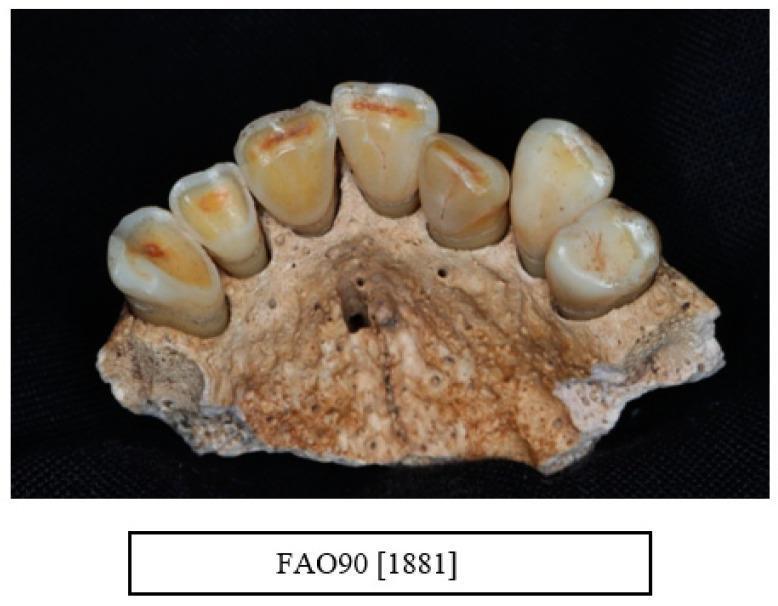
This clinical photograph displays a human dry bone specimen of a maxilla, specifically illustrating advanced attrition and tooth wear patterns in a male aged 36–45 years. The visual focuses on the anterior segment from the right canine (UR3) to the left lateral incisor (UL2). These teeth demonstrate severe wear (Raitapuro-Murray index code 3), characterized by the total loss of occlusal enamel and significant exposure of the underlying secondary dentine, appearing as yellowish-orange central craters on the biting surfaces. The left canine (UL3) shows a relatively less severe wear pattern with a flattened cusp (code 2). The surrounding alveolar process exhibits a mottled, porous texture with visible nutrient foramina and evidence of post-mortem mineralization. This image serves as an educational resource for dental anthropology and forensic odontology, highlighting the progression of chronic mechanical wear and its impact on dental anatomy, including the visible differentiation between enamel and dentine layers and the associated alveolar bone changes.

This clinical photograph displays a human dry bone specimen of a maxilla, specifically illustrating advanced attrition and tooth wear patterns in a male aged 36–45 years. The visual focuses on the anterior segment from the right canine (UR3) to the left lateral incisor (UL2). These teeth demonstrate severe wear (Raitapuro-Murray index code 3), characterized by the total loss of occlusal enamel and significant exposure of the underlying secondary dentine, appearing as yellowish-orange central craters on the biting surfaces. The left canine (UL3) shows a relatively less severe wear pattern with a flattened cusp (code 2). The surrounding alveolar process exhibits a mottled, porous texture with visible nutrient foramina and evidence of post-mortem mineralization. This image serves as an educational resource for dental anthropology and forensic odontology, highlighting the progression of chronic mechanical wear and its impact on dental anatomy, including the visible differentiation between enamel and dentine layers and the associated alveolar bone changes.

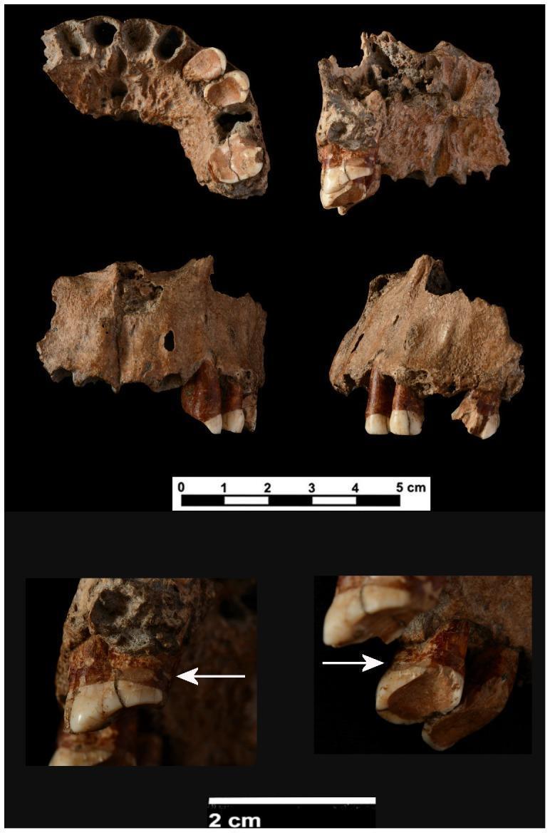
Anatomical and clinical photograph of a fragmented human maxilla featuring multiple perspectives: inferior, internal, frontal, and lateral. The specimen includes the alveolar process from the right canine to the second left molar, with the left canine (C), third premolar (Pm3), and first molar (M1) still in situ. The teeth exhibit severe dental attrition (Stage 7-8), characterized by extensive enamel loss and exposed secondary dentine on the occlusal surfaces. Notable pathology includes evidence of periodontal disease, specifically interproximal grooves located below the cervicoenamel junction on the distal surfaces of the left Pm3 and M1, highlighted by white arrows in detailed macro views. The M1 shows a significant buccal fracture with associated dentine loss. Anatomical landmarks present include the anterior nasal spine, the inferior nasal border, and the nasoalveolar clivus. The image serves as a clinical study of archaic dental health, age-related wear patterns, and antemortem dental modifications or pathologies.

Anatomical and clinical photograph of a fragmented human maxilla featuring multiple perspectives: inferior, internal, frontal, and lateral. The specimen includes the alveolar process from the right canine to the second left molar, with the left canine (C), third premolar (Pm3), and first molar (M1) still in situ. The teeth exhibit severe dental attrition (Stage 7-8), characterized by extensive enamel loss and exposed secondary dentine on the occlusal surfaces. Notable pathology includes evidence of periodontal disease, specifically interproximal grooves located below the cervicoenamel junction on the distal surfaces of the left Pm3 and M1, highlighted by white arrows in detailed macro views. The M1 shows a significant buccal fracture with associated dentine loss. Anatomical landmarks present include the anterior nasal spine, the inferior nasal border, and the nasoalveolar clivus. The image serves as a clinical study of archaic dental health, age-related wear patterns, and antemortem dental modifications or pathologies.

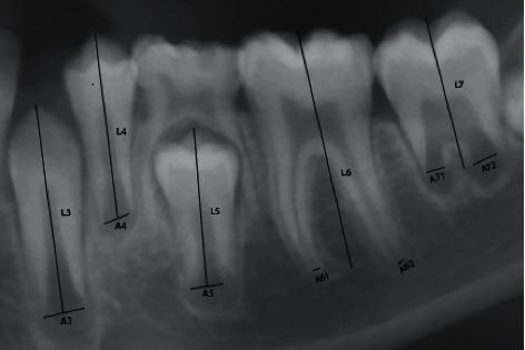
This diagnostic radiograph (panoramic detail) illustrates the Cameriere method for dental age estimation by measuring open apices in a developing permanent dentition. The image shows seven left mandibular teeth (canine through second molar) with superimposed measurement lines. For each tooth, the total length (Li) is measured from the incisal or occlusal edge to the apex. For single-rooted teeth (A3-A5), the width of the open apex (Ai) is measured as the distance between the inner walls of the root tip. For double-rooted molars (A6-A7), the apical width is the sum of measurements from both the mesial and distal roots (e.g., A71 + A72). This methodology uses the ratio xi = Ai/Li to normalize data against radiographic magnification and angulation variations. The presence of wide, radiolucent open apices indicates incomplete root development, a key clinical marker used in forensic odontology and pediatric dentistry to determine chronological age based on dental maturity stages.

This diagnostic radiograph (panoramic detail) illustrates the Cameriere method for dental age estimation by measuring open apices in a developing permanent dentition. The image shows seven left mandibular teeth (canine through second molar) with superimposed measurement lines. For each tooth, the total length (Li) is measured from the incisal or occlusal edge to the apex. For single-rooted teeth (A3-A5), the width of the open apex (Ai) is measured as the distance between the inner walls of the root tip. For double-rooted molars (A6-A7), the apical width is the sum of measurements from both the mesial and distal roots (e.g., A71 + A72). This methodology uses the ratio xi = Ai/Li to normalize data against radiographic magnification and angulation variations. The presence of wide, radiolucent open apices indicates incomplete root development, a key clinical marker used in forensic odontology and pediatric dentistry to determine chronological age based on dental maturity stages.

Reading File
I now have all the information needed. Let me compose the comprehensive answer.

Gustafson's Method / Gustafson's Changes in Teeth

Introduction

Gustafson's method (proposed by Gosta Gustafson in 1950, published in J Am Dent Assoc 41:45-54) is an objective, semi-quantitative forensic technique for estimating the age of an adult individual from teeth. It is applicable only in deceased individuals (cadavers/skeletal remains), since the procedure is destructive. It is useful for estimating age between 25 and 60 years, and is not applicable in persons below 15 years (changes are absent at that age).
The method involves microscopic examination of a longitudinal section of a tooth, assessing six progressive age-related changes.

The Six Criteria (Mnemonic: APSRTC)

The six changes are remembered by the mnemonic A P S R T C (like the name of a bus company!):
LetterCriterionWhat Happens with Age
AAttritionWearing of the occlusal surface
PPeriodontosisRegression of gums/periodontal tissue
SSecondary dentineDeposition in pulp cavity
RRoot resorptionResorption of root apex
TTransparency of rootTranslucency due to rarefaction of dentine
CCementum appositionIncreased cementum deposition on root surface

Detailed Description of Each Criterion (Scored 0 to 3)

1. Attrition (A) - Wearing of the Biting Surface

Progressive wearing of the occlusal (mastication) surface due to continuous friction between opposing teeth.
GradeDescription
A0No attrition
A1Attrition confined to enamel only
A2Attrition extends into dentine
A3Attrition exposes the soft pulp

2. Periodontosis (P) - Gum and Periodontal Regression

Regression of the gums and periodontal tissues surrounding the teeth, with gradual exposure of the root. Poor oral hygiene accelerates this. Hardened debris may also deposit over time.
GradeDescription
P0No periodontosis / no root exposure
P1Exposure of less than 1/3rd of the root (from crown)
P2Exposure of more than 1/3rd but less than 2/3rd of root
P3Periodontosis beyond 2/3rd of the root length near the crown
Note: The degree of periodontosis must be assessed before the tooth is extracted from the body.

3. Secondary Dentine (S) - Infilling of Pulp Cavity

With advancing age, secondary dentine is deposited within the pulp cavity, gradually reducing its size. In mandibular teeth, it starts from above (pulp chamber); in maxillary teeth, it starts from below. Also caused by pathological conditions (caries, periodontosis).
GradeDescription
S0No secondary dentine deposition
S1Deposition in the upper part of pulp cavity only
S2Deposition in the upper half of the pulp cavity
S3Almost the whole pulp cavity is filled

4. Root Resorption (R) - Decay of Root Apex

A destructive process involving both cementum and dentine. Characteristically shows sharp grooves. Begins at the apex and extends upward. Usually occurs in late age; may be due to pathological processes.
GradeDescription
R0No root resorption
R1Resorption seen only in some (small) parts
R2Seen over a large area
R3Involves both cementum and dentine extensively

5. Transparency of Root (T) - Most Reliable Criterion

Not seen until about 30 years of age. Occurs due to rarefaction (osteoporosis/mineralization) of dentinal tubules, which fills the wide canals with minerals, making the dentine translucent.
  • In the lower jaw: transparency progresses from below upwards (apex upward)
  • In the upper jaw: transparency progresses from above downwards
This is the most reliable of all six criteria for age estimation.
GradeDescription
T0No transparency anywhere
T1Transparency mostly in the apical region
T2Transparency up to 1/3rd from the apical region
T3Transparency up to 2/3rd of root length from apex

6. Cementum Apposition (C) - Increased Cementum

Cementum deposition increases with age due to changes in tooth position and surface near the root apex. It forms visible incremental lines.
GradeDescription
C0Only the normal layer of cementum is present
C1Slightly greater than normal
C2A thick layer has occurred
C3A very heavy layer is present

The Gustafson's Method Diagram

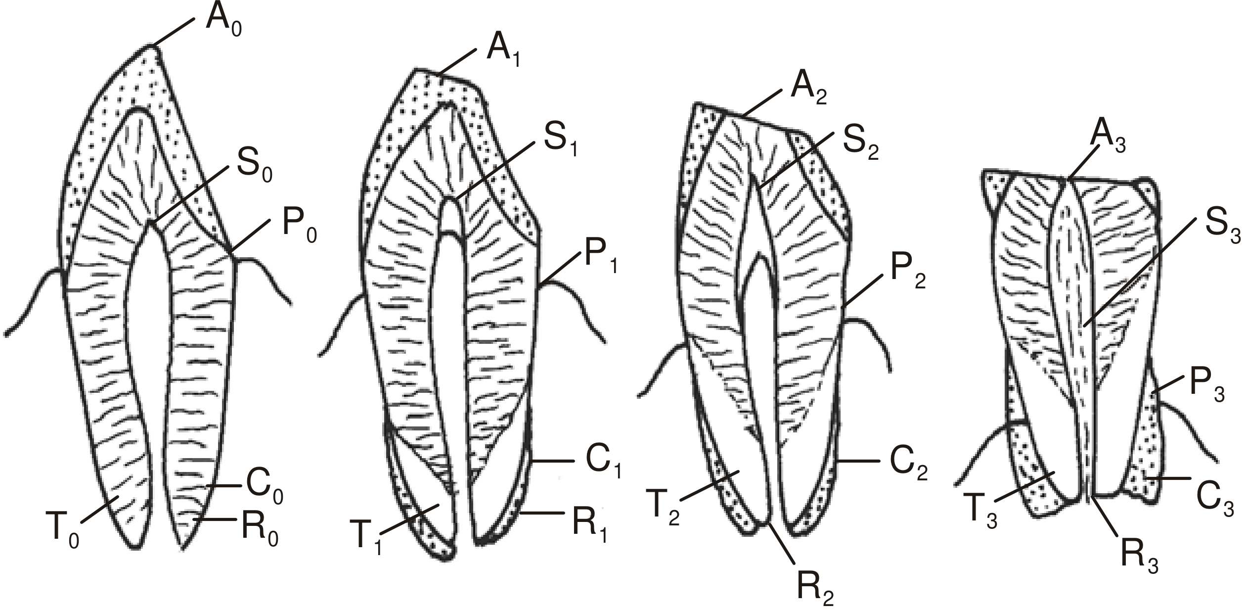
Here is the classic diagram showing all four grades (0-3) for each criterion side by side in longitudinal tooth sections:
Gustafson's Method - Six criteria graded 0-3 in tooth longitudinal sections
The four tooth diagrams show progressive grades 0 to 3. A = Attrition, S = Secondary dentine, P = Periodontosis, C = Cementum, T = Transparency, R = Root resorption.

Procedure

  1. Before extraction: Note and score the degree of periodontosis (P) while the tooth is still in the jaw.
  2. Extract the tooth from the jaw.
  3. Gross examination by naked eye: assess attrition, root resorption, cementum apposition.
  4. Grind down the tooth on a glass slab to about 1 mm thickness - this allows assessment of root transparency.
  5. Further grind to 0.25 mm (1/4 mm) for microscopic examination of secondary dentine, etc.

Scoring and Age Estimation Formula

Each of the six criteria is graded 0, 1, 2, or 3.
Total Score = An + Pn + Sn + Rn + Tn + Cn
(Maximum possible score = 18; minimum = 0)
The total score is then plotted on a regression graph comparing point values to teeth of accurately known age, with a regression line drawn. Age is read off the graph.
Gustafson's regression equation:
Age (years) = 11.43 + 4.56 × (Total points)
  • The error margin is ± 3.5 years (some sources quote ± 4 to 7 years, with the error increasing above 50 years of age).
  • Scoring is relative: 0 = no change; 1 = beginning of change; 2 = obvious change; 3 = maximum change.

Important Points to Remember

FeatureDetails
Applicable age range25-60 years (adults after dental development is complete)
Changes absent below15 years of age
Most reliable criterionTransparency of root (T)
Best teeth to useAnterior teeth (incisors > canines > premolars)
Worst tooth for this methodThird molar (quite unsuitable)
Nature of examinationDestructive - applicable only in dead bodies
Error of estimation± 3.5 years (increases after age 50)
Published byGosta Gustafson, 1950

Modifications and Related Methods

  • Johanson's modification of Gustafson's method is considered more accurate than the original.
  • Boyde's method: Uses incremental cross-striations in enamel (daily growth lines counted from the neonatal line) - most accurate for estimating age of dead infants.
  • Stack's method: Uses weight and height of erupting teeth to estimate age in infants/children.
  • Miles' method (1963): Specifically uses root transparency; the tooth is ground to 1 mm, placed on a dotted paper, and the percentage of visible dots through the transparent root is calculated for age estimation.

Sources:
  • The Essentials of Forensic Medicine and Toxicology, 36th Edition (Nagesh Kumar & Siddham), p. 82-83
  • P.C. Dikshit Textbook of Forensic Medicine and Toxicology, p. 68-69
  • Parikh's Textbook of Medical Jurisprudence, Forensic Medicine and Toxicology, p. 88

Write a short note for university exam

Gustafson's Method of Age Estimation from Teeth

Introduction

Gustafson (1950) described an objective method for estimating age in adults by assessing six progressive morphological changes that occur in teeth with advancing age. It is applicable only on deceased individuals, useful for ages 25-60 years, and the changes are absent below 15 years.

The Six Criteria — Mnemonic: APSRTC

Each criterion is graded on a scale of 0 to 3 (0 = absent; 1 = beginning; 2 = obvious; 3 = maximum).
#CriterionChange with AgeGrades
AAttritionWearing of occlusal surface; enamel → dentine → pulpA0–A3
PPeriodontosisGum regression exposing rootP0–P3
SSecondary DentineInfilling of pulp cavityS0–S3
RRoot ResorptionResorption starting at apex, moving upwardR0–R3
TTransparency of RootRarefaction of dentinal tubules; apex upward (lower jaw)T0–T3
CCementum AppositionThickening of cementum near root apexC0–C3
Transparency of root is the most reliable criterion.

Procedure

  1. Note periodontosis before extracting the tooth.
  2. Extract the tooth and examine by naked eye.
  3. Grind tooth to 1 mm - assess transparency.
  4. Grind further to 0.25 mm - microscopic examination.

Scoring and Formula

Total points = A + P + S + R + T + C (max = 18)
Gustafson's formula:
Age = 11.43 + 4.56 × (Total points)
The points are plotted on a regression graph; age is read off the line.
Error of estimation: ± 3.5 years (increases above 50 years).

Key Points

  • Best teeth: Anterior teeth (incisors most suitable; third molar least suitable)
  • Only on dead bodies (method is destructive)
  • Johanson's modification is considered more accurate
  • Boyde's method (incremental enamel lines) is used for infants

Source: Gustafson G. Age determination on teeth. J Am Dent Assoc 1950;41:45-54.

/visualizelearning

Generating Image

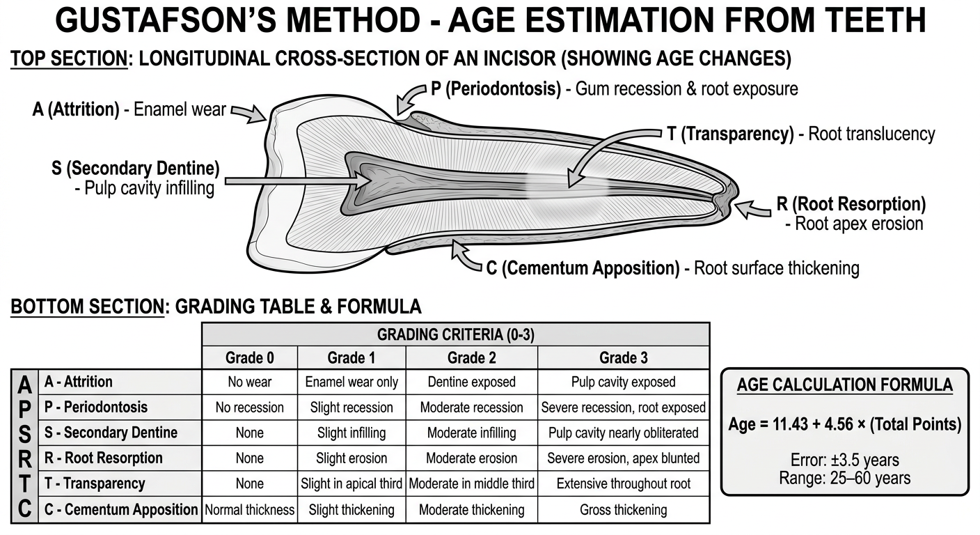
A clear, detailed medical education diagram titled "Gustafson's Method - Age Estimation from Teeth" for forensic medicine university exam study. The diagram should have two main sections: TOP SECTION: A large annotated longitudinal cross-section of a tooth (incisor) showing all 6 age changes labeled with arrows: - A (Attrition) - at the top/biting surface showing enamel wear - P (Periodontosis) - at the gum line showing gum recession and root exposure - S (Secondary Dentine) - inside the pulp cavity showing infilling - R (Root Resorption) - at the root apex showing erosion - T (Transparency) - in the root area showing translucency - C (Cementum Apposition) - on the root surface showing thickening BOTTOM SECTION: A grading table showing all 6 criteria with grades 0 to 3 in a clean grid layout. Include the mnemonic "APSRTC" prominently. Also include a small box showing the formula: Age = 11.43 + 4.56 × (Total Points), with "Error: ±3.5 years" and "Range: 25–60 years". Clean white background, black and labeled anatomy, bold headings, medical textbook style, high detail.

Gustafson's Method - Visual Learning Diagram
Here's your visual summary of Gustafson's Method. The diagram covers:
  • Annotated tooth cross-section showing where each of the 6 changes occurs
  • Mnemonic APSRTC with all six criteria
  • Grading scale 0-3 for each criterion
  • The age formula (Age = 11.43 + 4.56 × Total Points)
  • Key facts - applicable range, error margin, and most reliable criterion (root transparency)
Save this for quick revision before your exam!
This is a shared conversation. Sign in to Orris to start your own chat.